. 2015 Aug 24;212(9):1391-403.
doi: 10.1084/jem.20110575. Epub 2015 Jul 27.

CD68 acts as a major gateway for malaria sporozoite liver infection

Affiliations

CD68 acts as a major gateway for malaria sporozoite liver infection

Sung-Jae Cha et al. J Exp Med. .

Abstract

After being delivered by the bite from an infected mosquito, Plasmodium sporozoites enter the blood circulation and infect the liver. Previous evidence suggests that Kupffer cells, a macrophage-like component of the liver blood vessel lining, are traversed by sporozoites to initiate liver invasion. However, the molecular determinants of sporozoite-Kupffer cell interactions are unknown. Understanding the molecular basis for this specific recognition may lead to novel therapeutic strategies to control malaria. Using a phage display library screen, we identified a peptide, P39, that strongly binds to the Kupffer cell surface and, importantly, inhibits sporozoite Kupffer cell entry. Furthermore, we determined that P39 binds to CD68, a putative receptor for sporozoite invasion of Kupffer cells that acts as a gateway for malaria infection of the liver.


Figures

Figure 1.
Figure 1.
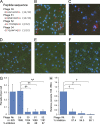
Selected phages inhibit sporozoite entry into Kupffer cells and mouse liver. (A) Predicted amino acid sequence of peptides displayed by phages selected for binding to Kupffer cells. Frequency of each of the four recovered peptides among the 32 phages sequenced is denoted in parenthesis. Asterisks indicate a stop codon. All peptides from this library have a cysteine residue (red) at positions 2 and 11. (B–F) Non-selected library phages (B), PBS buffer only (C), phage 39 (D), phage 61 (E), or phage 52 (F) was added to primary rat Kupffer cell cultures, followed by addition of P. berghei sporozoites. After 2-h incubation at 37°C, the cells were fixed, permeabilized, and then incubated with an anti-CSP antibody (red) and an anti-M13 phage antibody (green). Nuclei were stained with DAPI (blue). The anti-CSP antibody detects both attached and invaded sporozoites. Bar, 100 µm. (G) The mean number of sporozoites per cell was measured in 10 fields/well under 200-fold magnification. Data from three independent replicate experiments were combined. “n” indicates the number of cells assayed. The percent inhibition relative to the unselected library phage control group (Lib) is displayed. “No” indicates no phage (PBS buffer control). (H) Each selected phage (compare with A) or wild-type phage (Wt) was injected intravenously (1010 phages per mouse) into three mice, followed 5 min later by injection of 2 × 10⁴ P. berghei sporozoites. Parasite load in livers was determined 42 h later, and percent inhibition relative to the wild-type phage–treated group is displayed. Data from two replicate experiments were pooled, and each bar represents the mean of six mice. (G and H) P-values (*, P < 0.05; **, P < 0.01) were calculated using the one-way ANOVA test. Error bars indicate standard deviation.
Figure 2.
Figure 2.
GAG removal does not affect phage inhibition of Kupffer cell entry. (A and B) Primary rat Kupffer cells were either left untreated or treated with the enzymes indicated in the bottom of the figure. This was followed by the sequential addition of the selected phages and P. berghei sporozoites. PBS buffer or wild-type phages (Wt) were added as controls. After 2-h incubation at 37°C, unbound sporozoites were washed out and Kupffer cells were fixed for sporozoite counting. The number of attached (A) and intracellular (B) parasites per 20 microscopic fields/well at 400-fold magnification was determined separately. Data from three independent experiments were combined. The percent inhibition relative to the wild-type phage–treated group is displayed at the bottom of each panel. P-values (*, P < 0.05; **, P < 0.01; ***, P < 0.001) were calculated using the one-way ANOVA test. Error bars indicate standard deviation.
Figure 3.
Figure 3.
The P39 peptide binds to a macrophage-specific surface protein. (A) Primary Kupffer cells were incubated with heparinase I, chondroitinase ABC, or trypsin, as indicated. PBS buffer (no enzyme) served as a control. After enzyme treatment, 100 µg/ml biotinylated P39 peptide (green) and the anti-F4/80 antibody (red) were incubated for 1 h and visualized. The top row shows differential interference contrast (DIC) microscopy images merged with DAPI-stained nuclei (blue). P39 peptide binding was trypsin sensitive and unaffected by GAG-removing enzyme treatment. Bar, 20 µm. (B) Lysates from five different cell types were fractionated by gel electrophoresis, and binding of biotinylated P39 peptide to proteins on the blot was detected using alkaline phosphatase–conjugated streptavidin. KC, primary rat Kupffer cells; PtM, primary rat peritoneal macrophages; Raw, a mouse macrophage-like cell line; Hep, primary rat hepatocytes; ASM, primary rat airway smooth muscle cells. P39 peptide binding to an ∼110-kD protein band (arrows) was detected only for the macrophage-like cells (rat KC, rat PtM, and mouse Raw). (C) Kupffer cell fractions were tested for P39 peptide binding. M, membrane fraction; C, cytosolic fraction; I, insoluble fraction. P39 peptide bound only to an ∼110-kD membrane protein (arrow). (D) Activation of the human monocyte cell line THP-1 with PMA treatment for differentiation into a macrophage-like cell resulted in production of a protein recognized by the P39 peptide. No binding was detected for control THP-1 cells treated with DMSO carrier alone. An anti-actin antibody was used as a loading control. Immunofluorescence and far-Western blotting images are representative of two to three independent experiments.
Figure 4.
Figure 4.
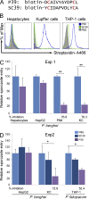
Specificity of P39 peptide binding and inhibition of sporozoite entry. (A) A scrambled peptide with the same amino acid composition as P39 (SC39) was designed to serve as a negative control. All peptides have a cysteine residue (red) at positions 2 and 11. (B) The cell type indicated at the top of each panel was incubated with buffer alone (PBS), with a biotinylated P39 peptide (P39), or with a biotinylated scrambled peptide (SC39), followed by incubation with fluorescent streptavidin and analysis by flow cytometry. The P39 peptide bound to macrophage-like cells (Kupffer, PMA-activated THP-1) but not to hepatocytes (Hep). The scrambled peptide did not bind appreciably to any of the cell types. All images are representative of two to three independent experiments. (C and D) Inhibition of sporozoite entry in vitro. (C) Each cell type was incubated with PBS alone or with the P39 peptide, before exposure to 2 × 10⁴ P. berghei sporozoites. The mean number of internalized sporozoites per 20 microscopic fields/well under 400-fold magnification was determined, and the percent inhibition relative to the PBS-treated group is shown at the bottom of the figure. (D) The SC39 peptide was tested for inhibition of P. berghei and P. falciparum sporozoite entry into each cell type. P39 peptide and PBS treatments were used as positive and negative controls, respectively. (C and D) Data from three independent biological experiments were combined. P-values (*, P < 0.05; **, P < 0.01) were calculated using the one-way ANOVA test. Error bars indicate standard deviation.
Figure 5.
Figure 5.
The P39 peptide binds to ∼110–120-kD Kupffer cell membrane proteins. (A) Kupffer cell membrane proteins were fractionated by 5% SDS-PAGE and either used for far-Western blotting with the biotinylated P39 peptide (left) or stained with silver (right). P39 bound to a broad band of ∼110–120 kD, suggesting that it may bind to a glycosylated protein. Size markers were fractionated in the left lanes. (B) The P39 peptide–binding area (left, arrow) was excised in three pieces from a Coomassie blue–stained gel of rat Kupffer cell membrane fraction (numbered in red on the right). Each piece was analyzed by mass spectrometry, and the identified proteins are listed below. Two candidate glycoproteins that were identified in two fractions, gp96 and CD68, are highlighted in bold.
Figure 6.
Figure 6.
The P39 peptide binds to CD68, a molecule that mediates sporozoite entry into Kupffer cells. (A) qRT-PCR assays of primary rat Kupffer cells (KC), primary rat hepatocytes (Hep), primary rat airway smooth muscle cells (ASM), and a monkey kidney cell line (Cos7) show that CD68 is strongly expressed in Kupffer cells, whereas gp96 is not. Data were pooled from three experiments. (B, top) CD68 is a macrophage-specific membrane protein. Western blots of whole cell lysates were incubated either with anti-gp96 antibody or anti-CD68 antibody. (bottom) Western blots of membrane fractions were incubated with an anti-CD68 antibody or with the biotinylated P39 peptide. PtM, rat primary peritoneal macrophages; RAW, mouse macrophage-like cell line; α-rCD68, rat anti-CD68 antibody; α-mCD68, mouse anti-CD68 antibody. (C) CD68 siRNA treatment of rat Kupffer cells for 6 d inhibits CD68 protein expression as well as P39 peptide binding activity. (left) Relative CD68 mRNA expression in Kupffer cell culture was determined by qRT-PCR after the number of days of siRNA treatment indicated at the bottom. Data were combined from three independent experiments. (right) Western blot of membrane fraction probed with an anti-CD68 antibody or with biotinylated P39 peptide as indicated. An anti-actin antibody served to control loading. siCont, control siRNA of sequence unrelated to that of CD68. (A and C) Error bars indicate standard deviation. (D) The 293T cell line was engineered to express rat CD68 protein lacking the C-terminal transmembrane domain for secretion into the culture supernatant. Culture supernatant of control (not transfected) 293T cells and 293T cells that secrete rat CD68 was tested for binding of anti-CD68 antibody, biotinylated P39 peptide, or scrambled peptide. Residual BSA that remained after cells were washed with serum-free medium was detected by Ponceau staining. (E) Western blots of a rat Kupffer cell membrane fraction treated either with N-glycosidase or with O-glycosidase were incubated with anti-CD68 antibody or P39 peptide, as indicated. CD68 mobility was sensitive to N-glycosidase but not to O-glycosidase treatment. Images represent two independent experiments. (F) Purified P. berghei sporozoites were incubated with supernatant from either control 293T cells or from 293T cells engineered to secrete rat CD68 (compare with D). CD68 binding to sporozoites was visualized with an anti–rat CD68 antibody. Immunofluorescence images are representative of two independent experiments with five replicates each, and flow cytometry of sporozoites incubated with control or CD68-containing supernatant is representative of three independent experiments. DIC, differential interference contrast. Bar, 10 µm.
Figure 7.
Figure 7.
Plasmodium sporozoites preferentially enter CD68-expressing cells. (A) Primary rat Kupffer cells were incubated with CD68 siRNA to inhibit CD68 expression (compare with Fig. 6 C). 6 d after siRNA treatment, P. berghei sporozoites were added to either CD68 siRNA– or control siRNA–treated Kupffer cells. Parasite entry into host cells was determined by flow cytometry. Sporozoite-positive cells decreased from 19.4% in the control Kupffer cell population (Cont siRNA) to 7.63% in the CD68 siRNA–treated cells. CD68 was detected with an anti-CD68 antibody, whereas sporozoites were detected with an anti-CSP antibody. (B) A Cos7 monkey kidney cell line (Cos7-Cont) was engineered to express rat CD68 on its surface (Cos7-rCD68) and tested for P. berghei sporozoite entry by flow cytometry. Sporozoite-positive cells increased from 1.91% in the control population to 2.23% in the transfected Cos7 population. (C) A mouse Raw macrophage-like cell line (Raw-Cont) was engineered to express rat CD68 (Raw-rCD68) and tested for P. berghei sporozoite entry by flow cytometry. (D) A human monocyte THP-1 cell line was treated with 0.1 µM PMA to induce differentiation into macrophage-like cells and tested for P. falciparum sporozoite entry by flow cytometry. (E) Anti-CD68 antibody inhibits sporozoite entry into Kupffer cells and other CD68-expressing macrophages. Rat Kupffer cells (KC), mouse peritoneal macrophages (PtM), or activated THP-1 cells were preincubated with anti-CD68 antibody or control antibody as indicated. Relative sporozoite entry into host cell was determined at 2 h after incubation with 2 × 10⁴ sporozoites per well. The percent inhibition of sporozoite entry compared with the control group (No antibody) is indicated. Data are from two independent experiments. *, P < 0.05; **, P < 0.01 (one-way ANOVA test). Error bars indicate standard deviation.
Figure 8.
Figure 8.
Characterization of wild-type and CD68 KO mice and their macrophages. (A) The genotype of the CD68 KO mice was confirmed using PCR with CD68-specific primers and mouse genomic DNA. +/+, wild-type mouse; +/−, heterozygous mouse; −/−, homozygous KO mouse. PCR with GAPDH primers served as a positive control. (B) Western blot of membrane proteins either stained (left) or incubated with an anti-CD68 antibody (right). Primary hepatocytes (Hep) served as a negative control. Arrow indicates CD68 signal in the +/+ lane. (C) Immunofluorescence assays of peritoneal macrophages from both wild-type and CD68 KO mice for anti-F4/80 expression. DIC, differential interference contrast. Bar, 20 µm. (D) Phagocytic activity of macrophages from CD68 KO mice. Flow cytometry results are shown for peritoneal macrophages incubated with two different fluorescent latex bead concentrations (Exp. 1 and Exp. 2 as indicated on the right of the figure). (E) KO macrophage recognition of the P39 peptide. Peritoneal macrophages from wild-type and CD68 KO mice were stained with the F4/80 antibody (recognizes macrophage-like cells) or an anti-CD68 antibody as indicated (top). The two cell types were also tested for uptake of the P39 peptide using the scrambled version, SC39, as a control (bottom). After incubation with the biotinylated peptides, cells were washed, fixed, and permeabilized for detection of intracellular peptide with Alexa Fluor 488–conjugated streptavidin. Data represent two independent experiments.
Figure 9.
Figure 9.
P. berghei sporozoite infection of CD68 KO mice is strongly impaired. (A) Primary cultures of peritoneal macrophages from wild-type mice (+/+) or from CD68 KO mice (−/−) were established in 8-well chamber slides, followed by incubation for 1 h with 2 × 10⁴ P. berghei sporozoites per well. Parasite entry was determined as described previously (Pradel and Frevert, 2001). The graph shows the mean number of sporozoites per 20 fields/well under 400-fold magnification. Data are from two independent experiments, with 4 wells per experiment. A total of 8 wells (160 microscopic fields) were assayed for each genotype in the two experiments. (B) A total of 2 × 10³ sporozoites were injected intravenously either into wild-type or CD68 KO mice. After 45 h, mice were sacrificed and liver parasite burden was determined by qRT-PCR. Data from three independent experiments were combined. (C) 2 × 10³ P. berghei–infected red blood cells were injected intravenously either into wild-type mice or CD68 KO mice, and parasitemia was determined after 6 d. Data from two experiments were combined. (B and C) The total number of mice assayed is listed in parentheses under the genotype. (D) Wild-type (+/+) and CD68 KO mice (−/−) were injected either with clodronate liposomes (CLD) or with control liposomes produced with PBS, and after 2 d, 2 × 10³ sporozoites were injected intravenously. 45 h after sporozoite injection, mice were sacrificed and the liver parasite burden was determined using qRT-PCR. The data from three independent experiments were combined. The total number of mice used for each type of assay is given in parentheses. (A–D) *, P < 0.05; **, P < 0.01. (E) Kupffer cell depletion by clodronate treatment was measured by relative expression of the F4/80 gene in the liver. Pearson correlation efficiency between parasite liver burden and Kupffer cell depletion indicates that Kupffer cells in CD68 KO mice act as an invasion barrier, whereas this is not the case for wild-type mice. Open circles in B–D represent out-group data points. The data from three independent experiments were combined.

Comment in

References

    1. Auwerx J. 1991. The human leukemia cell line, THP-1: a multifacetted model for the study of monocyte-macrophage differentiation. Experientia. 47:22–31. 10.1007/BF02041244 - DOI - PubMed
    1. Baer K., Roosevelt M., Clarkson A.B. Jr, van Rooijen N., Schnieder T., and Frevert U.. 2007. Kupffer cells are obligatory for Plasmodium yoelii sporozoite infection of the liver. Cell. Microbiol. 9:397–412. 10.1111/j.1462-5822.2006.00798.x - DOI - PubMed
    1. Bonnycastle L.L., Mehroke J.S., Rashed M., Gong X., and Scott J.K.. 1996. Probing the basis of antibody reactivity with a panel of constrained peptide libraries displayed by filamentous phage. J. Mol. Biol. 258:747–762. 10.1006/jmbi.1996.0284 - DOI - PubMed
    1. Bruña-Romero O., Hafalla J.C., González-Aseguinolaza G., Sano G., Tsuji M., and Zavala F.. 2001. Detection of malaria liver-stages in mice infected through the bite of a single Anopheles mosquito using a highly sensitive real-time PCR. Int. J. Parasitol. 31:1499–1502. 10.1016/S0020-7519(01)00265-X - DOI - PubMed
    1. Bykov I., Ylipaasto P., Eerola L., and Lindros K.O.. 2004. Functional differences between periportal and perivenous Kupffer cells isolated by digitonin-collagenase perfusion. Comp. Hepatol. 3:S34 10.1186/1476-5926-2-S1-S34 - DOI - PMC - PubMed

Publication types

MeSH terms

Associated data