. 2015 Aug;63(8):543-58.
doi: 10.1369/0022155415589119.

A Historical Perspective on the Identification of Cell Types in Pancreatic Islets of Langerhans by Staining and Histochemical Techniques

Affiliations

A Historical Perspective on the Identification of Cell Types in Pancreatic Islets of Langerhans by Staining and Histochemical Techniques

Denis G Baskin. J Histochem Cytochem. 2015 Aug.

Abstract

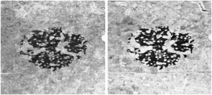
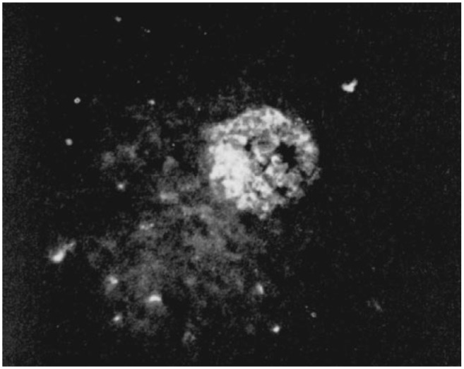
Before the middle of the previous century, cell types of the pancreatic islets of Langerhans were identified primarily on the basis of their color reactions with histological dyes. At that time, the chemical basis for the staining properties of islet cells in relation to the identity, chemistry and structure of their hormones was not fully understood. Nevertheless, the definitive islet cell types that secrete glucagon, insulin, and somatostatin (A, B, and D cells, respectively) could reliably be differentiated from each other with staining protocols that involved variations of one or more tinctorial techniques, such as the Mallory-Heidenhain azan trichrome, chromium hematoxylin and phloxine, aldehyde fuchsin, and silver impregnation methods, which were popularly used until supplanted by immunohistochemical techniques. Before antibody-based staining methods, the most bona fide histochemical techniques for the identification of islet B cells were based on the detection of sulfhydryl and disulfide groups of insulin. The application of the classical islet tinctorial staining methods for pathophysiological studies and physiological experiments was fundamental to our understanding of islet architecture and the physiological roles of A and B cells in glucose regulation and diabetes.

Keywords: beta cells; diabetes; glucagon; immunocytochemistry; immunohistochemistry; insulin; islet cells; pancreas; somatostatin; staining.


Conflict of interest statement

Competing Interests: The author declared no potential competing interests with respect to the research, authorship, and/or publication of this article.

Figures

Figure 1.
Figure 1.
Paul Langerhans 1878.
Figure 2.
Figure 2.
Hand-drawn figures from “The cytological characters of the areas of Langerhans,” by Lane, American Journal of Anatomy, Volume 7, Issue 3, Pages 409–422, 10 November 1907, illustrating guinea pig islets stained with Bensley’s neutral gentian following different fixations. Panel A represents an islet from a pancreas fixed in 70% alcohol. The islet A cells have a violet-stained cytoplasm, whereas the islet B cells are essentially unstained. Panel B represents an islet from a pancreas fixed in aqueous chrome-sublimate (no alcohol) and shows islet B cells filled with minute granules that stained violet, whereas the islet A cells are stained light orange. In panel B, some pancreatic acinar cells with clumps of violet-stained granules are depicted at the edge of the islet. Copyright John Wiley and Sons. Published with permission.
Figure 3.
Figure 3.
A drawing by Bloom from “A new type of granular cell in the islets of Langerhans of man,” from The Anatomical Record: Advances in Integrative Anatomy and Evolutionary Biology, 1931, illustrating a human islet stained with the Mallory-Heidenhain azan trichrome technique. A cells (A) are stained reddish brown, D cells (D) are blue, and B cells (B) are stained faintly orange. The figure shows pancreatic acinar cells (Pac) and reticular fibers (Ret). Copyright John Wiley and Sons. Published with permission.
Figure 4.
Figure 4.
George Gomori. Image courtesy Special Collections Research Center, University of Chicago Library. Published with permission.
Figure 5.
Figure 5.
Rat islet stained with Gomori’s aldehyde fuchsin after oxidation in periodic acid. Image from “A comparison of the staining affinities of aldehyde fuchsin and the Schiff reagent,” by Scott and Clayton, Journal of Histochemistry and Cytochemistry, 1953. Copyright Histochemical Society. Published with permission.
Figure 6.
Figure 6.
Rat islets stained with Gomori’s aldehyde fuchsin method. Top panel is an islet from a control rat, showing intensely stained granulated B cells. Bottom panel shows an islet from rat that was treated with a sulfonylurea to stimulate insulin secretion, showing depletion of staining. Image from “A portrait of the pancreatic B cell,” by Orci, Diabetologia, 1974. Published with kind permission kind permission from Springer Science and Business Media.
Figure 7.
Figure 7.
Reproduction of Figure 1 from “Staining of insulin with aldehyde fuchsin,” by Kvistberg, Lester, and Lazarow, Journal of Histochemistry and Cytochemistry, 1966, demonstrating staining of gels after disk electrophoresis. (A) Beef insulin stained with a dye for protein (aniline blue black) immediately after gel electrophoresis. (B) Beef insulin stained with Gomori’s aldehyde fuchsin after gel was oxidized with KMnO4-H2SO4. The aldehyde fuchsin stained the same band that contains insulin in gel A. (C) Beef insulin that was stained with aldehyde fuchsin without prior KMnO4-H2SO4 oxidation, showing a lack of stained insulin band in the absence of oxidation. Copyright Histochemical Society. Published with permission.
Figure 8.
Figure 8.
Staining of islet B cells in paraffin sections of rat pancreas for sulfhydryl and disulfide groups. Left panel shows absence of staining in a rat islet after extraction of insulin by fixation in acid ethanol. Right panel shows positively stained B cells after fixation in Zenker’s fluid (mercuric chloride, potassium dichromate, sodium sulfate, acetic acid). Image from “Histochemical demonstration of sulfhydryl and disulfide groups of protein,” by Barnnett and Seligman, Journal of the National Cancer Institute, 1954.
Figure 9.
Figure 9.
Staining of B cells in rat islet with pseudoisocyanin. Left panel shows a paraffin-embedded section stained with pseudoisocyanin, with intense staining of B cells. Right panel shows the same islet as that in the left panel after the pseudoisocyanin stain was removed from the section, and depicts B cells after the section was restained with Gomori’s aldehyde fuchsin. Image from “Pseudoisocyanin Staining of Insulin and Specificity of Empirical Islet Cell Stains,” by Coulson, Stain Technology, 1966. Copyright Informa Healthcare. Published with permission.
Figure 10.
Figure 10.
Islet from a human pancreas stained with the silver nitrate technique, showing black (argyrophilic) islet cells against unstained background. Image from “The argyrophil reaction in islet cells of adult human pancreas: studies with a new silver nitrate procedure,” by Grimelius, Acta Societatis Medicorum Upsaliensis, 1968. Copyright Informa Healthcare. Published with permission.
Figure 11.
Figure 11.
Original publication of immunofluorescent staining of islet B cells for insulin. Image from “Preliminary Studies on the Demonstration of Insulin in the Islets by the Fluorescent Antibody Technic,” by Lacy and Davies, Diabetes, 1957. Copyright American Diabetes Association. Published with permission.
Figure 12.
Figure 12.
Immunoperoxidase staining of insulin in islet B cells (B) of guinea pig pancreas. Other islet cells and exocrine (e) cells are unstained. Image from “Immunocytochemical identification of cells containing insulin, glucagon, somatostatin, and pancreatic polypetide in the islets of langerhans of the guinea pig pancreas with light and electron microscopy,” by Baskin, Gorray, and Fujimoto, The Anatomical Record: Advances in Integrative Anatomy and Evolutionary Biology, 1984. Copyright John Wiley and Sons. Published with permission.
Figure 13.
Figure 13.
Immunoperoxidase identification of classic islet cell types in rat pancreas. (1) B cells immunostained for insulin. (2) A cells immunostained for glucagon. (3) D cells immunostained for somatostatin. (4) F cells immunostained for pancreatic polypeptide. Image from “Pancreatic islet cell hormones distribution of cell types in the islet and evidence for the presence of somatostatin and gastrin within the D cell, by Erlandsen, Hegre, Parsons, McEvoy, and Elde, Journal of Histochemistry and Cytochemistry, 1976. Copyright Histochemical Society. Published with permission.

References

    1. Ahrén B, Wierup N, Sundler F. (2006). Neuropeptides and the regulation of islet function. Diabetes 55: S98-S107.
    1. Andralojc KM, Mercalli A, Nowak KW, Albarello L, Calcagno R, Luzi L, Bonifacio E, Doglioni C, Piemonti L. (2009). Ghrelin-producing epsilon cells in the developing and adult human pancreas. Diabetologia 52:486-493. - PubMed
    1. Andrew A. (1976). An experimental investigation into the possible neural crest origin of pancreatic APUD (islet) cells. J Embrol Exp Morphol 35:577-593. - PubMed
    1. Aye T, Toschi E, Sharma A, Sgroi D, Bonner-Weir S. (2010). Identification of markers for newly formed beta-cells in the perinatal period: a time of recognized beta-cell immaturity. J Histochem Cytochem 58:369-376. - PMC - PubMed
    1. Bangle R., Jr (1954). Gomori’s paraldehyde-fuchsin stain. I. Physico-chemical and staining properties of the dye. J Histochem Cytochem 2:62-67. - PubMed

Publication types

LinkOut - more resources