Review
. 2015:128:95-139.
doi: 10.1016/bs.acr.2015.04.002. Epub 2015 May 12.

Myeloid-Derived Suppressor Cells: Critical Cells Driving Immune Suppression in the Tumor Microenvironment

Affiliations
Review

Myeloid-Derived Suppressor Cells: Critical Cells Driving Immune Suppression in the Tumor Microenvironment

Katherine H Parker et al. Adv Cancer Res. 2015.

Abstract

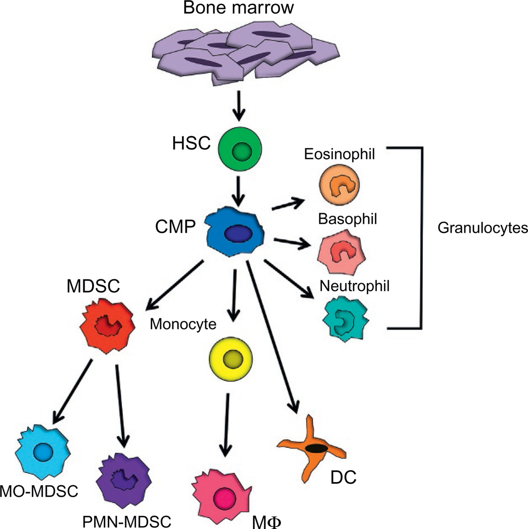
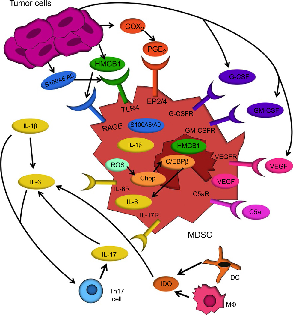
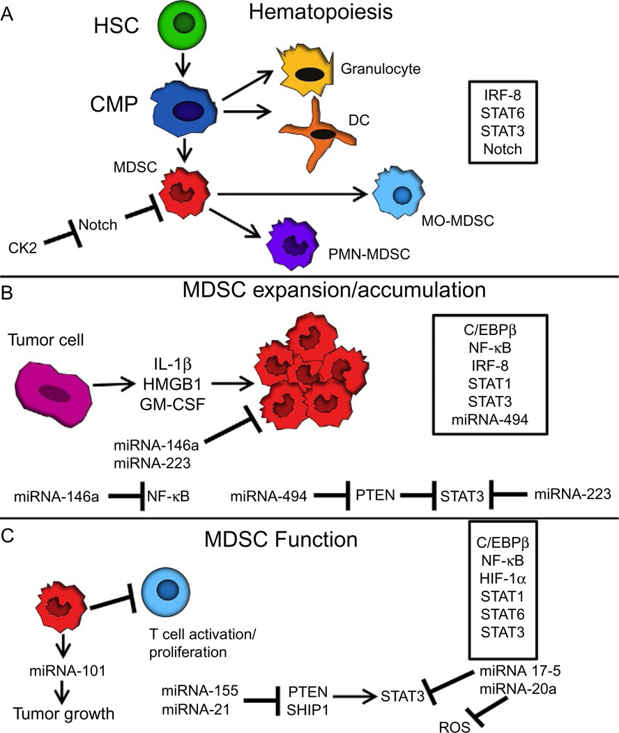
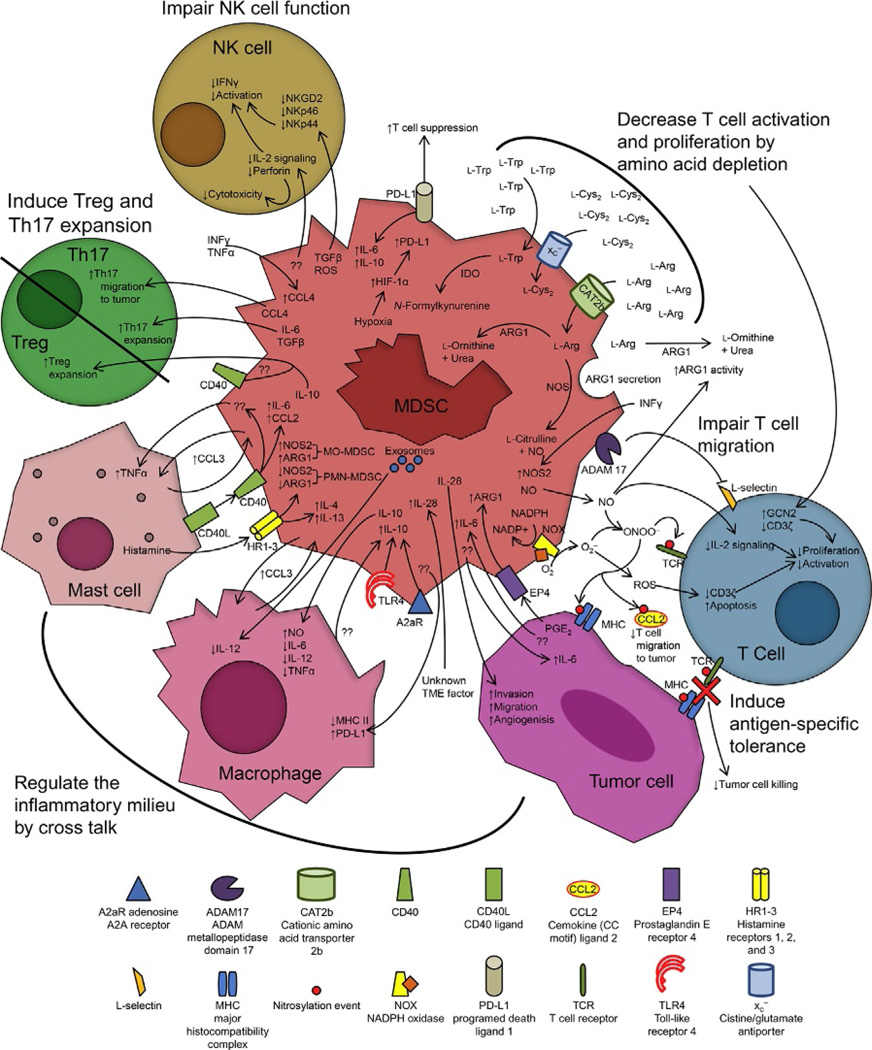
Myeloid-derived suppressor cells (MDSCs) are a heterogeneous population of immature myeloid cells that suppress innate and adaptive immunity. MDSCs are present in many disease settings; however, in cancer, they are a major obstacle for both natural antitumor immunity and immunotherapy. Tumor and host cells in the tumor microenvironment (TME) produce a myriad of pro-inflammatory mediators that activate MDSCs and drive their accumulation and suppressive activity. MDSCs utilize a variety of mechanisms to suppress T cell activation, induce other immune-suppressive cell populations, regulate inflammation in the TME, and promote the switching of the immune system to one that tolerates and enhances tumor growth. Because MDSCs are present in most cancer patients and are potent immune-suppressive cells, MDSCs have been the focus of intense research in recent years. This review describes the history and identification of MDSCs, the role of inflammation and intracellular signaling events governing MDSC accumulation and suppressive activity, immune-suppressive mechanisms utilized by MDSCs, and recent therapeutics that target MDSCs to enhance antitumor immunity.

Keywords: Cytokines; Immature myeloid cells; Immunotherapy; Inflammation; Macrophages; Myeloid cell cross talk; STAT3; Tumor-induced immune suppression; Tumor-infiltrating lymphocytes; cEBPbeta.


Figures

Figure 1
Figure 1
Myeloid cell differentiation under normal and tumor-induced conditions. Myeloid cells originate from bone marrow-derived hematopoietic stem cells (HSCs) that differentiate into common myeloid progenitors (CMPs). During normal myelopoiesis, CMPs differentiate into granulocytes including eosinophils, basophils, and neutrophils, as well as monocytes, macrophages, and dendritic cells. MDSCs also differentiate from CMPs and are categorized as MO-MDSCs or PMN-MDSCs. HSC, hematopoietic stem cell; CMP, common myeloid progenitor; DC, dendritic cell; MΦ, macrophage; MO-MDSCs, monocytic myeloid-derived suppressor cells; PMN-MDSCs, polymorphonuclear myeloid-derived suppressor cells.
Figure 2
Figure 2
Inflammation drives MDSC development and function. Chronic inflammation induces the production of HMGB1, S100A8/A9, IL-1β, IL-6, C5a, and IL-17, all of which induce the accumulation of MDSC. Induction of MDSCs by IL-1β is mediated through IL-17 and IL-6. IL-1β induces Th17 cells to produce IL-17 which induces the production of IL-6. IL-6 production is also upregulated by IDO produced by DCs and macrophages (MΦ). C/EBPβ, which is activated by chop following MDSC production of ROS, also induces IL-6. MDSCs also produce VEGF, IL-6, IL-1β, HMGB1, and S100A8/A9, thereby perpetuating their accumulation. Tumor cells may produce COX2, PGE2, VEGF, IL-6, G-CSF, GM-CSF, S100A8/A9, and HMGB1 all of which induce the accumulation of MDSCs and may increase the suppressive potency of MDSCs.
Figure 3
Figure 3
Multiple signal transduction pathways, transcription factors, and microRNAs regulate MDSC accumulation and function. (A) The differentiation of MDSCs from HSC and CMP is regulated by the transcription factors IRF-8, Notch, STAT6, and STAT3. IRF-8 and STAT6 regulate normal myelopoiesis and the differentiation of CMP to mature granulocytes and DCs. During abnormal myelopoiesis, which occurs in individuals with cancer, immature myeloid cells fail to terminally differentiate giving rise to elevated levels of MDSCs. Notch inhibits the differentiation of MDSCs, while CK2 blocks Notch and thereby increases MDSCs. STAT3 promotes MDSC development and suppressive potency. (B) Tumor and host cells produce multiple inflammatory molecules that perturb myelopoiesis and induce the expansion of MDSCs by activating or inactivating transcription factors. Pro-inflammatory mediators in the tumor microenvironment, such as IL-1β, HMGB1, and GM-CSF, drive the expansion of MDSCs by activating C/EBPβ, NF-κB, STAT1, STAT3, and miRNA-494 and downregulating IRF-8. Induction of miRNAs 146a and 223 prevents the expansion of MDSC. miRNA-494 promotes the expression of MMPs and inhibits PTEN function resulting in STAT3 induction. miRNA-146a inhibits NF-κB signaling, while miRNA-223 blocks C/EBPβ from binding to the c-myc promoter which downregulates STAT3 expression. (C) MDSC function is positively regulated by C/EBPβ, NF-κB, HIF-1α, STAT1, STAT6, and STAT3. The miRNAs 155 and 21 inhibit PTEN and SHIP1, negative regulators of STAT3, resulting in the activation of STAT3 and increased MDSC function. miRNAs 17-5 and 20a have the opposite effect by blocking STAT3 and ROS which negatively regulates MDSC function. MDSCs themselves also promote tumor growth by activating miRNA-101 in cancer cells.
Figure 4
Figure 4
MDSCs suppress T cells and regulate the inflammatory milieu by multiple mechanisms. MDSCs regulate antitumor immunity by (i) secretion of IL-10, which induces Tregs; (ii) secretion of IL-6 and TGFβ, which induces Th17 cells; (iii) production of ROS and TGFβ, which inhibits NK cell function; (iv) degradation of amino acids essential for T cell activation and proliferation; (v) production of NO and O2, which induces apoptosis and inhibits the activation and proliferation of T cells, and generates PNT that nitrates/nitrosylates MHC and TCR;and (vi) participation in cross talk with macrophages, tumor cells, and mast cells to generate a protumor environment. Question marks denote an unknown mechanism or signaling molecule.

References

    1. Adeegbe D, Serafini P, Bronte V, Zoso A, Ricordi C, Inverardi L. In vivo induction of myeloid suppressor cells and CD4(+)Foxp3(+) T regulatory cells prolongs skin allograft survival in mice. Cell Transplantation. 2011;20:941–954. - PubMed
    1. Alizadeh D, Trad M, Hanke NT, Larmonier CB, Janikashvili N, Bonnotte B, et al. Doxorubicin eliminates myeloid-derived suppressor cells and enhances the efficacy of adoptive T-cell transfer in breast cancer. Cancer Research. 2014;74:104–118. - PMC - PubMed
    1. Almand B, Clark JI, Nikitina E, van Beynen J, English NR, Knight SC, et al. Increased production of immature myeloid cells in cancer patients: A mechanism of immunosuppression in cancer. Journal of Immunology. 2001;166:678–689. - PubMed
    1. Annels NE, Shaw VE, Gabitass RF, Billingham L, Corrie P, Eatock M, et al. The effects of gemcitabine and capecitabine combination chemotherapy and of low-dose adjuvant GM-CSF on the levels of myeloid-derived suppressor cells in patients with advanced pancreatic cancer. Cancer Immunology, Immunotherapy. 2014;63:175–183. - PMC - PubMed
    1. Balkwill F, Mantovani A. Inflammation and cancer: Back to Virchow? Lancet. 2001;357:539–545. - PubMed