. 2015 Oct;197(19):3182-90.
doi: 10.1128/JB.00259-15. Epub 2015 Jul 27.

Acetate Dissimilation and Assimilation in Mycobacterium tuberculosis Depend on Carbon Availability

Affiliations

Acetate Dissimilation and Assimilation in Mycobacterium tuberculosis Depend on Carbon Availability

Nadine Rücker et al. J Bacteriol. 2015 Oct.

Abstract

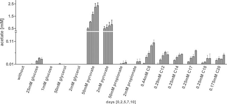
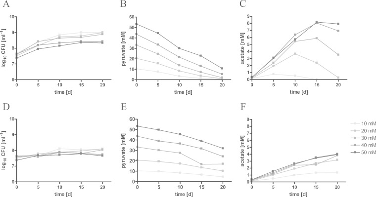
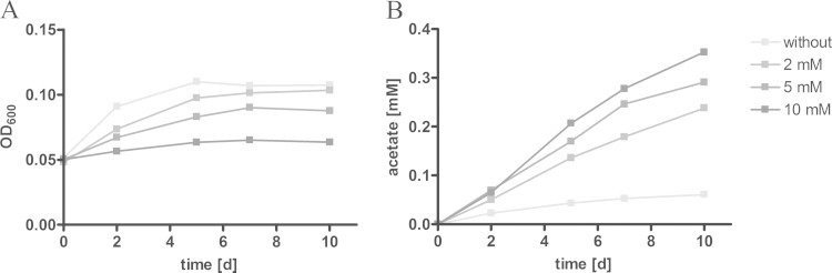
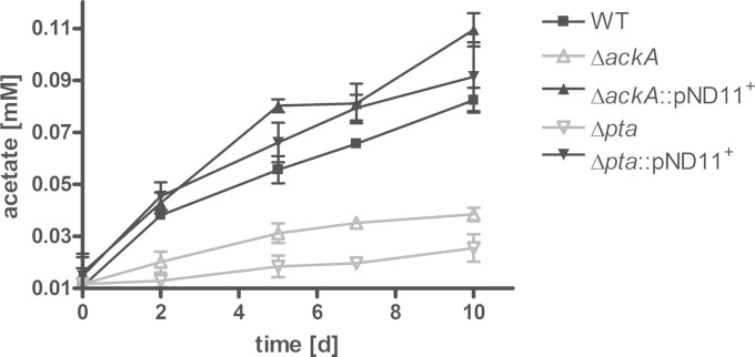
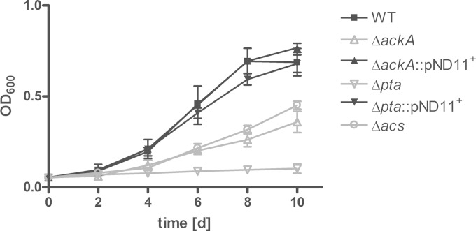
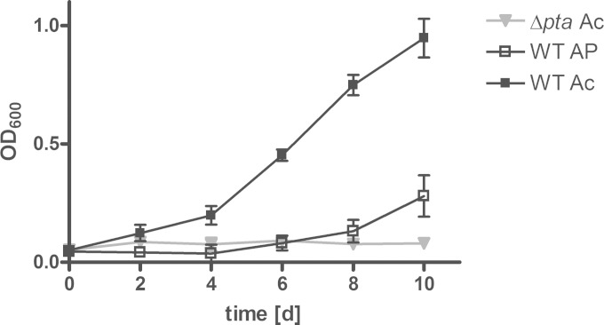
Mycobacterium tuberculosis persists inside granulomas in the human lung. Analysis of the metabolic composition of granulomas from guinea pigs revealed that one of the organic acids accumulating in the course of infection is acetate (B. S. Somashekar, A. G. Amin, C. D. Rithner, J. Troudt, R. Basaraba, A. Izzo, D. C. Crick, and D. Chatterjee, J Proteome Res 10:4186-4195, 2011, doi:http://dx.doi.org/10.1021/pr2003352), which might result either from metabolism of the pathogen or might be provided by the host itself. Our studies characterize a metabolic pathway by which M. tuberculosis generates acetate in the cause of fatty acid catabolism. The acetate formation depends on the enzymatic activities of Pta and AckA. Using actyl coenzyme A (acetyl-CoA) as a substrate, acetyl-phosphate is generated and finally dephosphorylated to acetate, which is secreted into the medium. Knockout mutants lacking either the pta or ackA gene showed significantly reduced acetate production when grown on fatty acids. This effect is even more pronounced when the glyoxylate shunt is blocked, resulting in higher acetate levels released to the medium. The secretion of acetate was followed by an assimilation of the metabolite when other carbon substrates became limiting. Our data indicate that during acetate assimilation, the Pta-AckA pathway acts in concert with another enzymatic reaction, namely, the acetyl-CoA synthetase (Acs) reaction. Thus, acetate metabolism might possess a dual function, mediating an overflow reaction to release excess carbon units and resumption of acetate as a carbon substrate.

Importance: During infection, host-derived lipid components present the major carbon source at the infection site. β-Oxidation of fatty acids results in the formation of acetyl-CoA. In this study, we demonstrate that consumption of fatty acids by Mycobacterium tuberculosis activates an overflow mechanism, causing the pathogen to release excess carbon intermediates as acetate. The Pta-AckA pathway mediating acetate formation proved to be reversible, enabling M. tuberculosis to reutilize the previously secreted acetate as a carbon substrate for metabolism.


Figures

FIG 1
FIG 1
Induction of acetate formation by fatty acids. M. tuberculosis H37Rv was grown in standing cultures for 10 days in the presence of different carbon sources. To analyze the impact of excess carbon substrates on acetate formation, substrates were supplied in low (0.175 to 2 mM) and high (25 to 50 mM) concentrations to the medium, and the resulting acetate production was detected in the supernatant at the indicated time points. Each bar represents the mean of three independent experiments; error bars indicate the standard deviation (SD).
FIG 2
FIG 2
Acetate formation depends on substrate concentration. M. tuberculosis H37Rv was cultivated for 20 days under fast-growth (A to C) or slow-growth (D to F) conditions. Pyruvate was provided in different concentrations (10 to 50 mM) to induce acetate formation. Growth was measured by colony counts (A and D). Pyruvate consumption (B and E) and acetate excretion (C and F) were detected in the supernatant by HPLC. The data represent one of two independent experiments.
FIG 3
FIG 3
Inhibition of the glyoxylate shunt increases acetate dissimilation. Standing cultures of M. tuberculosis H37Rv were grown for 10 days on 0.25 mM heptadecanoic acid as the carbon substrate. To limit carbon flow through the glyoxylate shunt, the isocitrate lyase was chemically inhibited by 2, 5, or 10 mM itaconate. The impact on growth was detected via optical density of the culture (A), and the resulting acetate formation was measured enzymatically in the supernatant (B). The data represent one of three independent experiments.
FIG 4
FIG 4
Acetate production is mediated by the Pta-AckA pathway. The M. tuberculosis H37Rv wild-type strain (WT), the ΔackA and Δpta mutant strains, and the complemented strains were grown in standing cultures for 10 days, and 0.25 mM heptadecanoic acid was supplied as carbon substrate. The influence of pta or ackA deletion on acetate dissimilation was analyzed by acetate detection in the supernatant of the knockout (KO) strains. The data represent the mean from three independent experiments; error bars indicate the SD.
FIG 5
FIG 5
The role of Acs and the Pta-AckA pathway in acetate assimilation. To analyze acetate utilization in M. tuberculosis, the wild type (WT), ΔackA, Δpta, and Δacs mutant strains, and complemented strains were grown for 10 days under fast-growth conditions. Acetate at 10 mM was supplied as the sole carbon substrate to the minimal medium, and growth was determined via optical density. The data represent the mean from three independent experiments; error bars indicate the SD.
FIG 6
FIG 6
Acetyl-phosphate accumulation results in restricted growth of M. tuberculosis. The M. tuberculosis H37Rv wild type (WT) and the Δpta mutant were cultivated under fast-growth conditions for 10 days. Growth was analyzed in the presence of 20 mM acetate (Ac) or 20 mM acetyl-phosphate (AP). The data represent the mean from three independent experiments; error bars indicate the SD.

Similar articles

Cited by

References

    1. Zumla A, George A, Sharma V, Herbert N, Baroness Masham of Ilton, Oxley A, Oliver M. 2015. The WHO 2014 global tuberculosis report—further to go. Lancet Glob Health 3:e10-2. doi:10.1016/S2214-109X(14)70361-4. - DOI - PubMed
    1. Rao SP, Alonso S, Rand L, Dick T, Pethe K. 2008. The protonmotive force is required for maintaining ATP homeostasis and viability of hypoxic, nonreplicating Mycobacterium tuberculosis. Proc Natl Acad Sci U S A 105:11945–11950. doi:10.1073/pnas.0711697105. - DOI - PMC - PubMed
    1. Bloch H, Segal W. 1956. Biochemical differentiation of Mycobacterium tuberculosis grown in vivo and in vitro. J Bacteriol 72:132–141. - PMC - PubMed
    1. Cole ST, Brosch R, Parkhill J, Garnier T, Churcher C, Harris D, Gordon SV, Eiglmeier K, Gas S, Barry CE III, Tekaia F, Badcock K, Basham D, Brown D, Chillingworth T, Connor R, Davies R, Devlin K, Feltwell T, Gentles S, Hamlin N, Holroyd S, Hornsby T, Jagels K, Krogh A, McLean J, Moule S, Murphy L, Oliver K, Osborne J, Quail MA, Rajandream MA, Rogers J, Rutter S, Seeger K, Skelton J, Squares R, Squares S, Sulston JE, Taylor K, Whitehead S, Barrell BG. 1998. Deciphering the biology of Mycobacterium tuberculosis from the complete genome sequence. Nature 393:537–544. doi:10.1038/31159. - DOI - PubMed
    1. Rohde KH, Veiga DF, Caldwell S, Balazsi G, Russell DG. 2012. Linking the transcriptional profiles and the physiological states of Mycobacterium tuberculosis during an extended intracellular infection. PLoS Pathog 8:e1002769. doi:10.1371/journal.ppat.1002769. - DOI - PMC - PubMed

Publication types

MeSH terms

LinkOut - more resources