. 2015 Aug 11;112(32):10002-7.
doi: 10.1073/pnas.1502159112. Epub 2015 Jul 27.

The isolation of an RNA aptamer targeting to p53 protein with single amino acid mutation

Affiliations

The isolation of an RNA aptamer targeting to p53 protein with single amino acid mutation

Liang Chen et al. Proc Natl Acad Sci U S A. .

Abstract

p53, known as a tumor suppressor, is a DNA binding protein that regulates cell cycle, activates DNA repair proteins, and triggers apoptosis in multicellular animals. More than 50% of human cancers contain a mutation or deletion of the p53 gene, and p53R175 is one of the hot spots of p53 mutation. Nucleic acid aptamers are short single-stranded oligonucleotides that are able to bind various targets, and they are typically isolated from an experimental procedure called systematic evolution of ligand exponential enrichment (SELEX). Using a previously unidentified strategy of contrast screening with SELEX, we have isolated an RNA aptamer targeting p53R175H. This RNA aptamer (p53R175H-APT) has a significantly stronger affinity to p53R175H than to the wild-type p53 in both in vitro and in vivo assays. p53R175H-APT decreased the growth rate, weakened the migration capability, and triggered apoptosis in human lung cancer cells harboring p53R175H. Further analysis actually indicated that p53R175H-APT might partially rescue or correct the p53R175H to function more like the wild-type p53. In situ injections of p53R175H-APT to the tumor xenografts confirmed the effects of this RNA aptamer on p53R175H mutation in mice.

Keywords: RNA aptamer; SELEX; contrast screening; p53; tumor.


Conflict of interest statement

Conflict of interest statement: G.S. and L.C. have an ownership interest in a patent related to this research.

Figures
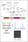
Fig. 1.
Fig. 1.
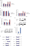
Contrast SELEX screening followed with ELISA verification yielded RNA aptamers for p53R175H. (A) Workflow of the contrast SELEX screening. WT p53 protein-coupled agarose beads and p53R175H-coupled magnetic beads were incubated with the in vitro transcribed RNA pool. After washing, magnetic beads and agarose beads were separated, and RNAs on the magnetic beads were isolated and subjected to RT-PCR and in vitro transcription. After five SELEX cycles, RNAs on p53R175H-coupled magnetic beads were isolated, cloned, and sequenced for further analysis. (B) The starting ssDNA library. This library consists of 1015 unique sequences, each containing a 25-base internal randomized region flanked by two fixed regions with a T7 promoter sequence. (C) Workflow of the ELISA affinity assay. WT p53 and p53R175H were coated, respectively, to individual wells. The biotinylated RNA aptamer was incubated with p53 WT and p53R175H, respectively. (D) Relative ratio of reading from p53R175H- and WT p53-coated wells for each individual RNA aptamer. (E) Gel shift assay confirmed that p53R175H-APT interacts specifically with the p53R175H mutant protein (MT) but not the WT p53 (WT). (–), without the protein; (+), with the protein.
Fig. S1.
Fig. S1.
Characterizing the RNA aptamers. (A) Gel image of the RNA pool in vitro transcribed from the DNA library. (B) Relative ratio of reading from p53R175H- and WT p53-coated wells for each individual RNA aptamer selected from round 3. (C) Relative ratio of reading from p53R175H- and WT p53-coated wells for each individual RNA aptamer selected from round 4. (D) Gel image of the p53R175H-APT and the scramble control RNA in vitro transcribed.
Fig. 2.
Fig. 2.
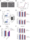
p53R175H-APT did not affect cells with WT p53 or p53R273H. (A) Images of HEK293T after transfection with either p53R175H-APT or the RNA control with scrambled sequences. (B) Cell growth curve plotted with MTT assay. Growth of HEK293T cells was unaffected by the application of p53R175H-APT. (C) Detection of apoptotic cells by FITC-annexin V staining and propidium iodide (PI) staining with FACS. Representative (n = 3) FACS data and bar graph summarizing the FACS data (Q2 + Q3 for early and late apoptosis) are shown. (D) HEK293 cells treated with various concentrations of nanoparticles with enclosed p53R175H-APT RNA or scramble control. Cell growth and apoptosis were measured with MTT assays and FACS, respectively. (E) p53R175H RIP followed with qPCR for p53R175H-APT RNA and the scramble control using cells transfected with either p53R175H-APT or scramble control plasmid. 7SL was examined as an irrelevant noncoding RNA as an assessment of nonspecific binding. (F) Cells with p53R273H expression (H1299-p53R273H cells) treated with various concentrations of nanoparticles with enclosed p53R175H-APT RNA or scramble control. Cell growth and apoptosis were measured with MTT assays and FACS, respectively. All data were from three repeats. Error bars represent SD. ns, difference between groups of data are not significant. P values were determined with two-tailed Student’s t test.
Fig. S2.
Fig. S2.
p53R175H-APT had no effect on HeLa cells. (A) Gross appearance of HeLa cells transfected with p53R175H-APT along with the scramble control. (B, Upper) No significant effect on cell growth and cell death with a higher concentration of transfected p53R175H-APT plasmid in HEK293T (a p53 WT cell line). (Lower) No significant effect on cell growth and cell death with various concentrations of transfected p53R175H-APT plasmid in HeLa (a p53 WT cell line). At a very high concentration of 16 μg/mL (10 times higher than the effective concentration of p53R175H-APT plasmid in H1299-p53R175H cells), p53R175H-APT plasmid somehow showed a significant effect on cell growth in HeLa. Cell growth and apoptosis were measured with MTT assays and FACS, respectively. (C) p53 RIP followed with qPCR for p53R175H-APT and the scramble control RNA. 7SL was examined as an irrelevant noncoding RNA as an assessment of nonspecific binding. All data were from three repeats. Error bars represent SD. ns, difference between groups of data are not significant. P values were determined with two-tailed Student’s t test.
Fig. S3.
Fig. S3.
Effects of p53R175H-APT on H1299-p53R175H cells. (A) FACS (apoptosis assay) at 1× concentration (1.6 μg/mL, confirmed effective concentration of the plasmid) after 24, 36, 48, and 60 h of transfection. (B) Cell growth and cell death with various concentrations of transfected p53R175H-APT plasmid in H1299-p53R175H cells. Lower concentrations had no significant effect compared with the effective concentration (1.6 μg/mL). The effect of p53R175H-APT plasmid on apoptosis of H1299-p53R175H cells does not seem dosage-dependent, but rather with a threshold at 16 μg/mL, it had a similar effect compared to 1.6 μg/mL. Cell growth and apoptosis were measured with MTT assays and FACS, respectively. (C) Cell death with various concentrations of nanoparticles with enclosed p53R175H-APT RNA in H1299-p53R175H cells. Lower concentrations had no significant effect compared with the effective concentration of 200 nM. Apoptosis was measured with FACS. (D and E) Western blot results of p53R175H from RIP assay shown in Fig. 3H to demonstrate efficient RIP pull-down of p53R175H protein. (F) Western blots of p53R175H from ChIP assay shown in Fig. 3J to demonstrate efficient ChIP pull-down of p53R175H protein. (G) RIP with a p53 fragment of 150 amino acids with the p53R175H mutation in the middle. This mutated part of p53 seems to interact with p53R175H-APT expressed in cells, although with less ability compared with the full mutant protein (Fig. 3H). (H) The PCR regions corresponding to the known binding sites of p53 for each of the genes examined in Fig. 3J are shown. All data were from three repeats. Error bars represent SD. ns, difference between groups of data are not significant. *P < 0.05; **P < 0.01; ***P < 0.001. P values were determined with a two-tailed Student’s t test.
Fig. 3.
Fig. 3.
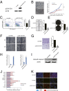
p53R175H-APT rescued p53R175H in cell cultures. (A) Western blot of p53R175H verified the successful establishment of the H1299-p53R175H stable cell line. (B) Gross appearance of H1299-p53R175H cells transfected with p53R175H-APT along with the scramble control. Relative cell growth of p53R175H-APT–transfected cells was determined by MTT assay. (C) Detection of apoptotic cells by FITC-annexin V staining and PI staining with FACS. Representative (n = 3) FACS data and bar graph summarizing the FACS data (Q2 + Q3 for early and late apoptosis) are shown. (D) Clonogenic assay and (E) soft agar assay with p53R175H-APT– and scramble control-transfected cells. (F) Representative images (n = 5) of scratch assay for cells transfected with either p53R175H-APT or the scramble control. (G) Transwell migration assay using H1299-p53R175H cells transfected with p53R175H-APT or the scramble control. Representative images (n = 3) of crystal violet staining of migrated cells and bar figure of relative migrating cells are shown. (H) p53R175H RIP assay followed with qPCR. IgG was the negative control of p53 antibody (α-P53) to show the relative pull-down efficiency of p53R175H-APT RNA by the α-P53 (Left). Scramble was the negative control of p53R175H-APT to show the relative pull-down efficiency of p53R175H-APT RNA by the α-P53 (Right). (I) Western blot of cleaved caspase 3 in p53R175H-APT– and the scramble control-transfected cells. (J) p53R175H ChIP assay followed by qPCR to examine 10 known p53 target genes. Promoter of tubulin (TUBB) was a negative control. The known binding sites of p53 for each of these genes examined are shown in Fig. S3H. (K) Immunostaining (red signal) of cells with the antibody PAb1620 specific for the WT p53 structure. HEK293T is a cell line with WT p53. p53R175H-APT, H1299-p53R175H cells treated with the p53R175H-APT aptamer; scramble, H1299-p53R175H cells treated with the scramble control. *P < 0.05; **P < 0.01; ***P < 0.001. P values were determined with two-tailed Student’s t test. All data were from at least three repeats. Error bars represent SD.
Fig. 4.
Fig. 4.
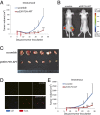
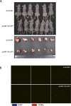
p53R175H-APT inhibited tumor growth. (A) Mice were treated with in vitro synthesized p53R175H-APT coated with nanoparticles at day 5 and day 8 by s.c. injection directly to the tumor. Tumor volumes were measured twice a week. (B) Bioluminescence imaging of representative tumors was shown. (C) Tumor dissected out from the p53R175H-APT– and the scramble control-treated groups. (D) TUNEL staining of tumor sections of p53R175H-APT– and the scramble control-treated groups. (E) Mice were treated with in vitro synthesized p53R175H-APT coated with nanoparticles at day 7 and day 17 by tail vein i.v. administration. Tumor volumes were measured twice a week. *P < 0.05; **P < 0.01; ***P < 0.001. P values were determined with two-tailed Student’s t test. All data were from at least three repeats. Error bars represent SD.
Fig. S4.
Fig. S4.
i.v.-administrated p53R175H-APT nanoparticles inhibited tumor growth. (A) Mice and tumor dissected out from the p53R175H-APT– and the scramble control-treated groups. (B) TUNEL staining of tumor sections of p53R175H-APT– and the scramble control-treated groups.

References

    1. Tuerk C, Gold L. Systematic evolution of ligands by exponential enrichment: RNA ligands to bacteriophage T4 DNA polymerase. Science. 1990;249(4968):505–510. - PubMed
    1. Ellington AD, Szostak JW. In vitro selection of RNA molecules that bind specific ligands. Nature. 1990;346(6287):818–822. - PubMed
    1. Bock LC, Griffin LC, Latham JA, Vermaas EH, Toole JJ. Selection of single-stranded DNA molecules that bind and inhibit human thrombin. Nature. 1992;355(6360):564–566. - PubMed
    1. Huizenga DE, Szostak JW. A DNA aptamer that binds adenosine and ATP. Biochemistry. 1995;34(2):656–665. - PubMed
    1. White RR, et al. Inhibition of rat corneal angiogenesis by a nuclease-resistant RNA aptamer specific for angiopoietin-2. Proc Natl Acad Sci USA. 2003;100(9):5028–5033. - PMC - PubMed

Publication types

MeSH terms