. 2015 Jul 7:3:43.
doi: 10.3389/fcell.2015.00043. eCollection 2015.

Cancer as a mitochondrial metabolic disease

Affiliations

Cancer as a mitochondrial metabolic disease

Thomas N Seyfried. Front Cell Dev Biol. .

Abstract

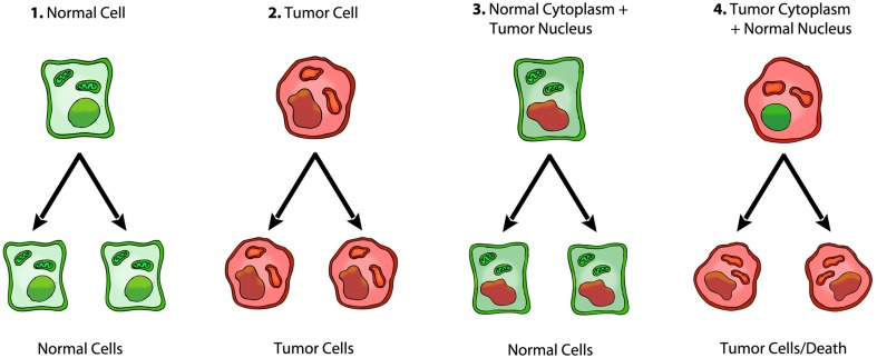
Cancer is widely considered a genetic disease involving nuclear mutations in oncogenes and tumor suppressor genes. This view persists despite the numerous inconsistencies associated with the somatic mutation theory. In contrast to the somatic mutation theory, emerging evidence suggests that cancer is a mitochondrial metabolic disease, according to the original theory of Otto Warburg. The findings are reviewed from nuclear cytoplasm transfer experiments that relate to the origin of cancer. The evidence from these experiments is difficult to reconcile with the somatic mutation theory, but is consistent with the notion that cancer is primarily a mitochondrial metabolic disease.

Keywords: Warburg effect; carcinogenesis; cybrids; fermentation; microenvironment; mitochondria; oxidative phosphorylation; tumorigenesis.


Figures

Figure 1
Figure 1
Nuclei from brain tumors support normal mouse embryonic development. (A) H&E staining of a mouse embryo (embryonic day, E-7.5) derived from a cell containing the transplanted “nucleus” from a medulloblastoma tumor. (B) the boxed area in (A) (at a higher magnification) showing the three germ layers; ecto-placental cone (pla); embryonic endoderm (end); embryonic mesoderm (mes,); embryonic ectoderm (ect), Scale bar, 20 μm. The cytoplasm will contain normal mitochondria. The results show that a nucleus derived from a brain tumor can direct normal embryonic development when implanted into normal cytoplasm. Reprinted with permission from Li et al. (2003).
Figure 2
Figure 2
Mouse embryo cloned from tumor cell nucleus. An E-9.5 mouse embryo cloned from a melanoma-derived R545-1 embryonic stem cell. The embryo expressed neural tube closure, a beating heart, and normal limb bud development consistent with regulated cell growth. The result shows that the nucleus of a malignant melanoma can direct early mouse development when placed into normal cytoplasm containing normal mitochondria. However, irreversible genetic alterations, from the melanoma donor genome, disrupted complete development similar to the situation found in Lucke frogs that were cloned from nuclei of renal tumors (McKinnell et al., 1969). Reprinted from in this figure (Hochedlinger et al., 2004) with permission.
Figure 3
Figure 3
Role of the nucleus and mitochondria in the origin of tumors. Summary of a role of the mitochondria in the origin of tumorigenesis, as we previously described (Seyfried, ; Seyfried et al., 2014). Normal cells are shown in green with nuclear and mitochondrial morphology indicative of normal gene expression and respiration, respectively. Tumor cells are shown in red with abnormal nuclear and mitochondrial morphology indicative of genomic instability and abnormal respiration, respectively. “(1) Normal cells beget normal cells. (2) Tumor cells beget tumor cells. (3) Transfer of a tumor cell nucleus into a normal cytoplasm begets normal cells, despite the presence of the tumor-associated genomic abnormalities. (4) Transfer of a normal cell nucleus into a tumor cell cytoplasm begets dead cells or tumor cells, but not normal cells. The results suggest that nuclear genomic defects alone cannot account for the origin of tumors, and that normal mitochondria can suppress tumorigenesis” (Seyfried, 2012d). Original diagram from Jeffrey Ling and Thomas N. Seyfried, with permission.

References

    1. Ackermann W. W., Kurtz H. (1952). The relation of herpes virus to host cell mitochondria. J. Exp. Med. 96, 151–157. 10.1084/jem.96.2.151 - DOI - PMC - PubMed
    1. Akimoto M., Niikura M., Ichikawa M., Yonekawa H., Nakada K., Honma Y., et al. . (2005). Nuclear DNA but not mtDNA controls tumor phenotypes in mouse cells. Biochem. Biophys. Res. Commun. 327, 1028–1035. 10.1016/j.bbrc.2004.12.105 - DOI - PubMed
    1. Allen N. J., Karadottir R., Attwell D. (2005). A preferential role for glycolysis in preventing the anoxic depolarization of rat hippocampal area CA1 pyramidal cells. J. Neurosci. 25, 848–859. 10.1523/JNEUROSCI.4157-04.2005 - DOI - PMC - PubMed
    1. Al Mamun A. A., Lombardo M. J., Shee C., Lisewski A. M., Gonzalez C., Lin D., et al. . (2012). Identity and function of a large gene network underlying mutagenic repair of DNA breaks. Science 338, 1344–1348. 10.1126/science.1226683 - DOI - PMC - PubMed
    1. Baker S. G., Kramer B. S. (2007). Paradoxes in carcinogenesis: new opportunities for research directions. BMC Cancer 7:151. 10.1186/1471-2407-7-151 - DOI - PMC - PubMed