. 2015 Jul 9:2:45.
doi: 10.3389/fmed.2015.00045. eCollection 2015.

Progressive Resistance Exercise with Eccentric Loading for the Management of Knee Osteoarthritis

Affiliations

Progressive Resistance Exercise with Eccentric Loading for the Management of Knee Osteoarthritis

Haniel J Hernandez et al. Front Med (Lausanne). .

Abstract

Introduction: The patient was a 58-year-old African-American male with radiographic evidence of bilateral knee osteoarthritis (OA). He participated in a standardized 12-week eccentric strengthening program within a Veterans Affairs (VA) medical center.

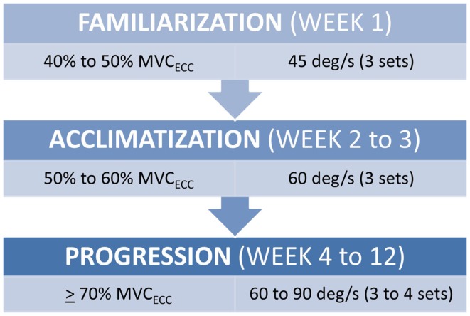
Background: The use of an eccentric training paradigm may prove to be beneficial for older adults with knee OA since eccentric muscle actions are involved in the energy absorption at the knee joint during gait and controlled movement during stair descent. Furthermore, in comparison to standard muscle actions, eccentric muscle actions result in higher torque generation and a lower rate of oxygen consumption at a given level of perceived exertion. Therefore, this mode of progressive resistance exercise may be ideal for older adults.

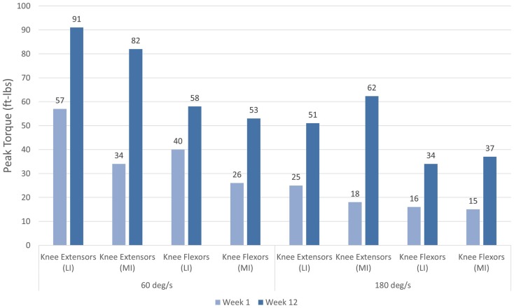
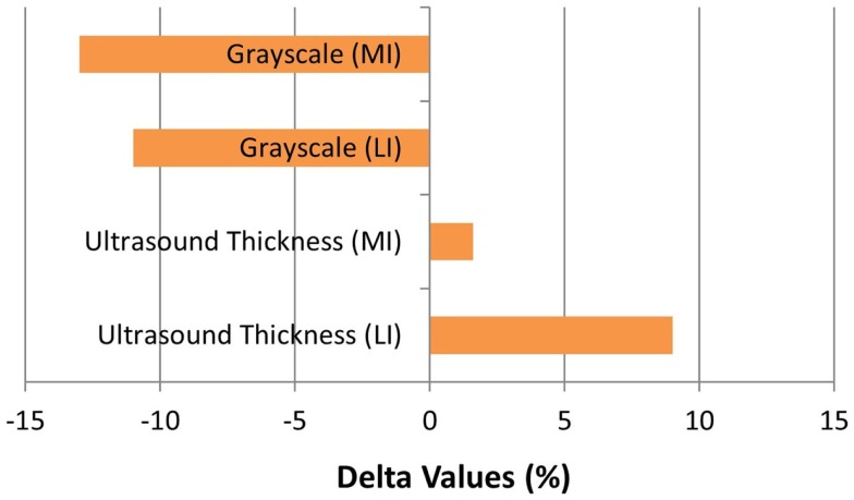
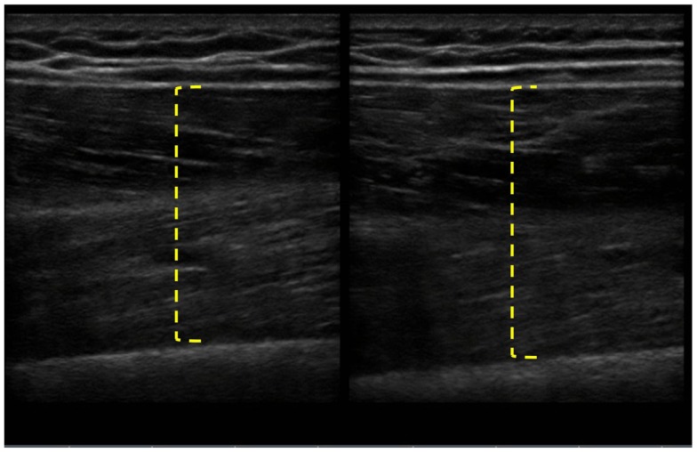
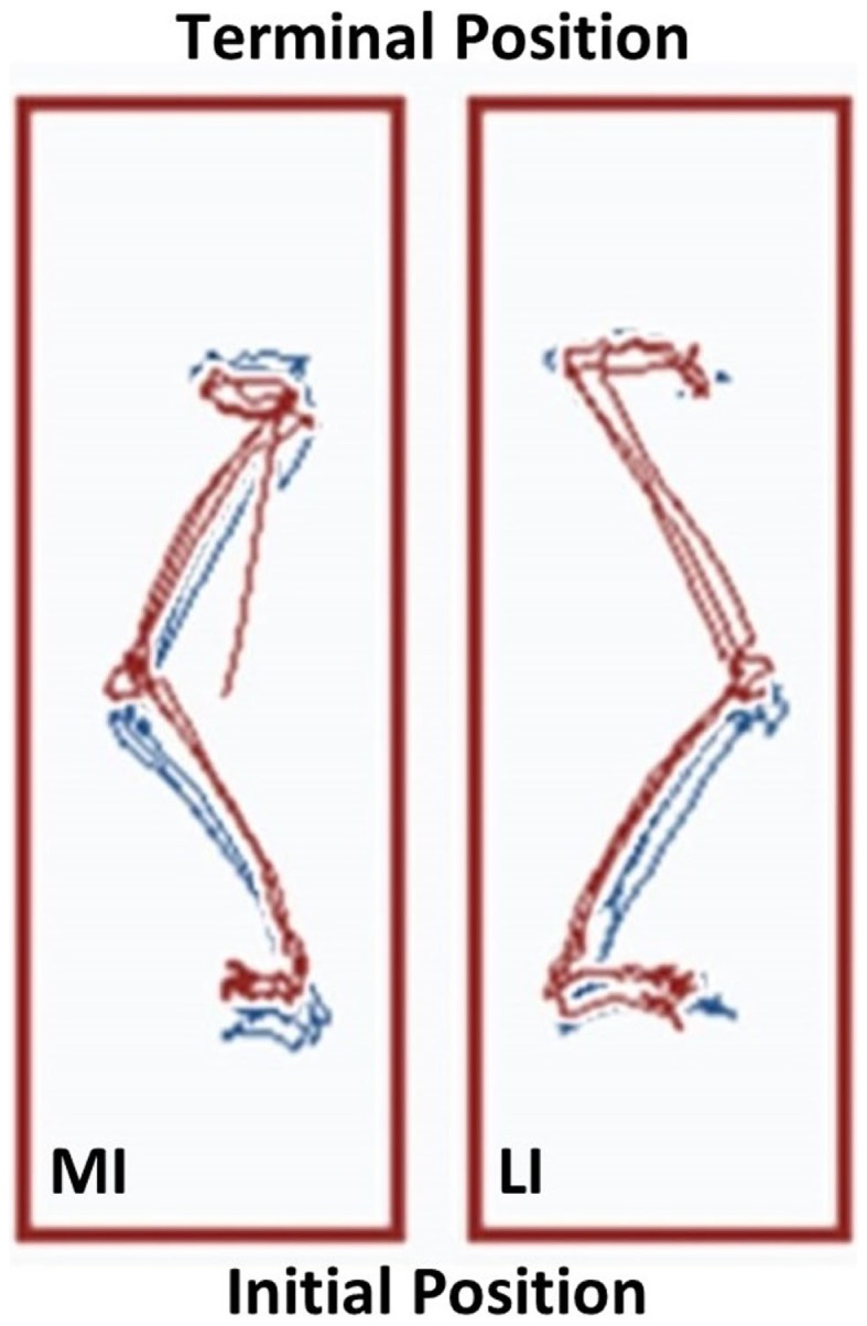
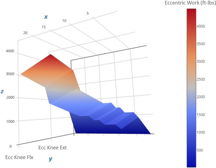
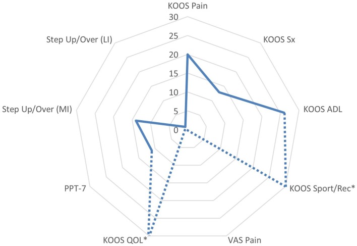
Discussion: The patient completed an eccentric strengthening regimen for the knee flexors and extensors twice per week without an exacerbation of knee pain. Muscle morphology measures of the rectus femoris were measured using diagnostic ultrasound. Isokinetic measures of muscle peak torque were obtained at 60°/s and 180°/s. Functional performance was assessed using a physical performance battery and stair-step performance was assessed from the linear displacement of the center of gravity trajectories obtained with a force plate. Visual analog scale pain ratings and self-reported global disease status were also documented. Post-exercise assessments revealed improvements in sonographic muscle size and tissue composition estimates, peak knee extensor torque (ranging from 60 to 253%), functional performance, and global disease status.

Concluding remarks: The patient exhibited improvements in muscle morphology, muscle strength, functional performance, pain, and global disease status after 12 weeks of an eccentric strengthening regimen. The intervention and outcomes featured in this case were feasible to implement within a VA medical center and merit further investigation.

Keywords: eccentric exercise; knee arthritis; strengthening; ultrasound; veterans.


Figures

Figure 1
Figure 1
Submaximal eccentric progressive resistance exercise phases. The training regimen features structured phases of submaximal eccentric exercise designed to safely progress the strength training stimulus in people with musculoskeletal impairments. (MVCECC, estimated eccentric maximum voluntary contraction, i.e., concentric MVC at 60°/s × 1.35).
Figure 2
Figure 2
Pre and post-intervention concentric knee extensor and flexor peak torque values. Increases in concentric knee extensor and flexor performance were noted following the 12-week eccentric progressive resistance exercise regimen. The adaptations to the eccentric strength training stimulus were transferable to the concentric muscle actions across both muscle groups and movement speeds. The greatest magnitude of strength change was observed in the more involved limb (left side).
Figure 3
Figure 3
Muscle morphology adaptations following the 12-week intervention period. The grayscale delta percentage values shown at the top of the graph depict the decreased echogenicity estimates from the ultrasound images of the rectus femoris, which is suggestive of improved tissue composition following the eccentric exercise regimen. The ultrasound muscle thickness delta percentage values depicted at the bottom of the graph suggest that muscle hypertrophy occurred, primarily in the less involved limb (right side), following the 12-week intervention period (MI, more involved limb; LI, less involved limb).
Figure 4
Figure 4
Pre and post-intervention ultrasound images of the rectus femoris following the 12-week intervention period. The longitudinal B-mode ultrasound images of the less involved limb (right side) were obtained with a 13–6 MHz linear array transducer and using the averaged digital caliper values from three measurements. The rectus femoris of the less involved limb had an increase in muscle thickness from 3.97 to 4.32 cm, whereas the more involved limb experienced a change in muscle thickness of <0.1 cm.
Figure 5
Figure 5
The Neurocom® Step Up and Over test performance following the 12-week intervention period. The center of gravity (COG) displacement trajectories derived from the Step Up and Over test are displayed for the more involved limb (left side) and the less involved limb (right side). The Week 12 trajectories are in blue and overlaid with the Week 1 trajectories in red. The trajectories on the right have a maximum linear COG displacement that differs by <2% for the less involved limb. In contrast, the trajectories on the left show that the linear COG displacement of the more involved limb increased by 13.7% after the intervention period. This observation is consistent with a longer stepping length for the more involved limb, which resulted in improved symmetry between sides (MI, more involved limb; LI, less involved limb).
Figure 6
Figure 6
Progression of the estimated eccentric work volume produced during the 12-week intervention period. The 3-D surface plot depicts the estimated unilateral eccentric (Ecc) work produced by the less involved limb (right side) knee extensors over the course of the exercise regimen. The work produced is a function of the isokinetic torque produced during knee excursion and the exercise volume (in repetitions and sets). The x-axis features exercise sessions (two per week, for a total of 24 sessions), the y-axis includes the agonist muscle groups (knee flexors and extensors), and the z-axis includes the estimated work yield per exercise session (ft-lbs). Note that the lower work volume at the start of the regimen (the dark blue region on the right side of the plot) corresponds to the lower torque targets that characterize the “Familiarization” and “Acclimatization” phases of the eccentric exercise regimen, and may have afforded a protective adaptation against excessive muscle damage during the subsequent exercise bouts marked by higher work volumes and torque levels during the final 4 weeks of the “Progression” phase (the red region on the left side of the plot).
Figure 7
Figure 7
Changes in functional assessments and pain scores following the submaximal eccentric progressive resistance exercise regimen. The radar graph features the delta percentage values of the Knee Injury and Osteoarthritis Outcome Score (KOOS) Subscale Scores, the center of gravity linear displacement trajectories derived from the Step Up and Over test, and the visual analog scale pain rating. The largest post-intervention responses were observed in KOOS QOL and Sport/Rec subscales (*note: the change in these values exceeded 100%, and are estimated on the graph in order to maintain the graph scale and allow for comparisons among the remaining outcomes). Additionally, the VAS Pain rating was stable throughout the 12-week intervention period, whereas the KOOS Pain subscale improved by 20% (ADL, activities of daily living; QOL, quality of life; sport/rec, sports and recreation; Sx, symptom; PPT-7, Physical Performance Test; MI, more involved limb; LI, less involved limb; VAS, visual analog scale).

References

    1. Nguyen U-SDT, Zhang Y, Zhu Y, Niu J, Zhang B, Felson DT. Increasing prevalence of knee pain and symptomatic knee osteoarthritis: survey and cohort data. Ann Intern Med (2011) 155:725–32.10.7326/0003-4819-155-11-201112060-00004 - DOI - PMC - PubMed
    1. March L, Smith EUR, Hoy DG, Cross MJ, Sanchez-Riera L, Blyth F, et al. Burden of disability due to musculoskeletal (MSK) disorders. Best Pract Res Clin Rheumatol (2014) 28:353–66.10.1016/j.berh.2014.08.002 - DOI - PubMed
    1. Roos EM, Herzog W, Block JA, Bennell KL. Muscle weakness, afferent sensory dysfunction and exercise in knee osteoarthritis. Nat Rev Rheumatol (2011) 7:57–63.10.1038/nrrheum.2010.195 - DOI - PubMed
    1. Hurley MV, Newham DJ. The influence of arthrogenous muscle inhibition on quadriceps rehabilitation of patients with early, unilateral osteoarthritic knees. Br J Rheumatol (1993) 32:127–31.10.1093/rheumatology/32.2.127 - DOI - PubMed
    1. Doré AL, Golightly YM, Mercer VS, Shi XA, Renner JB, Jordan JM, et al. Lower limb osteoarthritis and the risk of falls in a community-based longitudinal study of adults with and without osteoarthritis. Arthritis Care Res (2015) 67(5):633–9.10.1002/acr.22499 - DOI - PMC - PubMed