Review
. 2015 Jul 7:2:38.
doi: 10.3389/fmolb.2015.00038. eCollection 2015.

Time-resolved infrared spectroscopic techniques as applied to channelrhodopsin

Affiliations
Review

Time-resolved infrared spectroscopic techniques as applied to channelrhodopsin

Eglof Ritter et al. Front Mol Biosci. .

Abstract

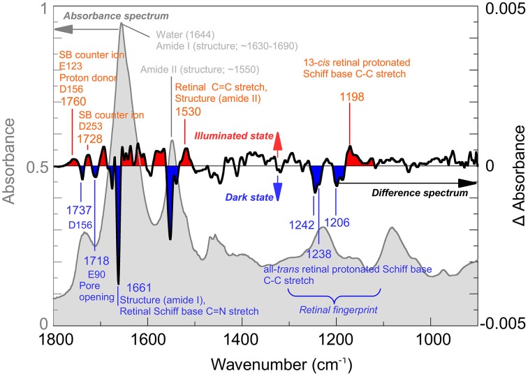
Among optogenetic tools, channelrhodopsins, the light gated ion channels of the plasma membrane from green algae, play the most important role. Properties like channel selectivity, timing parameters or color can be influenced by the exchange of selected amino acids. Although widely used, in the field of neurosciences for example, there is still little known about their photocycles and the mechanism of ion channel gating and conductance. One of the preferred methods for these studies is infrared spectroscopy since it allows observation of proteins and their function at a molecular level and in near-native environment. The absorption of a photon in channelrhodopsin leads to retinal isomerization within femtoseconds, the conductive states are reached in the microsecond time scale and the return into the fully dark-adapted state may take more than minutes. To be able to cover all these time regimes, a range of different spectroscopical approaches are necessary. This mini-review focuses on time-resolved applications of the infrared technique to study channelrhodopsins and other light triggered proteins. We will discuss the approaches with respect to their suitability to the investigation of channelrhodopsin and related proteins.

Keywords: FTIR; IR-spectrometer; channelrhodopsin; infrared spectroscopy; retinal proteins; time-resolved spectroscopy.


Figures

Figure 1
Figure 1
Infrared spectroscopy of Channelrhodopsin. The absorption spectrum (gray) of retinal proteins like Channelrhodopsin-2 reconstituted in lipid vesicles shows bands associated with the lipid environment and protonated carboxyl groups (~1700–1800 cm−1), water (1644 cm−1) and the overall helical structure of the protein (amide I ~1650 cm−1; amide II ~1550 cm−1). Note, that the lipid vesicles allow a very dense packing of the protein in the cuvette thus reducing the water content. Light induced alterations are represented by the difference spectrum (black), where negative bands (blue) occur due to the dark state while positive bands (red) are due to the illuminated state, achieved by illumination with blue (480 nm) light. The spectrum was recorded at cryogenic conditions where a mixture of species, including the Schiff base deprotonated state and the conducting state is observed. Note that, while total absorbance is in the order of 0.9 OD (left scale, gray), largest changes in the difference spectrum are within 0.004 OD (right scale, black). In the picture, some bands assigned so far to their structural counterparts are marked. For details of the band assignments, see (Eisenhauer et al., ; Lórenz-Fonfría et al., , ; Kuhne et al., 2015).
Figure 2
Figure 2
Different types of IR spectrometers. (A) Basic concept of a typical Fourier-transform infrared spectrometer showing light source (Globar), beam splitter, fixed and movable mirrors and single element infrared detector. Conformational changes in the sample are initiated by the trigger laser. The conversion of the sample can then be followed with a time resolution either determined by the sliding mirror movement (rapid-scan) or by the rise-time of the detector (step-scan). (B) Concept of a recently proposed dispersive device (Schade et al., 2014) with Synchrotron light source, dispersive prism and focal-plane array detector. (C) Laser based pump-probe setup. A first pulse from the pump laser starts the photoreaction. A subsequent short pulse from the probe laser probes the system. The probe pulse can be dispersed to obtain spectra; however, spectral bandwidth is determined by the duration of the probe pulse.

Similar articles

Cited by

References

    1. Alexandre M. T., Domratcheva T., Bonetti C., Van Wilderen L. J., Van Grondelle R., Groot M. L., et al. . (2009). Primary reactions of the LOV2 domain of phototropin studied with ultrafast mid-infrared spectroscopy and quantum chemistry. Biophys. J. 97, 227–237. 10.1016/j.bpj.2009.01.066 - DOI - PMC - PubMed
    1. Amenabar I., Poly S., Nuansing W., Hubrich E. H., Govyadinov A. A., Huth F., et al. . (2013). Structural analysis and mapping of individual protein complexes by infrared nanospectroscopy. Nat. Commun. 4, 2890. 10.1038/ncomms3890 - DOI - PMC - PubMed
    1. Andrews S. S., Boxer S. G. (2001). Analysis of noise for rapid-scan and step-scan methods of FT-IR difference spectroscopy. Appl. Spectrosc. 55, 1161–1165. 10.1366/0003702011953414 - DOI
    1. Bandekar J., Krimm S. (1979). Vibrational analysis of peptides, polypeptides, and proteins: characteristic amide bands of ß-turns. Proc. Natl. Acad. Sci. U.S.A. 76, 774–777. - PMC - PubMed
    1. Barth A. (2007). Infrared spectroscopy of proteins. Biochim. Biophys. Acta 1767, 1073–1101. 10.1016/j.bbabio.2007.06.004 - DOI - PubMed