

Evaluation of rigid registration methods for whole head imaging in diffuse optical tomography
- PMID: 26217675
- PMCID: PMC4509792
- DOI: 10.1117/1.NPh.2.3.035002
Evaluation of rigid registration methods for whole head imaging in diffuse optical tomography
Abstract
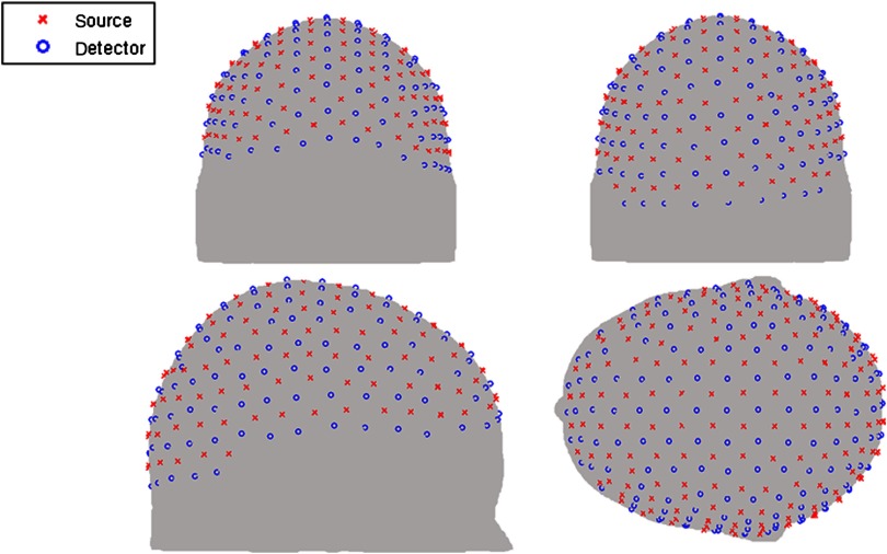
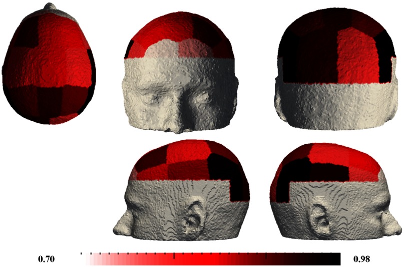
Functional brain imaging has become an important neuroimaging technique for the study of brain organization and development. Compared to other imaging techniques, diffuse optical tomography (DOT) is a portable and low-cost technique that can be applied to infants and hospitalized patients using an atlas-based light model. For DOT imaging, the accuracy of the forward model has a direct effect on the resulting recovered brain function within a field of view and so the accuracy of the spatially normalized atlas-based forward models must be evaluated. Herein, the accuracy of atlas-based DOT is evaluated on models that are spatially normalized via a number of different rigid registration methods on 24 subjects. A multileveled approach is developed to evaluate the correlation of the geometrical and sensitivity accuracies across the full field of view as well as within specific functional subregions. Results demonstrate that different registration methods are optimal for recovery of different sets of functional brain regions. However, the "nearest point to point" registration method, based on the EEG 19 landmark system, is shown to be the most appropriate registration method for image quality throughout the field of view of the high-density cap that covers the whole of the optically accessible cortex.
Keywords: atlas-based tomography; diffuse optical tomography; functional connectivity brain imaging; registration; sensitivity analyses; whole head imaging.
Figures


Similar articles
-
Atlas-based head modeling and spatial normalization for high-density diffuse optical tomography: in vivo validation against fMRI.Neuroimage. 2014 Jan 15;85 Pt 1(0 1):117-26. doi: 10.1016/j.neuroimage.2013.03.069. Epub 2013 Apr 8. Neuroimage. 2014. PMID: 23578579 Free PMC article.
-
Quantitative evaluation of atlas-based high-density diffuse optical tomography for imaging of the human visual cortex.Biomed Opt Express. 2014 Oct 13;5(11):3882-900. doi: 10.1364/BOE.5.003882. eCollection 2014 Nov 1. Biomed Opt Express. 2014. PMID: 25426318 Free PMC article.
-
Brain-Wide Diffuse Optical Tomography Based on Cap-Based, Whole-Head fNIRS Recording.Annu Int Conf IEEE Eng Med Biol Soc. 2021 Nov;2021:3609-3612. doi: 10.1109/EMBC46164.2021.9630249. Annu Int Conf IEEE Eng Med Biol Soc. 2021. PMID: 34892019
-
Overview of diffuse optical tomography and its clinical applications.J Biomed Opt. 2016 Sep;21(9):091312. doi: 10.1117/1.JBO.21.9.091312. J Biomed Opt. 2016. PMID: 27420810 Review.
-
Review of recent progress toward a fiberless, whole-scalp diffuse optical tomography system.Neurophotonics. 2018 Jan;5(1):011012. doi: 10.1117/1.NPh.5.1.011012. Epub 2017 Sep 26. Neurophotonics. 2018. PMID: 28983490 Free PMC article. Review.
Cited by
-
Effects of atlas-based anatomy on modelled light transport in the neonatal head.Phys Med Biol. 2023 Jul 3;68(13):135019. doi: 10.1088/1361-6560/acd48c. Phys Med Biol. 2023. PMID: 37167982 Free PMC article.
-
Detector location selection based on VIP analysis in near-infrared detection of dural hematoma.Saudi J Biol Sci. 2018 Mar;25(3):452-456. doi: 10.1016/j.sjbs.2017.11.044. Epub 2017 Nov 16. Saudi J Biol Sci. 2018. PMID: 29692647 Free PMC article.
-
High-density diffuse optical tomography for imaging human brain function.Rev Sci Instrum. 2019 May;90(5):051101. doi: 10.1063/1.5086809. Rev Sci Instrum. 2019. PMID: 31153254 Free PMC article.
-
Video-based motion-resilient reconstruction of three-dimensional position for functional near-infrared spectroscopy and electroencephalography head mounted probes.Neurophotonics. 2020 Jul;7(3):035001. doi: 10.1117/1.NPh.7.3.035001. Epub 2020 Jul 20. Neurophotonics. 2020. PMID: 32704521 Free PMC article.
-
Robust photogrammetric scalp morphology estimation for functional optical neuroimaging.Neurophotonics. 2025 Jul;12(3):035002. doi: 10.1117/1.NPh.12.3.035002. Epub 2025 Jul 28. Neurophotonics. 2025. PMID: 40735542 Free PMC article.
References
Grants and funding
LinkOut - more resources
Full Text Sources
Miscellaneous
