. 2015 Oct;21(19-20):2559-71.
doi: 10.1089/ten.TEA.2015.0105. Epub 2015 Sep 10.

HLA Class I Depleted hESC as a Source of Hypoimmunogenic Cells for Tissue Engineering Applications

Affiliations

HLA Class I Depleted hESC as a Source of Hypoimmunogenic Cells for Tissue Engineering Applications

Zaruhi Karabekian et al. Tissue Eng Part A. 2015 Oct.

Abstract

Background: Rapidly improving protocols for the derivation of autologous cells from stem cell sources is a welcome development. However, there are many circumstances when off-the-shelf universally immunocompatible cells may be needed. Embryonic stem cells (ESCs) provide a unique opportunity to modify the original source of differentiated cells to minimize their rejection by nonautologous hosts.

Hypothesis: Immune rejection of nonautologous human embryonic stem cell (hESC) derivatives can be reduced by downregulating human leukocyte antigen (HLA) class I molecules, without affecting the ability of these cells to differentiate into specific lineages.

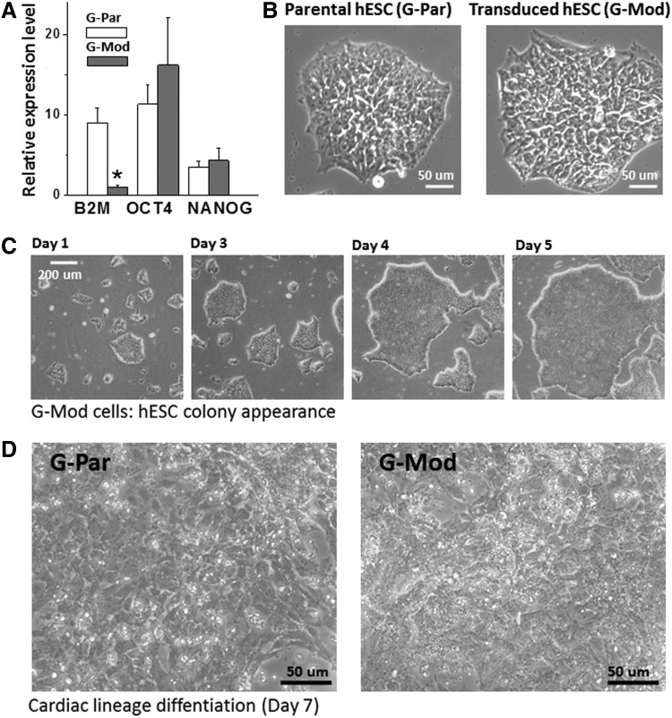
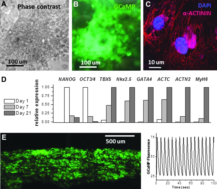
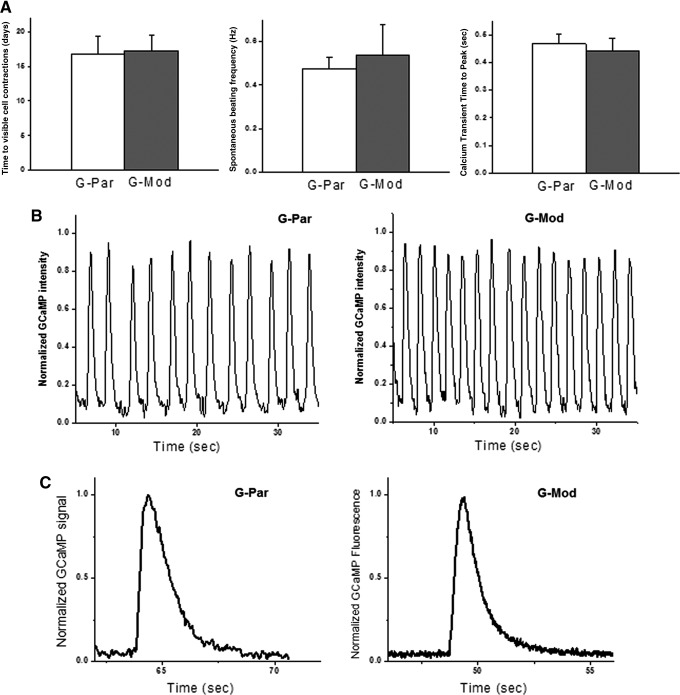
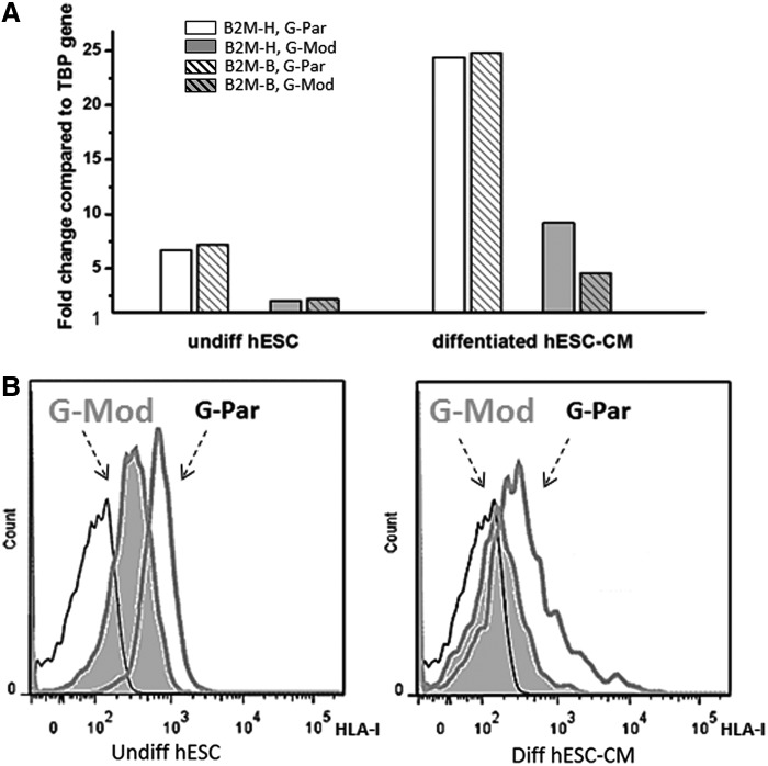
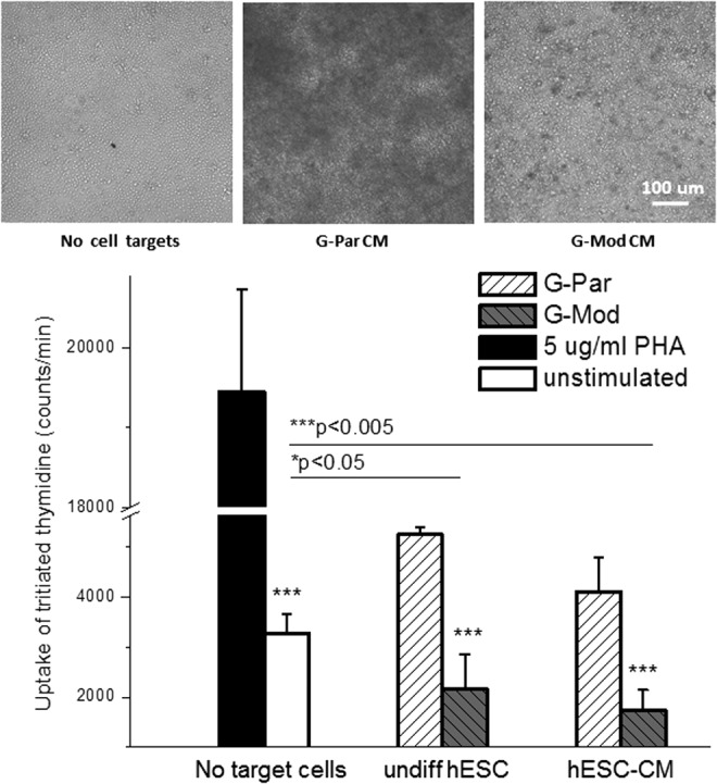
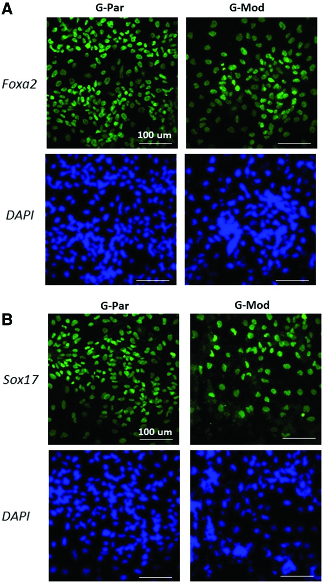
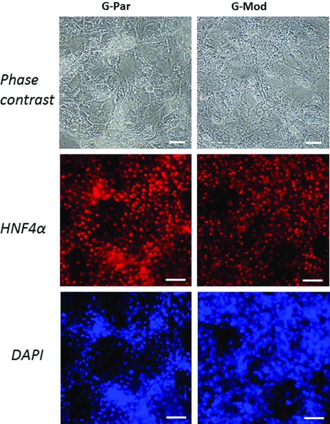
Methods and results: Beta-2-microglobulin (B2M) expression was decreased by lentiviral transduction using human anti-HLA class I light-chain B2M short hairpin RNA. mRNA levels of B2M were decreased by 90% in a RUES2-modified hESC line, as determined by quantitative real time-polymerase chain reaction analysis. The transduced cells were selected under puromycin pressure and maintained in an undifferentiated state. The latter was confirmed by Oct4 and Nanog expression, and by the formation of characteristic round-shaped colonies. B2M downregulation led to diminished HLA-I expression on the cell surface, as determined by flow cytometry. When used as target cells in a mixed lymphocyte reaction assay, transduced hESCs and their differentiated derivatives did not stimulate allogeneic T-cell proliferation. Using a cardiac differentiation protocol, transduced hESCs formed a confluent layer of cardiac myocytes and maintained a low level of B2M expression. Transduced hESCs were also successfully differentiated into a hepatic lineage, validating their capacity to differentiate into multiple lineages.

Conclusions: HLA-I depletion does not preclude hESC differentiation into cardiac or hepatic lineages. This methodology can be used to engineer tissue from nonautologous hESC sources with improved immunocompatibility.


Figures

<b>FIG. 1.</b>
FIG. 1.
Transduced human embryonic stem cells (hESCs) (G-Mod) in an undifferentiated state are phenotypically similar to the parental (G-Par) cell line. (A) The expression levels of beta-2-microglobulin (B2M) are decreased by 80–90% in undifferentiated G-Mod cells compared to controls, while the expression levels of undifferentiated markers such as OCT4 or NANOG remained unchanged. G-Par and G-Mod samples were run in triplicate. Average fold-change in gene expression levels compared to two different housekeeping genes is shown. *p < 0.05. (B) Transduced cells formed round-shaped colonies undistinguishable from G-Par cells. (C) G-Mod cells grew at a similar rate compared to G-Par cells, with a similar number of days required to passage cell colonies (4–5 days). (D) When subjected to cardiac differentiation protocol, G-Mod underwent visible changes in cell morphology, similarly to those observed in G-Par cell layers.
<b>FIG. 2.</b>
FIG. 2.
Properties of cardiac myocytes derived from hESCs with diminished B2M expression. The top row shows a typical appearance of a differentiated G-Mod cardiomyocyte layer in a phase-contrast (A) and a fluorescence mode (B). The presence of GCaMP sensor in G-Mod cells enables direct monitoring of rhythmical cell contractions upon cardiac lineage differentiation seen as transient increases in endogenous cell fluorescence. The panel (C) shows sarcomeric alpha-actinin immunostaining with regular striations indicative of proper structural development. The panel (D) illustrates changes in relative expression levels of pluripotency (NANOG, Oct3/4), mesodermal (TBX5), early and late (alpha-actin, GATA4, NkX2.5, alpha-2 actinin, myosin heavy-chain 6) markers in samples acquired on day 1, 7, and 21 of cardiac differentiation protocol (n = 3). The panel (E) shows 3D cardiac fiber composed of G-Mod cardiac myocytes with the trace of calcium transients acquired from it to illustrate regular spontaneous contractions.
<b>FIG. 3.</b>
FIG. 3.
Functional assessment of cardiac myocytes derived from hESCs with diminished B2M expression. (A) There were no detectable functional differences between cardiac myocytes differentiated from G-Par and G-Mod cell lines. Neither the number of days to develop spontaneous contractions nor the average beating frequency or calcium transient time to peak was different between the two cell lines. (B) Typical recordings of calcium transients from spontaneously beating confluent cell monolayers. Frequency of spontaneous beating ranged from 0.1 to 1 Hz for individual coverslips with an average frequency of 0.45 Hz for both cell types. (C) Sparsely plated individual cells contracted less frequently with longer calcium transients, consistent with frequency-dependent acceleration of relaxation frequency.
<b>FIG. 4.</b>
FIG. 4.
Levels of major costimulatory molecules remained unchanged in hESCs with diminished B2M expression in either undifferentiated or fully differentiated states. Gene expression levels of major costimulatory molecules, including CD80, CD81, CD83, CD86, FAS, and FAS-Ligand, after normalization to housekeeping gene TBP (human TATA box binding protein). No significant differences between G-Par and G-Mod cells were found in either undifferentiated (day 1) or fully differentiated (day 21) states.
<b>FIG. 5.</b>
FIG. 5.
Expression levels of B2M versus the presence of functional human leukocyte antigen (HLA)-I on cell surface. (A) Data show decreased levels of B2M expression in a single round of parallel differentiation of G-Par and G-Mod cells to cardiac lineage. Two different primers (patterned vs. solid bars) mapping two different regions on the B2M gene: the 3′ end B2M-H (589–674) and the two exons spanning B2M-B (322–435) were used in this experiment. (B) Flow cytometry analysis confirmed a significant loss of HLA-I on the cell surface in both undifferentiated and differentiated G-Mod cells, compared to their G-Par counterparts. Thin line corresponds to unstained cell sample.
<b>FIG. 6.</b>
FIG. 6.
T-lymphocyte activation by parental versus transduced hESCs with diminished B2M expression and their differentiated derivatives. Mixed lymphocyte reaction assay was used to test whether G-Mod cells escaped recognition by allogeneic T lymphocytes. The graph also shows data from positive (phytohemagglutinin [PHA]) and negative (no target cells) controls that were run in parallel with G-Par and G-Mod samples. Images on the top illustrate the appearance of the wells with a large amount of proliferating allogeneic T lymphocytes (the latter are darker in appearance) that were present in G-Par cardiomyocyte samples, but not in the wells with G-Mod cells or negative control.
<b>FIG. 7.</b>
FIG. 7.
Induction of parental versus transduced hESCs with diminished B2M expression into definitive endoderm. Immunostaining images of differentiated G-Par and G-Mod cells for Foxa2 and Sox17 at 9 days in differentiation media. Nuclei were stained with DAPI (blue). Scale bar: 100 μm. Color images available online at www.liebertpub.com/tea
<b>FIG. 8.</b>
FIG. 8.
Induction of parental versus transduced hESCs with diminished B2M expression into hepatocytes. Brightfield and immunofluorescent staining images of differentiated G-Par and G-Mod for HNF4α (red) at 21 days. Nuclei were stained with DAPI (blue). Scale bar: 100 μm. Color images available online at www.liebertpub.com/tea

References

    1. Pearl J.I., Kean L.S., Davis M.M., and Wu J.C. Pluripotent stem cells: immune to the immune system? Sci Transl Med 4, 164ps25, 2012 - PMC - PubMed
    1. Zhao T., Zhang Z.-N., Rong Z., and Xu Y. Immunogenicity of induced pluripotent stem cells. Nature 474, 212, 2011 - PubMed
    1. De Almeida P.E., Ransohoff J.D., Nahid A., and Wu J.C. Immunogenicity of pluripotent stem cells and their derivatives. Circ Res 112, 549, 2013 - PMC - PubMed
    1. Batista L.F.Z., Pech M.F., Zhong F.L., et al. . Telomere shortening and loss of self-renewal in dyskeratosis congenita induced pluripotent stem cells. Nature 474, 399, 2011 - PMC - PubMed
    1. Fatima A., Kaifeng S., Dittmann S., et al. . The disease-specific phenotype in cardiomyocytes derived from induced pluripotent stem cells of two long QT syndrome type 3 patients. PLoS One 8, e83005, 2013 - PMC - PubMed

Publication types

MeSH terms

Substances