. 2015 Jul 28;10(7):e0134259.
doi: 10.1371/journal.pone.0134259. eCollection 2015.

Orally Administered Enoxaparin Ameliorates Acute Colitis by Reducing Macrophage-Associated Inflammatory Responses

Affiliations

Orally Administered Enoxaparin Ameliorates Acute Colitis by Reducing Macrophage-Associated Inflammatory Responses

Qi Ying Lean et al. PLoS One. .

Abstract

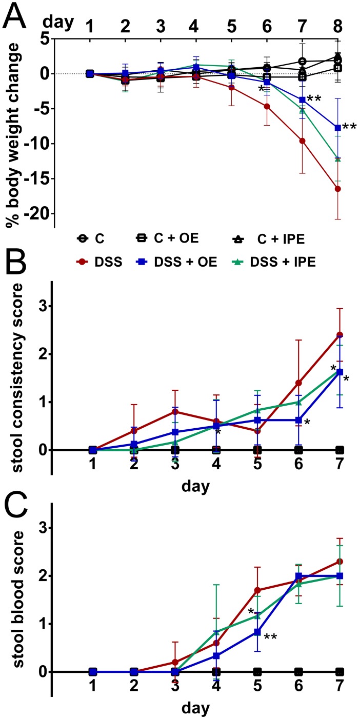
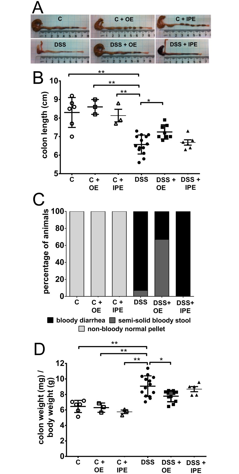
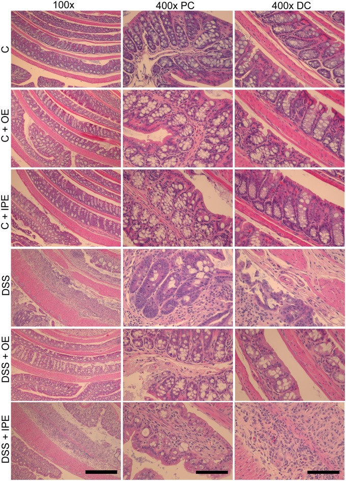
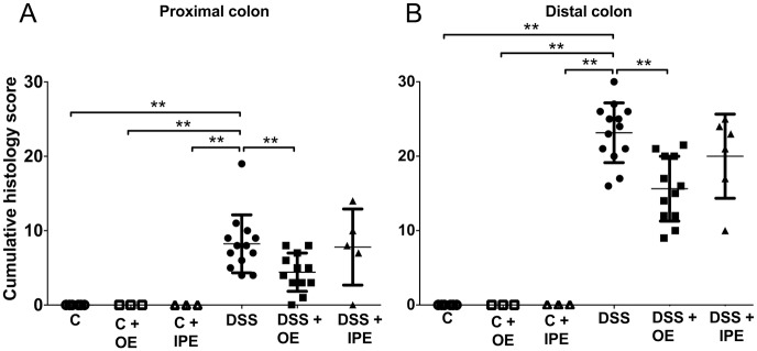
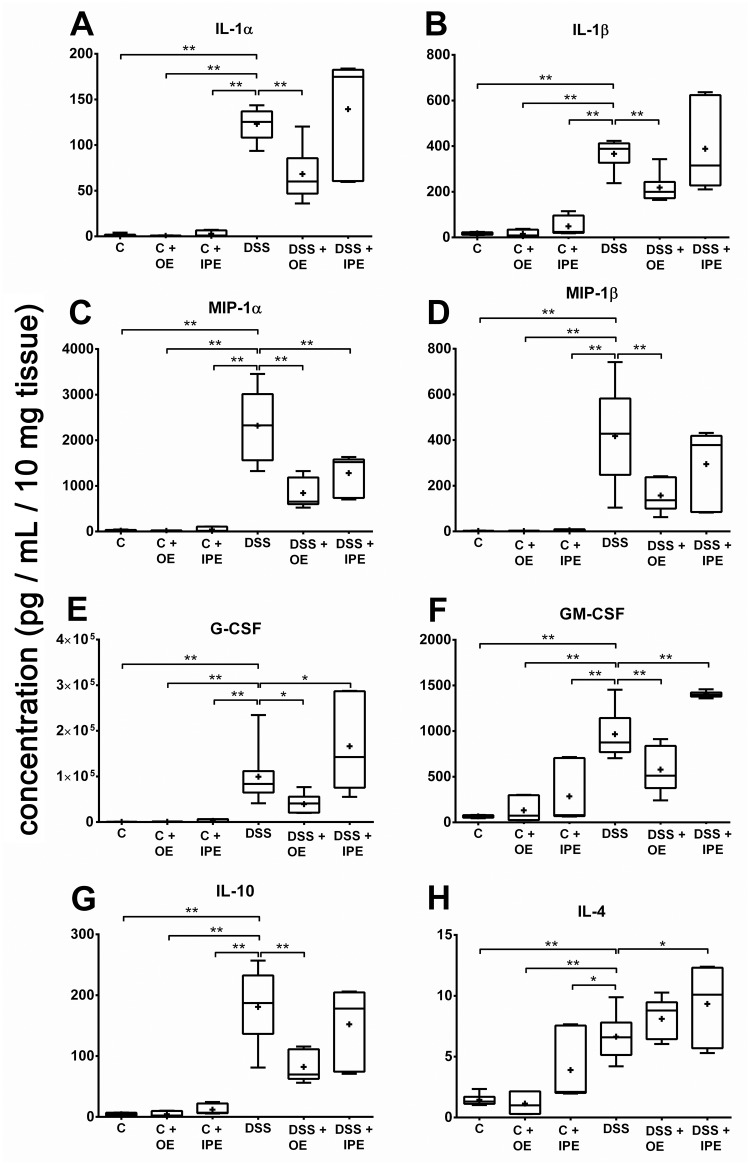
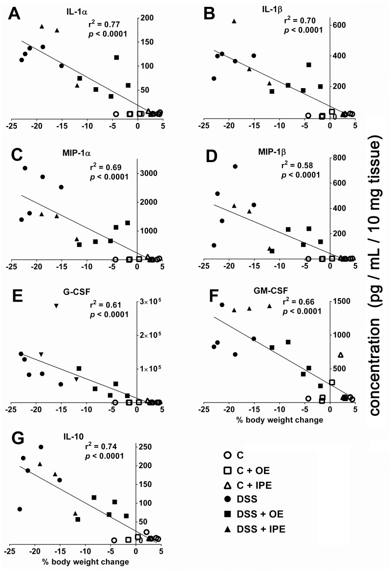
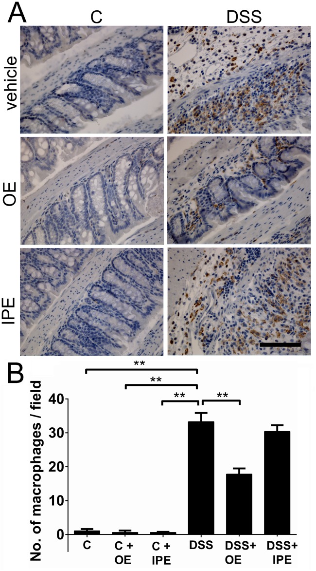
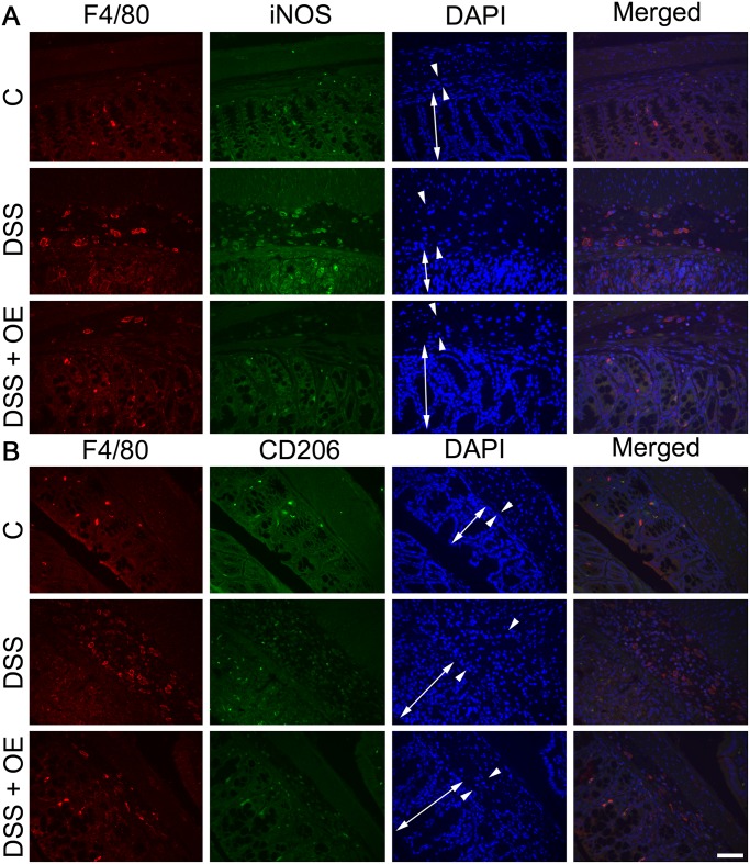
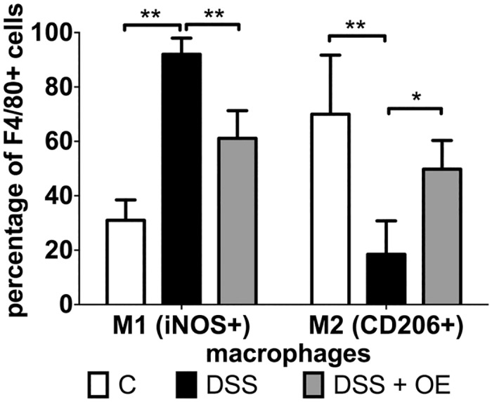
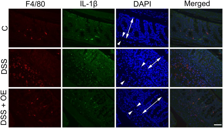
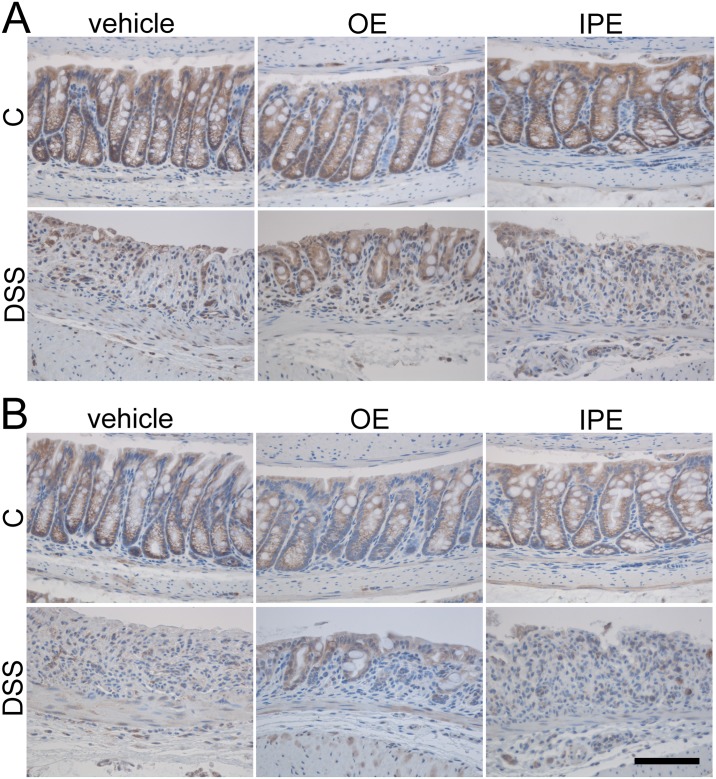
Inflammatory bowel diseases, such as ulcerative colitis, cause significant morbidity and decreased quality of life. The currently available treatments are not effective in all patients, can be expensive and have potential to cause severe side effects. This prompts the need for new treatment modalities. Enoxaparin, a widely used antithrombotic agent, is reported to possess anti-inflammatory properties and therefore we evaluated its therapeutic potential in a mouse model of colitis. Acute colitis was induced in male C57BL/6 mice by administration of dextran sulfate sodium (DSS). Mice were treated once daily with enoxaparin via oral or intraperitoneal administration and monitored for colitis activities. On termination (day 8), colons were collected for macroscopic evaluation and cytokine measurement, and processed for histology and immunohistochemistry. Oral but not intraperitoneal administration of enoxaparin significantly ameliorated DSS-induced colitis. Oral enoxaparin-treated mice retained their body weight and displayed less diarrhea and fecal blood loss compared to the untreated colitis group. Colon weight in enoxaparin-treated mice was significantly lower, indicating reduced inflammation and edema. Histological examination of untreated colitis mice showed a massive loss of crypt architecture and goblet cells, infiltration of immune cells and the presence of edema, while all aspects of this pathology were alleviated by oral enoxaparin. Reduced number of macrophages in the colon of oral enoxaparin-treated mice was accompanied by decreased levels of pro-inflammatory cytokines. Oral enoxaparin significantly reduces the inflammatory pathology associated with DSS-induced colitis in mice and could therefore represent a novel therapeutic option for the management of ulcerative colitis.


Conflict of interest statement

Competing Interests: Nuri Gueven acts as academic editor for PLOS ONE. This does not alter the authors’ adherence to PLOS ONE policies on sharing data and materials.

Figures

Fig 1
Fig 1. Effect of enoxaparin during acute colitis.
(A) Daily body weight changes during colitis induction in C57BL/6 mice with and without enoxaparin compared to healthy control. Stool samples collected from mice were scored for (B) consistency and the presence of (C) occult blood on a daily basis during experiment. Results are expressed as mean ± SD of n = 3–14 mice. * p < 0.05 and ** p < 0.01. Control, C; control with oral enoxaparin, C+OE; control with intaperitoneal injection of enoxaparin, C+IPE; untreated colitis, DSS; colitis with oral enoxaparin, DSS+OE; colitis with intraperitoneal injection of enoxaparin, DSS+IPE.
Fig 2
Fig 2. Effect of enoxaparin on macroscopic appearance of colon.
(A) Representative images of colons from mice treated with and without enoxaparin. (B) The colons were measured for their length. (C) The appearance of colon luminal content was evaluated before the measurement of colon weight. (D) The relative colon weight was presented as colon weight divided by body weight. Results are expressed as mean ± SD of n = 3–14 mice. * p < 0.05 and ** p < 0.01. Control, C; control with oral enoxaparin, C+OE; control with intraperitoneal injection of enoxaparin, C+IPE; untreated colitis, DSS; colitis with oral enoxaparin, DSS+OE; colitis with intraperitoneal injection of enoxaparin, DSS+IPE.
Fig 3
Fig 3. Effect of enoxaparin on histological changes of colon.
Representative hematoxylin and eosin stained colon sections of healthy and colitis mice with and without enoxaparin. Scale bars = 100 μm for 400 × and 400 μm for 100 × magnification. Control, C; control with oral enoxaparin, C+OE; control with intraperitoneal injection of enoxaparin, C+IPE; untreated colitis, DSS; colitis with oral enoxaparin, DSS+OE; colitis with intraperitoneal injection of enoxaparin, DSS+IPE.
Fig 4
Fig 4. Effect of enoxaparin on histological changes of colon.
Cumulative histology damage scores for (A) proximal colon and (B) distal colon. Results are expressed as mean ± SD of n = 3–14 mice. ** p < 0.01. Control, C; control with oral enoxaparin, C+OE; control with intraperitoneal injection of enoxaparin, C+IPE; untreated colitis, DSS; colitis with oral enoxaparin, DSS+OE; colitis with intraperitoneal injection of enoxaparin, DSS+IPE.
Fig 5
Fig 5. Effect of enoxaparin on colonic cytokine levels.
Distal colon tissues of mice were cultured for 24 hours. Supernatants were collected and cytokine levels were measured using Bio-Plex assay. Cytokine levels in the supernatant were normalized to tissue weight to obtain pg / mL of cytokines/ 10 mg of tissue. Results are expressed as minimum, 25th percentile, median, mean, 75th percentile and maximum of cytokine levels of 3–5 mice. * p < 0.05 and ** p < 0.01. Interleukin, IL; macrophage inflammatory protein, MIP; granulocyte colony-stimulating factor, G-CSF; granulocyte–macrophage colony-stimulating factor, GM-CSF; Control, C; control with oral enoxaparin, C+OE; control with intraperitoneal injection of enoxaparin, C+IPE; untreated colitis, DSS; colitis with oral enoxaparin, DSS+OE; colitis with intraperitoneal injection of enoxaparin, DSS+IPE.
Fig 6
Fig 6. Correlation between colonic cytokine levels and body weight changes during acute colitis.
Changes in cytokine levels were correlated with the changes of body weight of individual mice. The value of the Pearson correlation coefficient (r2) is reported and significance is indicated by p value. Interleukin, IL; macrophage inflammatory protein, MIP; granulocyte colony-stimulating factor, G-CSF; granulocyte–macrophage colony-stimulating factor, GM-CSF; Control, C; control with oral enoxaparin, C+OE; control with intraperitoneal injection of enoxaparin, C+IPE; untreated colitis, DSS; colitis with oral enoxaparin, DSS+OE; colitis with intraperitoneal injection of enoxaparin, DSS+IPE.
Fig 7
Fig 7. Effect of enoxaparin on macrophage infiltration into the inflamed colon.
(A) Representative immunostaining of F4/80-positive macrophages in in the distal colon from healthy and colitic mice treated with and without enoxaparin. (B) Quantification of macrophages (F4/80+ cells) in the colons. Results are expressed as mean ± SD of ten representative high-power fields per tissue section of 3–5 mice each. ** p < 0.01. Scale bar = 100 μm for 400 × magnification. Control, C; untreated colitis, DSS; oral enoxaparin, OE; intraperitoneal injection of enoxaparin, IPE.
Fig 8
Fig 8. Effect of enoxaparin on macrophage phenotypes.
Co-immunostaining of macrophages and their phenotypes. Representative images of (A) M1 macrophages (F4/80+ and iNOS+) and (B) M2 macrophages (F4/80+ and CD206+) using colon tissue from n = 3–5 mice. F4/80 positive cells were visualized using Alexa Fluor 594-conjugated goat anti-rat IgG (red) and iNOS or CD206 positive cells using Alexa Fluor 488-conjugated goat anti-rabbit IgG (green). Nuclei were stained with 4’,6-diamidino-2-phenylindole (DAPI, blue). Localization of mucosa (double-headed arrow) and submucosa (arrowheads) is indicated. Scale bar = 50 μm for 400 × magnification. Control, C; untreated colitis, DSS; colitis with oral enoxaparin, DSS+OE.
Fig 9
Fig 9. Effect of enoxaparin on macrophage phenotypes.
Quantification of macrophages expressing M1 and M2 phenotype markers in the colon. Results are expressed as percentage of double-positive macrophages from total macrophages ± SD of five representative high-power fields per tissue section of n = 3–5 mice each. * p < 0.05 and ** p < 0.01.
Fig 10
Fig 10. Effect of enoxaparin on IL-1β expression on colon tissues.
Co-immunostaining of macrophages and IL-1β. Representative images of IL-1β and F4/80 staining of colons from n = 3–5 mice. F4/80 positive cells were visualized using Alexa Fluor 594-conjugated goat anti-rat IgG (red) and IL-1β positive cells using Alexa Fluor 488-conjugated goat anti-rabbit IgG (green). Nuclei were stained with 4’,6-diamidino-2-phenylindole (DAPI, blue). Localization of mucosa (double-headed arrow) and submucosa (arrowheads) is indicated. Scale bar = 50 μm for 400 × magnification. Control, C; untreated colitis, DSS; colitis with oral enoxaparin, DSS+OE.
Fig 11
Fig 11. Effect of enoxaparin on epithelial tight junctions of colon.
Immunostaining of (A) occludin and (B) claudin-4 in the distal colon of healthy and colitis mice treated with and without enoxaparin. Scale bar = 100 μm for 400 × magnification. Control, C; untreated colitis, DSS; oral enoxaparin, OE; intraperitoneal injection of enoxaparin, IPE.

References

    1. Cosnes J, Gower-Rousseau C, Seksik P, Cortot A. Epidemiology and natural history of inflammatory bowel diseases. Gastroenterology. 2011;140: 1785–1794. 10.1053/j.gastro.2011.01.055 - DOI - PubMed
    1. Ordás I, Eckmann L, Talamini M, Baumgart DC, Sandborn WJ. Ulcerative colitis. Lancet. 2012;380: 1606–1619. 10.1016/S0140-6736(12)60150-0 - DOI - PubMed
    1. Triantafillidis JK, Merikas E, Georgopoulos F. Current and emerging drugs for the treatment of inflammatory bowel disease. Drug Des Devel Ther. 2011;5: 185–210. 10.2147/DDDT.S11290 - DOI - PMC - PubMed
    1. Sands BE, Kaplan GG. The role of TNFα in ulcerative colitis. J Clin Pharmacol. 2007;47: 930–941. - PubMed
    1. Ferrante M, Vermeire S, Fidder H, Schnitzler F, Noman M, Van Assche G, et al. Long-term outcome after infliximab for refractory ulcerative colitis. J Crohns Colitis. 2008;2: 219–225. 10.1016/j.crohns.2008.03.004 - DOI - PubMed

Publication types

MeSH terms