

Expression and clinical implication of cyclooxygenase-2 and E-cadherin in oral squamous cell carcinomas
- PMID: 26218314
- PMCID: PMC7537792
- DOI: 10.1080/15384047.2015.1071741
Expression and clinical implication of cyclooxygenase-2 and E-cadherin in oral squamous cell carcinomas
Abstract
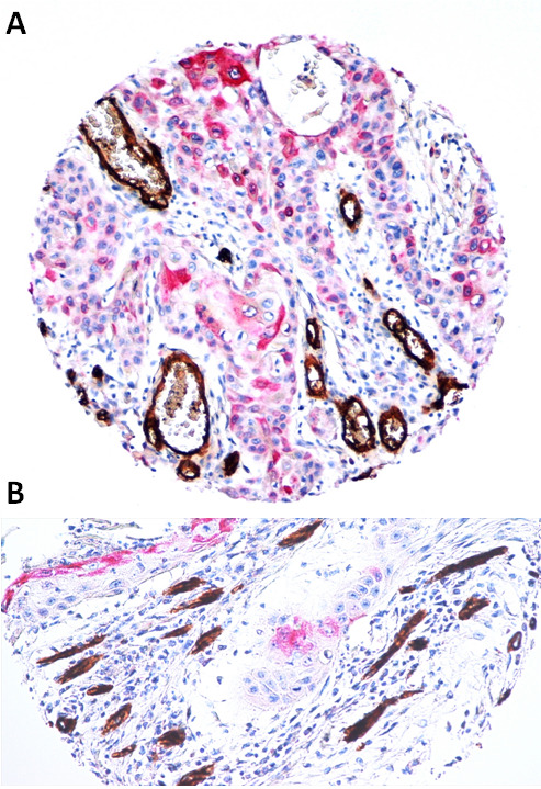
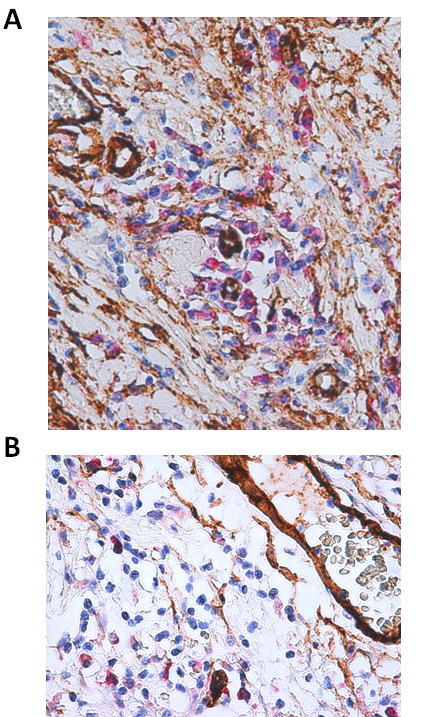
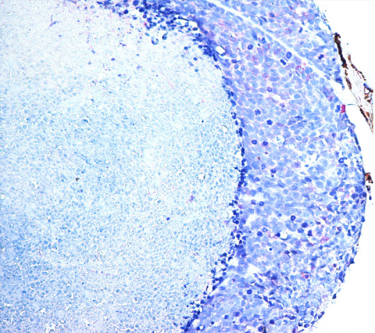
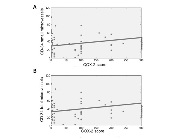
Epithelial-Mesenchymal Transition (EMT) and angiogenesis are crucial events for development of aggressive and often fatal Oral Squamous Cell Carcinomas (OSCCs). Both promote cancer progression and metastasis development, but while the former induces the loss of E-cadherin expression and, hence cadherin switching; the latter produces hematic blood vessel neo-formation and contribute to OSCC cell growth, tumor mass development, and dissemination. Cyclooxygenase-2 (COX-2) has an important role, not only in angiogenic mechanisms, but also in favoring cancer invasion. Indeed it decreases the expression of E-cadherin and leads to phenotypic changes in epithelial cells (EMT) enhancing their carcinogenic potential. Our aim is to evaluate the interplay between E-cadherin cytoplasmic delocalization, COX-2 up-regulation and COX-2 induced neo-angiogenesis in 120 cases of OSCC. We have analyzed the distribution and the number of neo-formed endothelial buds surrounding infiltrating cells that express COX-2, as well as the neo-formed vessels in chronic inflammatory infiltrate, which surround the tumor. A double immunostaining method was employed in order to verify co-localization of endothelial cell marker (CD34) and COX-2. IHC has also been used to assess E-cadherin expression. Our data demonstrate that the OSCC cells, which lose membranous E-cadherin staining, acquiring a cytoplasmic delocalization, overexpress COX-2. Moreover, we find a new CD34+ vessel formation (sprouting angiogenesis). Only basaloid type of OSCC showes low level of COX-2 expression together with very low level of neo-angiogenesis and consequent tumor necrosis. The well-known anti-metastatic effect of certain COX-2 inhibitors suggests that these molecules might have clinical utility in the management of advanced cancers.
Keywords: Prostaglandins; CD-34; Cox-2; E-cadherin; OSCC; TMA; neo-angiogenesis; tumor microenvironment.
Figures
Similar articles
-
Restoration of E-cadherin expression by selective Cox-2 inhibition and the clinical relevance of the epithelial-to-mesenchymal transition in head and neck squamous cell carcinoma.J Exp Clin Cancer Res. 2014 May 10;33(1):40. doi: 10.1186/1756-9966-33-40. J Exp Clin Cancer Res. 2014. PMID: 24887090 Free PMC article.
-
The role of E-cadherin down-regulation in oral cancer: CDH1 gene expression and epigenetic blockage.Curr Cancer Drug Targets. 2014;14(2):115-27. doi: 10.2174/1568009613666131126115012. Curr Cancer Drug Targets. 2014. PMID: 24274398
-
Inactivation of homeodomain-interacting protein kinase 2 promotes oral squamous cell carcinoma metastasis through inhibition of P53-dependent E-cadherin expression.Cancer Sci. 2021 Jan;112(1):117-132. doi: 10.1111/cas.14691. Epub 2020 Nov 9. Cancer Sci. 2021. PMID: 33063904 Free PMC article.
-
Epithelial-to-mesenchymal transition in oral squamous cell carcinoma: Challenges and opportunities.Int J Cancer. 2021 Apr 1;148(7):1548-1561. doi: 10.1002/ijc.33352. Epub 2020 Oct 29. Int J Cancer. 2021. PMID: 33091960 Review.
-
A Bloody Conspiracy- Blood Vessels and Immune Cells in the Tumor Microenvironment.Cancers (Basel). 2022 Sep 21;14(19):4581. doi: 10.3390/cancers14194581. Cancers (Basel). 2022. PMID: 36230504 Free PMC article. Review.
Cited by
-
The Role of Prostaglandins in Different Types of Cancer.Cells. 2021 Jun 13;10(6):1487. doi: 10.3390/cells10061487. Cells. 2021. PMID: 34199169 Free PMC article. Review.
-
Loss of E-Cadherin Expression Correlates With Ki-67 in Head and Neck Squamous Cell Carcinoma.In Vivo. 2022 May-Jun;36(3):1150-1154. doi: 10.21873/invivo.12814. In Vivo. 2022. PMID: 35478157 Free PMC article.
-
Sex Hormones and Inflammation Role in Oral Cancer Progression: A Molecular and Biological Point of View.J Oncol. 2020 Jun 27;2020:9587971. doi: 10.1155/2020/9587971. eCollection 2020. J Oncol. 2020. PMID: 32684934 Free PMC article. Review.
-
Expression of PTGS2 along with genes regulating VEGF signalling pathway and association with high-risk factors in locally advanced oral squamous cell carcinoma.Cancer Med. 2024 Feb;13(3):e6986. doi: 10.1002/cam4.6986. Cancer Med. 2024. PMID: 38426619 Free PMC article.
-
Staphylococcus aureus induces COX-2-dependent proliferation and malignant transformation in oral keratinocytes.J Oral Microbiol. 2019 Jul 22;11(1):1643205. doi: 10.1080/20002297.2019.1643205. eCollection 2019. J Oral Microbiol. 2019. PMID: 31448061 Free PMC article.
References
-
- Pannone G, Santoro A, Feola A, Bufo P, Papagerakis P, Lo Muzio L, Staibano S, Ionna F, Longo F, Franco R, et al. The role of E-cadherin down-regulation in oral cancer: CDH1 gene expression and epigenetic blockage. Curr Cancer Drug Targets. 2014;14(2):115–127. doi:10.2174/1568009613666131126115012. - DOI - PubMed
-
- Higgins DF, Kimura K, Bernhardt WM, Shrimanker N, Akai Y, Hohenstein B, Saito Y, Johnson RS, Kretzler M, Cohen CD, et al. Hypoxia promotes fibrogenesis in vivo via HIF-1 stimulation of epithelial to-mesenchymal transition. J Clin Invest. 2007;117(12):3810–3820. doi:10.1172/JCI30487. - DOI - PMC - PubMed
Publication types
MeSH terms
Substances
LinkOut - more resources
Full Text Sources
Other Literature Sources
Medical
Research Materials