. 2016 Dec;31(12):1720-1730.
doi: 10.1002/tox.22174. Epub 2015 Jul 27.

The impairment of learning and memory and synaptic loss in mouse after chronic nitrite exposure

Affiliations

The impairment of learning and memory and synaptic loss in mouse after chronic nitrite exposure

Yongfang Chen et al. Environ Toxicol. 2016 Dec.

Abstract

The objective of this study is to understand the impairment of learning and memory in mouse after chronic nitrite exposure. The animal model of nitrite exposure in mouse was created with the daily intubation of nitrite in adult healthy male mice for 3 months. Furthermore, the mouse's learning and memory abilities were tested with Morris water maze, and the expression of Synaptophysin and γ-Synuclein was visualized with immunocytochemistry and Western blot. Our results showed that nitrite exposure significantly prolonged the escape latency period (ELP) and decreased the values of the frequency across platform (FAP) as well as the accumulative time in target quadrant (ATITQ) compared to control, in dose-dependent manner. In addition, after nitrite exposure, synaptophysin (SYN) positive buttons in the visual cortex was reduced, in contrast the increase of γ-synuclein positive cells. The results above were supported by Western blot as well. We conclude that nitrite exposure could lead to a decline in mice's learning and memory. The overexpression of γ-synuclein contributed to the synaptic loss, which is most likely the cause of learning and memory impairment. © 2015 Wiley Periodicals, Inc. Environ Toxicol 31: 1720-1730, 2016.

Keywords: learning and memory; nitrite exposure; synapse; γ-synuclein.


Figures

Figure 1
Figure 1
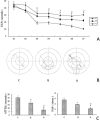
Learning and memory of mice after nitrite exposure. A: Escape latency period (ELP) was measured in various groups at different time. During the first 2 days, there was no difference among various groups (P > 0.05). After day 3, ELP was longer in nitrite exposure groups compared to control with dose dependency (P < 0.05), and from day 7 onward, the differentiation became more significant (P < 0.01). B shows the orbits of platform seeking in various groups at day 7. C: the frequency of target quadrant search (FAP) and the accumulative time in target quadrant (ATITQ) decreased after nitrate exposure with dose dependency (P < 0.05). *: P < 0.05 for nitrate treatments versus control; #: P < 0.05 for high‐ versus moderate‐dose treatment. Control, moderate and high dose treatment are marked with C, M, and H, respectively.
Figure 2
Figure 2
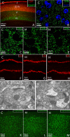
Synapses and γ‐synuclein positive cells in visual cortex (immunolabeling). Photo A shows the lamination of cortex with CDP (red) and FOXP2 immunolabeling. CDP was mainly expressed in layer II–IV, and FOXP2 was expressed in layer V and VI which was selected as target area. Photo B shows the high magnification of presynaptic buttons (green, synaptophysin immunolabeling) with DAPI counterstaining (blue). After nitrate exposure, the Synaptophysin positive buttons decreased with dose dependency (C–E). In the meantime, the dendritic spines were also changed after nitrite exposure, and nitrite exposure could cause dendrites to appear sparser and longer with dose dependency (F–H). I–J: Changes in synaptic ultrastructure following nitrite exposure. The typical synaptic ultrastructure with normal presynaptic membrane, synaptic cleft and postsynaptic membrane can be seen in control (I). After high dose exposure, dark and thick postsynaptic density appears prominently in the postsynaptic membrane, accompanied by a narrowing of the synaptic cleft (J). On the other hand, the γ‐Synuclein positive cells increased after nitrite treatment (K–M). Control, moderate‐ and high‐dose treatment are marked with C, M, and H, respectively.
Figure 3
Figure 3
Histogram and statistical analysis of synaptophysin positive buttons and γ‐synuclein positive cells. Nitrite exposure can induce the loss of presynaptic buttons (P < 0.01, A) and the increase of γ‐synuclein positive cells with dose dependency (P < 0.05, B). Columns are the mean values ± standard deviation (SD). Statistical analysis was performed with one‐way analysis of variance (ANOVA) q tests. ∗: P < 0.05, if nitrite treatments versus control; #: P < 0.05, if high‐ versus moderate‐dose treatment. Control, moderate‐ and high‐dose treatment are marked with C, M, and H, respectively.
Figure 4
Figure 4
Synaptophysin expression in visual cortex by Western blot. A: the expression bands of Synaptophysin in various groups, and β‐actin was used as internal control. B: Semi‐quantitative analysis of Synaptophysin was made in various groups. ∗P < 0.05, if nitrite treatments versus control. #: P < 0.05, if high‐ versus moderate‐dose treatment. Control, moderate‐ and high‐dose treatment are marked with C, M, and H, respectively.
Figure 5
Figure 5
Dendritic spine length and spine density following nitrite exposure. A: Nitrite exposure could lead to a reduction in dendritic spine density with dose dependency. B: Nitrite exposure induces the elongation of dendritic spines with dose dependency. Columns are the mean values ± standard deviation (SD). Statistical analysis was made with one‐way analysis of variance (ANOVA) q tests. ∗P < 0.05, if nitrate treatments versus control. #: P < 0.05, if high‐ versus moderate‐dose treatment. Control, moderate‐ and high‐dose treatment are marked with C, M, and H, respectively.

References

    1. Bankiewicz KS, Plunkett RJ, Jacobowitz DM, Porrino L, di Porzio U, London WT, Kopin IJ, Oldfield EH. 1990. The effect of fetal mesencephalon implants on primate MPTP‐induced parkinsonism: Histochemical and behavioral studies. J Neurosurg 72:231–244. - PubMed
    1. Bliss TVP, Lømo T. 1973. Long‐lasting potentiation of synaptic transmission in the dentate area of the anaesthetized rabbit following stimulation of the perforant path. J Physiol 232:331–356. - PMC - PubMed
    1. Brender JD, Olive JM, Felkner M, Suarez L, Marckwardt W, Hendricks KA. 2004. Dietary nitrites and nitrates, nitrosatable drugs, and neural tube defects. Epidemiology 15:330–336. - PubMed
    1. Buchman VL, Hunter HJ, Pinõn LG, Thompson J, Privalova EM, Ninkina NN, Davies AM. 1998. Persyn, a member of the synuclein family, has a distinct pattern of expression in the developing nervous system. J Neurosci 18:9335–9341. - PMC - PubMed
    1. Calkins MJ, Reddy PH. 2011. Amyloid beta impairs mitochondrial anterograde transport and degenerates synapses in Alzheimer's disease neurons. Biochim Biophys Acta 1812:507–513. - PMC - PubMed