. 2015 Jul 10;6(19):17016-30.
doi: 10.18632/oncotarget.4849.

Deletion of Smad3 improves cardiac allograft rejection in mice

Affiliations

Deletion of Smad3 improves cardiac allograft rejection in mice

Ying-Ying Wang et al. Oncotarget. .

Abstract

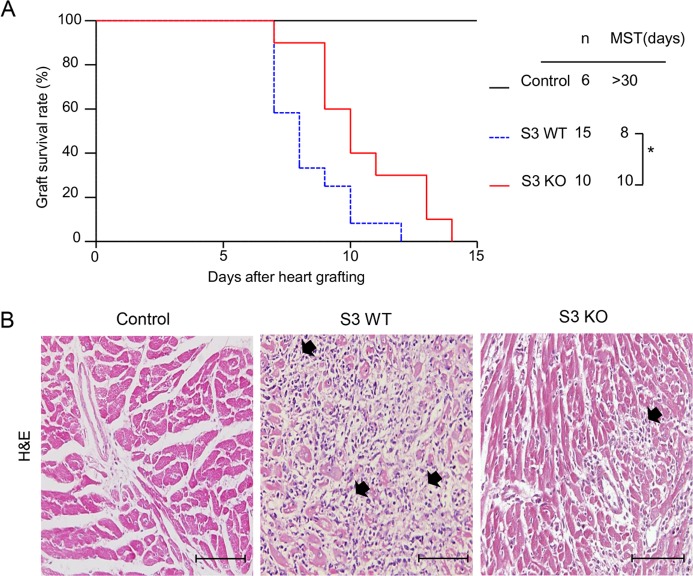
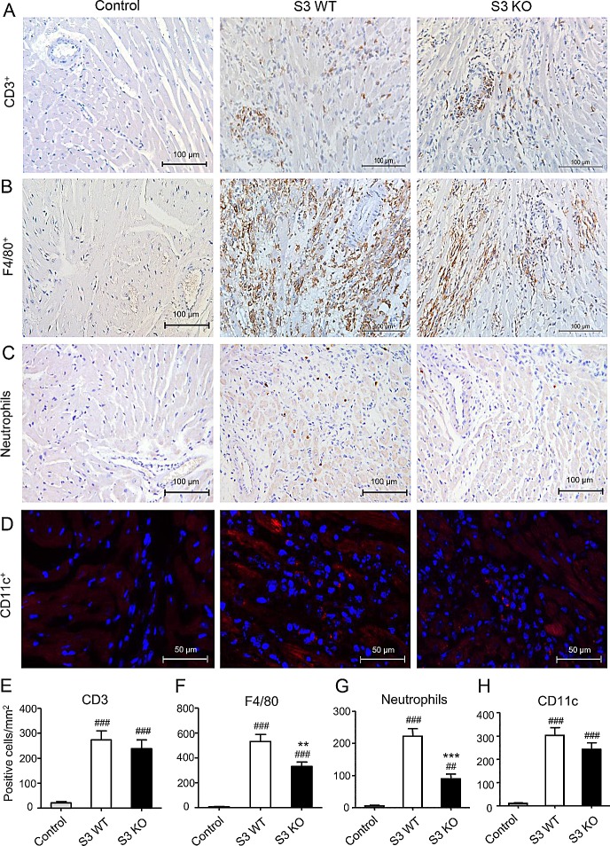
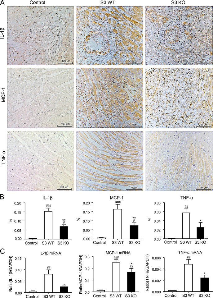
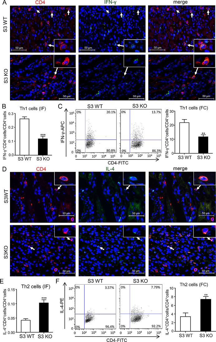
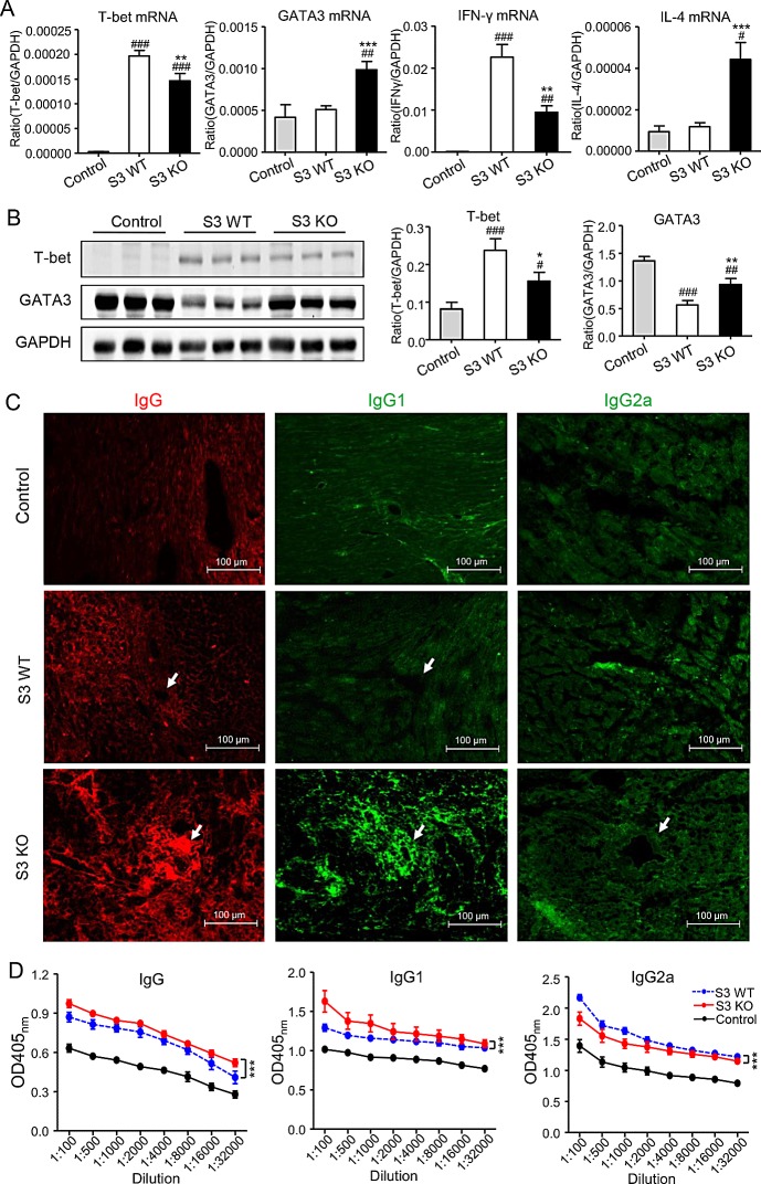
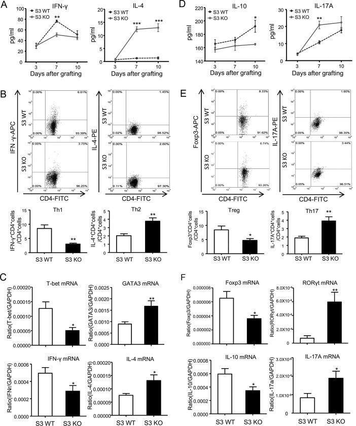
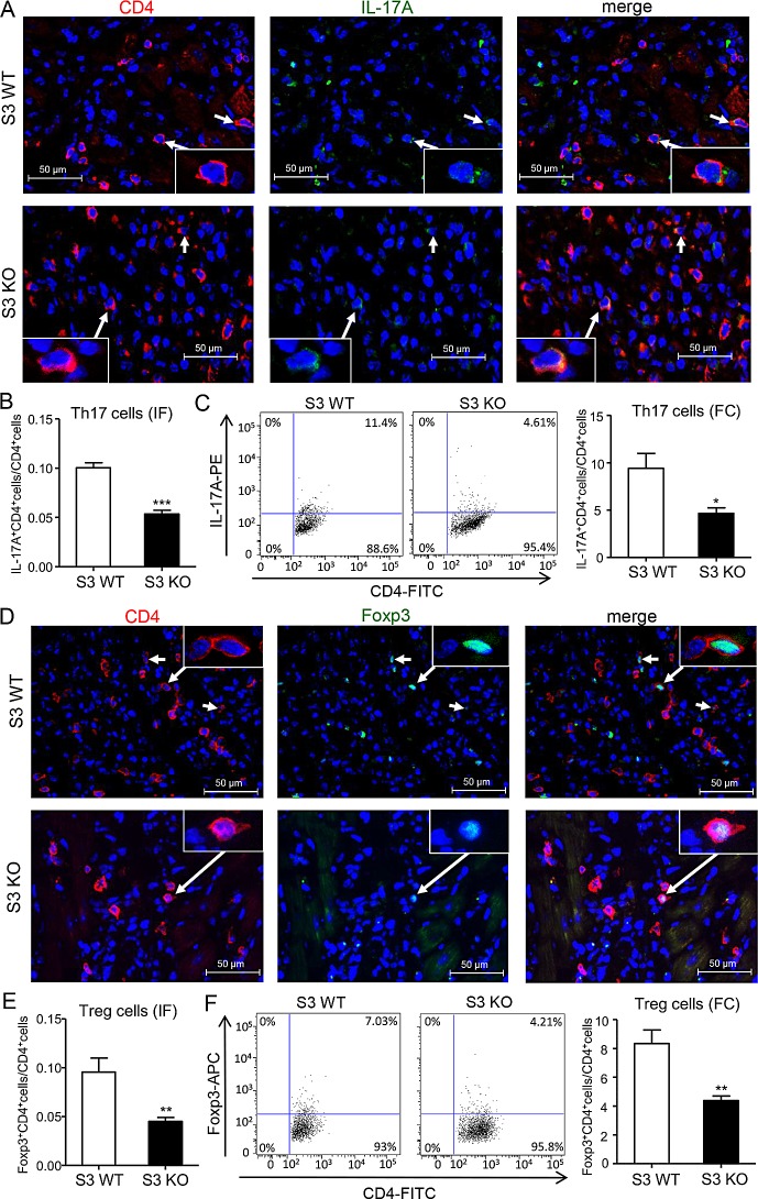
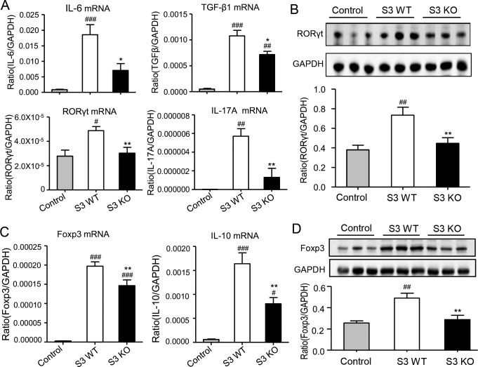
T cells play a critical role in acute allograft rejection. TGF-β/Smad3 signaling is a key pathway in regulating T cell development. We report here that Smad3 is a key transcriptional factor of TGF-β signaling that differentially regulates T cell immune responses in a mouse model of cardiac allograft rejection in which donor hearts from BALB/c mice were transplanted into Smad3 knockout (KO) and wild type (WT) mice. Results showed that the cardiac allograft survival was prolonged in Smad3 KO recipients. This allograft protection was associated with a significant inhibition of proinflammatory cytokines (IL-1β, TNF-α, and MCP-1) and infiltration of neutrophils, CD3+ T cells, and F4/80+ macrophages. Importantly, deletion of Smad3 markedly suppressed T-bet and IFN-γ while enhancing GATA3 and IL-4 expression, resulting in a shift from the Th1 to Th2 immune responses. Furthermore, mice lacking Smad3 were also protected from the Th17-mediated cardiac injury, although the regulatory T cell (Treg) response was also suppressed. In conclusion, Smad3 is an immune regulator in T cell-mediated cardiac allograft rejection. Loss of Smad3 results in a shift from Th1 to Th2 but suppressing Th17 immune responses. Thus, modulation of TGF-β/Smad3 signaling may be a novel therapy for acute allograft rejection.

Keywords: Pathology Section; Smad3; Th1; Th17; Th2; cardiac allograft rejection.


Conflict of interest statement

CONFLICTS OF INTEREST

The authors declare that there are no conflicts of interest.

Figures

Figure 1
Figure 1. Deletion of Smad3 from the recipients significantly improves cardiac allograft rejection
A. Cardiac allograft survival rate. B. Histologic changes (H&E staining) in cardiac allografts on postoperative days (POD) 7, black arrows show the loci of myocyte damage. Results show that deletion of Smad3 from the recipients results in prolonged allograft survival and less histological damage. *p < 0.05 versus Smad3 WT mice; MST, median survival time; Scale bar = 100 μm.
Figure 2
Figure 2. Deletion of Smad3 inhibits infiltration of inflammatory cells in cardiac allograft rejection
A. Immunohistochemistry shows CD3+ T cells, B. F4/80+ macrophages, C. neutrophils, and D. CD11c+ dendritic cells infiltration in cardiac allografts on POD 7. (E-H) Quantitative data of CD3+ cells, F4/80+ cells, neutrophils and CD11c+ cells infiltrating the allograft tissues. Data represent mean ± SEM for groups of 6 mice. **p < 0.01, ***p < 0.01 versus Smad3 WT mice; ##p < 0.001, ###p < 0.001 versus control mice (BALB/c mice received cardiac allografts from BALB/c mice).
Figure 3
Figure 3. Improvement of cardiac allograft rejection in Smad3 deficient recipients is associated with inhibition of upregulation of proinflammatory cytokines
A. Expression levels of IL-1β, MCP-1 and TNF-α in cardiac allografts on POD 7 by immunohistochemistry. B. Quantitative data of IL-1β, MCP-1 and TNF-α from immunohistochemically stained tissues. C. Real-time PCR for IL-1β, MCP-1 and TNF-α mRNA expression in cardiac allografts on POD 7. Data are expressed as the mean ± SEM. *p < 0.05, **p < 0.01 versus Smad3 WT mice; #p < 0.05, ##p < 0.01, ###p < 0.001 versus control mice.
Figure 4
Figure 4. Deletion of Smad3 suppresses Th1 but enhances Th2 cells infiltrating the cardiac allografts
A. Two-color immunofluorescence for detecting Th1 cells (CD4+IFN-γ+) infiltrating the allografts on POD 7. B. Quantitative data of Th1 cells on two-color immunofluorescence-staining sections. C. Quantitative analysis of CD4+IFN-γ+ cells by two-color flow cytometry in cardiac allografts of Smad3 KO and WT recipients on POD 7. D. Two-color immunofluorescence for detecting Th2 cells (CD4+IL-4+) infiltrating the allografts on POD 7. B. Quantitative data of Th2 cells on two-color immunofluorescence-staining sections. C. Quantitative analysis of CD4+IL-4+ cells by two-color flow cytometry in cardiac allografts of Smad3 KO and WT recipients on POD 7. Data are expressed as the mean ± SEM. **p < 0.01, ***p < 0.001 versus Smad3 WT mice.
Figure 5
Figure 5. Smad3-deficient mice are more likely to suffer a Th2-type immune response, while suppressing Th1-type immune response in cardiac allografts
A. Real-time PCR for T-bet, GATA3, IFN-γ and IL-4 mRNA expression in cardiac allografts on POD 7. B. Western blot analysis shows the expression of T-bet and GATA3 in cardiac allografts in control, Smad3 KO and WT recipients on POD 7. C. Immunofluorescence shows the total IgG, IgG1 and IgG2a deposition within the cardiac allograft tissues on POD 7. D. Serum levels of IgG, IgG1 and IgG2a in control, Smad3 KO and WT recipients on POD 7 detected by ELISA. Data are expressed as the mean ± SEM. *p < 0.05, **p < 0.01, ***p < 0.001 versus Smad3 WT mice; #p < 0.05, ##p < 0.01, ###p < 0.001 versus control mice.
Figure 6
Figure 6. Deletion of Smad3 differentially regulates T cell differentiation during cardiac allograft rejection
A., D. Serum levels of IFN-γ, IL-4, IL-10 and IL-17A in Smad3 KO and WT recipients on POD 7 detected by ELISA. B., E. Quantitative analysis of CD4+IFN-γ+ cells, CD4+IL-4+ cells, CD4+Foxp3+ cells and CD4+IL-17A+ cells by two-color flow cytometry analysis in the spleen of Smad3 KO and WT recipients on POD 7. C., F. Real-time PCR analysis of T-bet, GATA3, Foxp3, RORγt, IFN-γ, IL-4, IL-10 and IL-17A mRNA expression in the spleen of Smad3 KO and WT recipients on POD 7. Data are expressed as the mean ± SEM. *p < 0.05, **p < 0.01, ***p < 0.001 versus Smad3 WT mice.
Figure 7
Figure 7. Deletion of Smad3 suppresses Th17 and Treg cells infiltrating the cardiac allografts
A. Two-color immunofluorescence for detecting Th17 cells (CD4+IL17A+) infiltrating the allografts on POD 7. B. Quantitative data of Th17 cells on two-color immunofluorescence-staining sections. C. Quantitative analysis of CD4+IL17A+ cells by two-color flow cytometry in cardiac allografts of Smad3 KO and WT recipients on POD 7. D. Two-color immunofluorescence for detecting Treg cells (CD4+Foxp3+) infiltrating the allografts on POD 7. B. Quantitative data of Treg cells on two-color immunofluorescence-staining sections. C. Quantitative analysis of CD4+Foxp3+ cells by two-color flow cytometry in cardiac allografts of Smad3 KO and WT recipients on POD 7. Data are expressed as the mean ± SEM. *p < 0.05, **p < 0.01, ***p < 0.001 versus Smad3 WT mice.
Figure 8
Figure 8. Deletion of Smad3 from the recipients suppresses Th17 and Treg immune response in cardiac allografts
A. Real-time PCR for IL-6, TGF-β1, RORγt, and IL-17A mRNA expression in cardiac allografts on POD 7. B. Western blot analysis shows the expression of RORγt in cardiac allografts in control, Smad3 KO and WT recipients on POD 7. C. Real-time PCR for Foxp3 and IL-10 mRNA expression in cardiac allografts on POD 7. D. Western blot analysis shows the expression of RORγt in cardiac allografts in control, Smad3 KO and WT recipients on POD 7. Data are expressed as the mean ± SEM. *p < 0.05, **p < 0.01 versus Smad3 WT mice; #p < 0.05, ##p < 0.01, ###p < 0.001 versus control mice.

References

    1. Wood KJ, Bushell A, Hester J. Regulatory immune cells in transplantation. Nature Reviews Immunology. 2012;12:417–430. - PubMed
    1. Krieger NR, Yin D, Fathman CG. CD4+ but not CD8+ cells are essential for allorejection. The Journal of experimental medicine. 1996;184:2013–2018. - PMC - PubMed
    1. Plenter RJ, Grazia TJ, Doan AN, Gill RG, Pietra BA. CD4 T cells mediate cardiac xenograft rejection via host MHC Class II. The Journal of Heart and Lung Transplantation. 2012;31:1018–1024. - PMC - PubMed
    1. Li MO, Flavell RA. TGF-β: a master of all T cell trades. Cell. 2008;134:392–404. - PMC - PubMed
    1. Li MO, Wan YY, Sanjabi S, Robertson A-KL, Flavell RA. Transforming growth factor-β regulation of immune responses. Annu Rev Immunol. 2006;24:99–146. - PubMed

Publication types