. 2015 Jul 10;6(19):17777-91.
doi: 10.18632/oncotarget.4397.

Purification and identification of a polysaccharide from medicinal mushroom Amauroderma rude with immunomodulatory activity and inhibitory effect on tumor growth

Affiliations

Purification and identification of a polysaccharide from medicinal mushroom Amauroderma rude with immunomodulatory activity and inhibitory effect on tumor growth

Honghui Pan et al. Oncotarget. .

Abstract

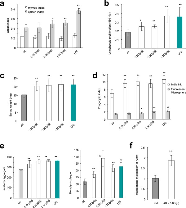
Medicinal mushrooms in recent years have been the subject of many experiments searching for anticancer properties. We previously screened thirteen mushrooms for their potential in inhibiting tumor growth, and found that the water extract of Amauroderma rude exerted the highest activity. Previous studies have shown that the polysaccharides contained in the water extract were responsible for the anticancer properties. This study was designed to explore the potential effects of the polysaccharides on immune regulation and tumor growth. Using the crude Amauroderma rude extract, in vitro experiments showed that the capacities of spleen lymphocytes, macrophages, and natural killer cells were all increased. In vivo experiments showed that the extract increased macrophage metabolism, lymphocyte proliferation, and antibody production. In addition, the partially purified product stimulated the secretion of cytokines in vitro, and in vivo. Overall, the extract decreased tumor growth rates. Lastly, the active compound was purified and identified as polysaccharide F212. Most importantly, the purified polysaccharide had the highest activity in increasing lymphocyte proliferation. In summary, this molecule may serve as a lead compound for drug development.

Keywords: amauroderma; cytokine; herbal medicine; medicinal mushroom; tumor growth.


Figures

Figure 1
Figure 1. Effects of AR on cellular activities of mouse spleen lymphocytes and macrophages
a. Procedure for preparation of water extract from Amauroderma rude. b. Mouse spleen lymphocytes were incubated with AR extract at 37°C for 44 h, followed by MTT assay. Incubation with AR extract increased lymphocyte proliferation. Inclusion of ConA and LPS displayed additive effect on enhancing lymphocyte proliferation. Asterisks indicate significant differences. *p < 0.05, **p < 0.01. Error bars, SD (n = 5). All experiments were repeated three times. c. In energy metabolism assay, mouse macrophages were incubated at 37°C for 2 h, followed by addition of drugs or AR extract. The cultures were then incubated at 37°C for 20 h followed by MTT assays. Treatment with AR extract improved energy metabolism. Asterisks indicate significant differences. **p < 0.01. Error bars, SD (n = 5). d. In the neutral red engulfment assay, cells in the 96-well plates were incubated at 37°C for 24 h. After centrifugation (1800 rpm, 5 min), 100 μl of neutral red in physiological saline solution (0.1%) were added to each well. The plates were incubated at 37°C for 30 min. After centrifugation (1800 rpm, 5 min), each well was washed with 200 μl PBS twice, followed by addition of 100 μl cytolysate (acetic acid:anhydrous alcohol = 50:50). The cells were incubated at room temperature overnight followed by MTT assay. Incubation with AR extract increased engulfment of neutral red by the cells. Asterisks indicate significant differences. **p < 0.01. Error bars, SD (n = 5).
Figure 2
Figure 2. Effects of AR on macrophages and natural killer cells
a. Mouse macrophages were incubated at 37°C for 2 h. Drugs and samples were added to the cultures at the concentrations indicated. The cultures were incubated at 37°C for 48 h followed by Griess assay to measure nitric oxide concentrations. Incubation with AR extract increased the production of nitric oxide by macrophages. Asterisks indicate significant differences. *p < 0.05, **p < 0.01. Error bars, SD (n = 5). All experiments were repeated three times. b. In NK cell assay, the harvested lymphocytes were incubated at 37°C for 2 h followed by treatment with AR extract. The effect of NK cells was assayed as described in the Methods. Incubation with AR extract stimulated and increased the activity of NK cells in killing breast cancer cells. Asterisks indicate significant differences. *p < 0.05, **p < 0.01. Error bars, SD (n = 5). c. Lymphocytes were inoculated and incubated at 37°C for 2 h, followed by addition of drugs and samples. The cultures were then incubated at 37°C for 48 h before flow cytometry analysis for CD45 expression. Incubation with AR extract increased CD45 expression. Asterisks indicate significant differences. *p < 0.05, **p < 0.01. Error bars, SD (n = 5). Right, typical measurements of CD45 levels by flow cytometry.
Figure 3
Figure 3. The immunomodulatory activity of Amauroderma rude in normal animals
a. The in vivo experiments were described in detail in the Methods. Injection of normal mice with AR extract increased sizes of spleens, but had little effect on thymus. Similarly, injection with LPS only increased the size of spleen but not the thymus. Asterisks indicate significant differences. *p < 0.05, **p < 0.01. Error bars, SD (n = 5). All experiments were repeated three times. b. Injection of mice with AR extract increased the proliferation rates of the spleen lymphocytes. Asterisks indicate significant differences. *p < 0.05, **p < 0.01. Error bars, SD (n = 5). c. Mice injected with AR extract displayed increased weight of the earlaps. Asterisks indicate significant differences. *p < 0.05, **p < 0.01. Error bars, SD (n = 5). d. Engulfment of Fluorescent Microspheres and Indian ink by the cells was increased by treatment with AR extract. Asterisks indicate significant differences. *p < 0.05, **p < 0.01. Error bars, SD (n = 5). e. Antiserum isolated from mice injected with AR extract displayed hemolysis plaques (left) and antibody aggregation in the agglutination assay (right). Asterisks indicate significant differences. *p < 0.05, **p < 0.01. Error bars, SD (n = 5). f. Injection of mice with AR extract increased energy metabolism of macrophages. Asterisks indicate significant differences. *p < 0.05, **p < 0.01. Error bars, SD (n = 5).
Figure 4
Figure 4. AR induced cytokine expression
a. After tumor cell implantation, the mice were orally given AR extract (2.4 mg/mouse). 7 h later, the levels of secreted IL-2 (left) and IFN-γ (right) were measured. Asterisks indicate significant differences. *p < 0.05, Error bars, SD (n = 5). All experiments were repeated three times. b-c. AR extract was applied to the cultures of mouse spleen lymphocytes in the concentrations indicated. 24 h later, the levels of secreted IL-2 (b) and TNF-α (c) were measured. Asterisks indicate significant differences. *p < 0.05, **p < 0.01. Error bars, SD (n = 5).
Figure 5
Figure 5. AR inhibited tumor growth by inducing tumor cell death and inhibiting Ki67 and CD34 expression
a-b. After tumor cell implantation, the mice were injected with AR extract and monitored for tumor growth. Injection with AR extract inhibited tumor growth by decreasing tumor volumes (a, upper, tumor growth curve, lower, typical photo of tumor sizes) and decreased tumor weight (b). Greater dose of AR extract displayed stronger inhibitory effect on tumor growth. Asterisks indicate significant differences. *p < 0.05, **p < 0.01. Error bars, SD (n = 12 mice). All experiments were repeated three times. c. The injection of AR had little effect on the body weight of the mice. d. Tumor sections were subject to H&E staining and examined under a light microscope. Extensive cell death was detected in mice injected with both doses of polysaccharides. Tumor-C, control group; Tumor-L, low dose group; Tumor-H, high dose group; S, stromal tissue; T, tumor; Scale bars, 50 μm. e. Tumor sections (control, low dose, and high dose) were subject to Ki67 and CD34 staining followed by examination and photograph under a light microscope. Tumors formed in the mice injected with the polysaccharides, and displayed a decreased number of Ki67 positive cells and CD34 positive cells. Scale bars, 50 μm.
Figure 6
Figure 6. Partial purification of the polysaccharides and their effects on lymphocyte activity and tumor growth
a. Procedure for purification of polysaccharides from the water extract of Amauroderma rude. b. The effect of four different components purified from AR extract on the proliferation of mouse spleen lymphocytes. It appeared that the fraction eluted with 0.5 M (F0.5) NaCl displayed the strongest effect on increasing lymphocyte proliferation. c. After tumor cell implantation, the mice were injected with the partially purified polysaccharides F0.5 at different time points as indicated in the Methods section, and monitored for tumor growth. Injection with F0.5 inhibited tumor growth in both doses used. Asterisks indicate significant differences. *p < 0.05, **p < 0.01. Error bars, SD (n = 12 mice). d. The body weight of the mice was not affected by tumor cell implantation or by polysaccharide injection.
Figure 7
Figure 7. Purification of polysaccharides and their effects on cell proliferation GC-MS
a. Diagram showing two components (F211 and F212) after chromatography from F1 fraction using SephacrylTM S100 High Resolution. b. Identification of the molecular structure and composition of F212. The peaks were of polysaccharide characteristics, and are labeled with numbers. The table inset provides the names and composition of the monosaccharides. The molecular number and molecular weight of the purified polysaccharide are shown underneath the table. c. Incubation of the purified polysaccharide (F212) with mouse cell line RAW264.7 increased cell proliferation. At the concentrations used, the AR crude extract and the partially purified polysaccharides (F0.5) showed little effect on cell proliferation as compared with the control (ctrl). Asterisks indicate significant differences. *p < 0.05, **p < 0.01. Error bars, SD (n = 5).

Similar articles

Cited by

References

    1. Horng CT, Huang JK, Wang HY, Huang CC, Chen FA. Antioxidant and antifatigue activities of Polygonatum Alte-lobatum Hayata rhizomes in rats. Nutrients. 2014;6:5327–5337. - PMC - PubMed
    1. Smiderle FR, Baggio CH, Borato DG, Santana-Filho AP, Sassaki GL, Iacomini M, Van Griensven LJ. Anti-inflammatory properties of the medicinal mushroom Cordyceps militaris might be related to its linear (1—>3)-beta-D-glucan. PLoS One. 2014;9:e110266. - PMC - PubMed
    1. Vo TS, Ngo DH, Kang KH, Jung WK, Kim SK. The beneficial properties of marine polysaccharides in alleviation of allergic responses. Mol Nutr Food Res. 2015;59:129–138. - PubMed
    1. Hung JS, Huang J, Lin YC, Huang MJ, Lee PH, Lai HS, Liang JT, Huang MC. C1GALT1 overexpression promotes the invasive behavior of colon cancer cells through modifying O-glycosylation of FGFR2. Oncotarget. 2014;5:2096–2106. - PMC - PubMed
    1. Hsu HY, Lin TY, Wu YC, Tsao SM, Hwang PA, Shih YW, Hsu J. Fucoidan inhibition of lung cancer in vivo and in vitro: role of the Smurf2-dependent ubiquitin proteasome pathway in TGFbeta receptor degradation. Oncotarget. 2014;5:7870–7885. - PMC - PubMed

Publication types

LinkOut - more resources