. 2015 Jul 29:6:7887.
doi: 10.1038/ncomms8887.

Legionella suppresses the host unfolded protein response via multiple mechanisms

Affiliations

Legionella suppresses the host unfolded protein response via multiple mechanisms

Sean Treacy-Abarca et al. Nat Commun. .

Abstract

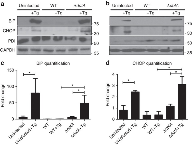
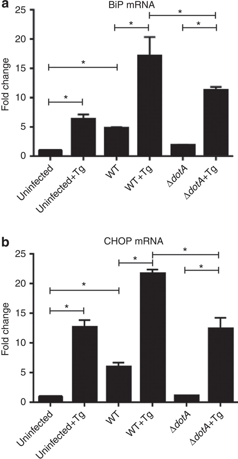
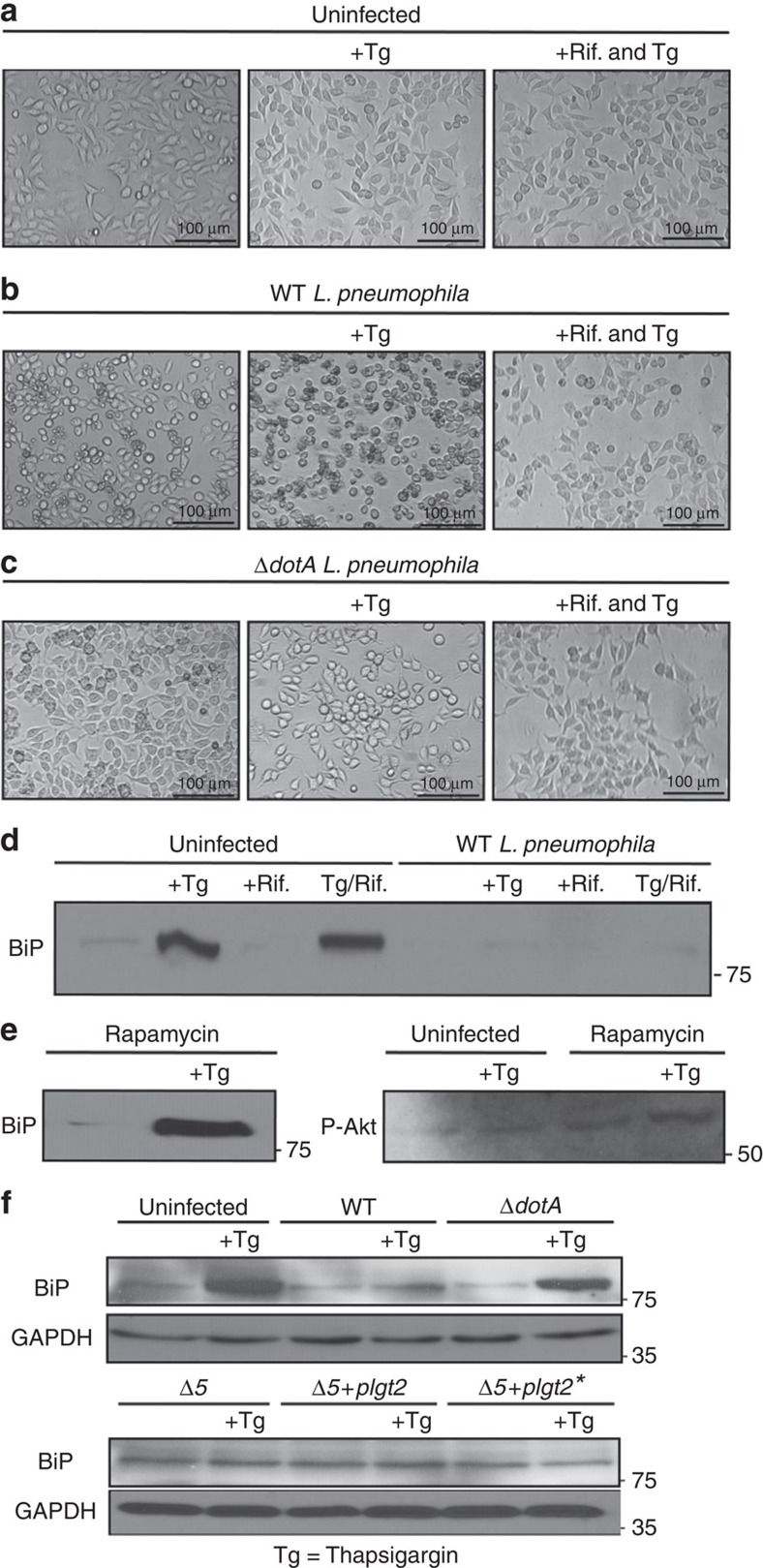
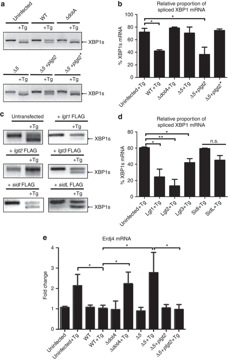
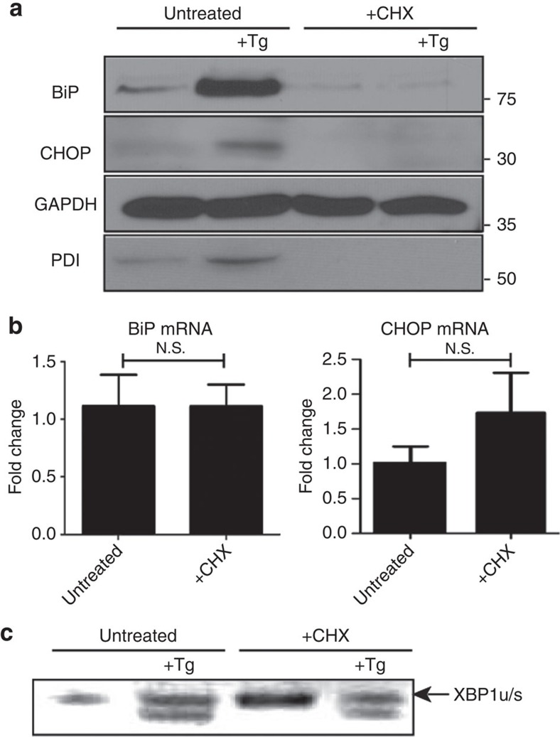
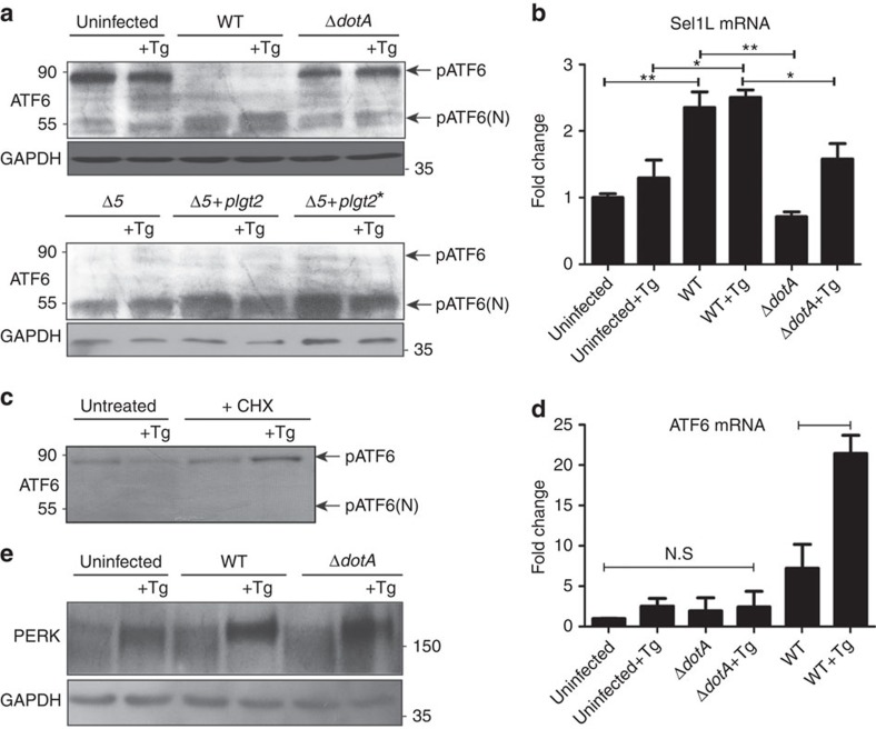
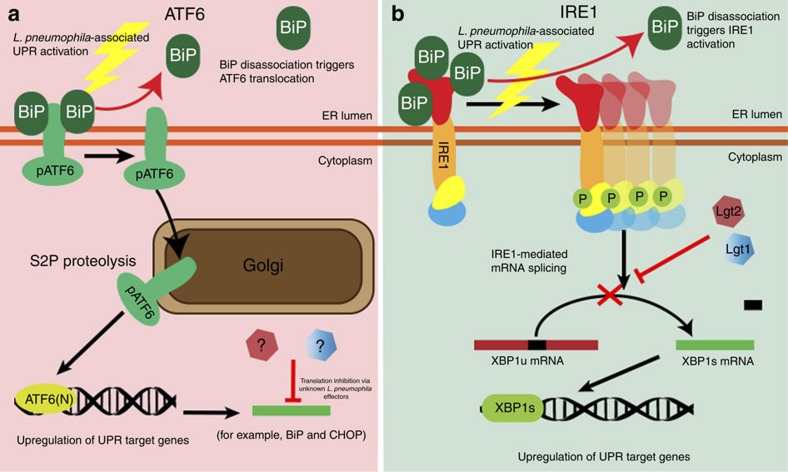
The intracellular pathogen, Legionella pneumophila, secretes ∼300 effector proteins to modulate the host environment. Given the intimate interaction between L. pneumophila and the endoplasmic reticulum, we investigated the role of the host unfolded protein response (UPR) during L. pneumophila infection. Interestingly, we show that the host identifies L. pneumophila infection as a form of endoplasmic reticulum stress and the sensor pATF6 is processed to generate pATF6(N), a transcriptional activator of downstream UPR genes. However, L. pneumophila is able to suppress the UPR and block the translation of prototypical UPR genes, BiP and CHOP. Furthermore, biochemical studies reveal that L. pneumophila uses two effectors (Lgt1 and Lgt2) to inhibit the splicing of XBP1u mRNA to spliced XBP1 (XBP1s), an UPR response regulator. Thus, we demonstrate that L. pneumophila is able to inhibit the UPR by multiple mechanisms including blocking XBP1u splicing and causing translational repression. This observation highlights the utility of L. pneumophila as a powerful tool for studying a critical protein homeostasis regulator.


Figures

Figure 1
Figure 1. Expression of two critical components of the UPR, BiP and CHOP are blocked by L. pneumophila
(a) Caspase 1/11 (−/−) bone marrow derived macrophages or (b) HEK-293 FCγ cells-expressing Fc gamma (HEK-293 FCγ) were either left uninfected or infected with WT or ΔdotA strains of L. pneumophila at an MOI of 150. Cells were then left untreated or treated with thapsigargin (Tg; 1 μg μl−1) for 6 h. BiP, CHOP and PDI protein expression were monitored via immunoblot. GAPDH was used as a loading control. Full blots are provided for Fig. 1a,b in Supplementary Fig. 5. (c,d) BiP and CHOP levels were quantified respectively from three biological replicates using the same experimental conditions in HEK-293 FCγ cells. Data are depicted as the fold change normalized to uninfected levels. Data represent three independent experiments. Values in all graphs are means±s.e.m. *P<0.05; Student's t-test.
Figure 2
Figure 2. BiP and CHOP expression are not blocked transcriptionally.
(a,b) Raw 264.7 cells were infected at an MOI of 150 with either WT L. pneumophila, ΔdotA L. pneumophila, or were left uninfected. Cells were then either untreated or treated with thapsigargin (Tg; 1 μg μl−1) for 6 h and mRNA was harvested and qRT–PCR was performed to assay abundance of BiP and CHOP mRNA transcripts. GAPDH was used for the endogenous normalization of gene expression. In both cases two biological replicates were used each containing three technical replicates. Values in all graphs are means±s.e.m. *P<0.05; Student's t-test.
Figure 3
Figure 3. Suppression of ER stress is an effector driven process that is independent of the mTOR pathway.
(ac) HEK-293 FCγ cells were either left uninfected or infected with WT L. pneumophila or ΔdotA L. pneumophila at an MOI of 150. Cells were then either left untreated or treated with thapsigargin (Tg; 1 μg μl−1) or treated with a combination of Tg and the antibiotic Rifampicin (Rif.; 2 μg μl−1) for 6 h. After 6 h phase contrast images were taken. (d) HEK-293 FCγ cells were infected at an MOI of 150 with WT L. pneumophila, or left uninfected. Cells were then treated with Rif. alone (2 μg μl−1), Rif. with Tg (1 μg μl−1), Tg (1 μg μl−1) alone or left untreated for 6 h. BiP protein levels were monitored via immunoblot. (e) HEK-293 FCγ cells were untreated or treated with rapamycin (10 nM) or rapamycin with Tg (1 μg μl−1) for 6 h. BiP protein levels were then monitored via immunoblot and levels of phosphorylated Akt at serine 473 (P-Akt) were monitored from these lysates via immunoblot. (f) RAW 264.7 cells were infected with WT L. pneumophila, dotA, or Δ5 strains of L. pneumophila at an MOI of 150. BiP protein expression was monitored via immunoblot. GAPDH was used as a loading control. Full blots for Fig. 3d–f are provided in Supplementary Fig. 5.
Figure 4
Figure 4. The L. pneumophilaeffector Lgt2 is necessary and sufficient to block XBP1 mRNA splicing.
(a) RAW 264.7 cells were infected with WT L. pneumophila, ΔdotA, Δ5 mutant, Δ5 +plgt2 complement, or Δ5 +plgt2* a strain secreting a catalytically dead Lgt2 effector at an MOI of 150. Cells were then treated for 6 h with thapsigargin (Tg; 1 μg μl−1). The level of spliced XBP1 (XBP1s) mRNA was assayed via RT–PCR. (b) Three biological replicates of XBP1 mRNA splicing during L. pneumophila infection and Tg treatment were quantified. (c) HEK-293 FCγ cells were transfected with (1.5 μg) DNA in 24-well dishes with each of the five bacterial effectors Lgt1-FLAG, Lgt2-FLAG, Lgt3-FLAG, SidI-FLAG and SidL-FLAG. Twenty-four hours post-transfection cells were left untreated or treated with Tg (1 μg μl−1) for 6 h and RT–PCR was performed to assay for XBP1s mRNA. (d) Three biological replicates of XBP1 mRNA splicing in cells transfected with one of the five bacterial effectors Lgt1-FLAG, Lgt2-FLAG, Lgt3-FLAG, SidI-FLAG and SidL-FLAG were quantified. (e) RAW267.4 cells were infected with WT L. pneumophila, ΔdotA, Δ5 mutant, Δ5 +plgt2 complement, or Δ5 +plgt2* at an MOI of 150. Cells were then either untreated or treated with Tg (1 μg μl−1) for 6 h. Erdj4 mRNA was monitored via qRT–PCR. Values in all graphs are means±s.e.m. *P<0.05; Student's t-test. Full blots of Fig. 3a–d are provided in Supplementary Fig. 5.
Figure 5
Figure 5. Global protein translation inhibition does not the affect the UPR in the same manner as L. pneumophila infection.
(a) RAW 264.7 cells were left untreated or treated with either thapsigargin (Tg; 1 μg μl−1), CHX (1 μg μl−1), or a combination of both for 6 h. Protein levels of UPR targets BiP, CHOP and PDI were monitored via immunoblot. (b) RAW267.4 macrophage cells were treated with CHX or left untreated and BiP and CHOP mRNA levels were monitored via qRT–PCR. Three internal replicates were performed, values in all graphs are means±s.e.m. *P<0.05; Student's t-test. (c) HEK-293 FCγ cells were treated with CHX (1 μg μl−1), Tg (1 μg μl−1), Tg (1 μg μl−1) and CHX (1 μg μl−1), or left untreated for 6 h. XBP1u mRNA splicing was monitored via RT–PCR. Full blots for Fig. 5a,b are provided in Supplementary Fig. 5.
Figure 6
Figure 6. ATF6 is processed during L. pneumophilainfection.
(a) RAW 264.7 cells were left uninfected or infected with either WT L. pneumophila, ΔdotA, B. Δ5, Δ5+plgt2, or Δ5 plgt2* (catalytically dead lgt2) at an MOI of 150. Cells were then left untreated or treated with thapsigargin (Tg; 1 μg μl−1) for 6 h. Full-length ATF6 (pATF6) and cleaved pATF6(N) were monitored via immunoblot. Full blots for Fig. 6a are provided in Supplementary Fig. 5. (b) HEK-293 FCγ cells infected with either WT L. pneumophila or ΔdotA and then treated for 6 h with Tg (1 μg μl−1). Sel1L mRNA levels were then monitored via qRT–PCR, with GAPDH serving as an endogenous control. Two biological replicates were used each with three technical replicates using the identical experimental conditions. Values in all graphs are means±s.e.m. *P<0.05; Student's t-test. (c) Cleaved and uncleaved ATF6 levels were similarly monitored in cells treated with CHX; 1 μg μl−1), Tg (1 μg μl−1), Tg and CHX, or left untreated for 6 h. Full blots for Fig. 6b are provided in Supplementary Fig. 5. (d) RAW267.4 cells were infected with either WT L. pneumophila or the ΔdotA strain at an MOI of 150 and cells were then treated with Tg (1 μg μl−1) or left untreated for 6 h. ATF6 mRNA transcript levels were monitored via qRT–PCR. Two biological replicates were used each with three technical replicates using the identical experimental conditions. Values in all graphs are means±s.e.m. *P<0.05; Student's t-test. (e) RAW267.4 cells were infected with either WT L. pneumophila or the ΔdotA strain at an MOI of 150 and cells were then treated with Tg (1 μg μl−1) or left untreated for 6 h. PERK phosphorylation was monitored via immunoblot. GDH served as loading control. Full blots for Fig. 6d are provided in Supplementary Fig. 5.
Figure 7
Figure 7. Legionella specifically blocks the ATF6 arm and IRE1 arms of the UPR.
(a) ATF6 is processed from the inactive form pATF6 to ATF6(N) via SP2-mediated proteolysis at the Golgi, in the presence of WT L. pneumophila and this leads to the upregulation of unfolded protein response genes BiP and CHOP. Currently unknown L. pneumophila effectors can block the translation of these genes to inhibit the UPR. (b) The L. pneumophila effectors Lgt1 and Lgt2 are necessary and sufficient to block the UPR endonuclease protein IRE1-mediated XBP1 mRNA splicing during infection. Under normal conditions XBP1 is spliced to the XBP1s form with is translated to the XBP1 transcription factor that targets UPR related genes.

Similar articles

Cited by

References

    1. Segal G., Purcell M. & Shuman H. A. Host cell killing and bacterial conjugation require overlapping sets of genes within a 22-kb region of the Legionella pneumophila genome. Proc. Natl Acad. Sci. USA 95, 1669–1674 (1998) . - PMC - PubMed
    1. Vogel J. P., Andrews H. L., Wong S. K. & Isberg R. R. Conjugative transfer by the virulence system of Legionella pneumophila. Science 279, 873–876 (1998) . - PubMed
    1. Mori K. Signalling pathways in the unfolded protein response: development from yeast to mammals. J. Biochem. 146, 743–750 (2009) . - PubMed
    1. Walter P. & Ron D. The unfolded protein response: from stress pathway to homeostatic regulation. Science 334, 1081–1086 (2011) . - PubMed
    1. Harding H. P., Zhang Y. & Ron D. Protein translation and folding are coupled by an endoplasmic-reticulum-resident kinase. Nature 397, 271–274 (1999) . - PubMed

Publication types

MeSH terms