. 2015 Sep;46(9):2616-27.
doi: 10.1161/STROKEAHA.115.009854. Epub 2015 Jul 28.

Intravenous Bone Marrow Stem Cell Grafts Preferentially Migrate to Spleen and Abrogate Chronic Inflammation in Stroke

Affiliations

Intravenous Bone Marrow Stem Cell Grafts Preferentially Migrate to Spleen and Abrogate Chronic Inflammation in Stroke

Sandra A Acosta et al. Stroke. 2015 Sep.

Abstract

Background and purpose: Adult stem cell therapy is an experimental stroke treatment. Here, we assessed homing and anti-inflammatory effects of bone marrow stromal cells (hBMSCs) in chronic stroke.

Methods: At 60 days post stroke, adult Sprague-Dawley rats received intravenous hBMSCs (4×10(6) labeled or nonlabeled cells) or vehicle (saline). A sham surgery group served as additional control. In vivo imaging was conducted between 1 hour and 11 days post transplantation, followed by histological examination.

Results: Labeled hBMSCs migrated to spleen which emitted significantly higher fluorescent signal across all time points, especially during the first hour, and were modestly detected in the head region at the 12 hours and 11 days, compared with nonlabeled hBMSCs and vehicle-infused stroke animals, or sham (P<0.05). At 11 days post transplantation, ex vivo imaging confirmed preferential hBMSC migration to the spleen over the brain. Hematoxylin and eosin staining revealed significant 15% and 30% reductions in striatal infarct and peri-infarct area, and a trend of rescue against neuronal loss in the hippocampus. Unbiased stereology showed significant 75% and 60% decrements in major histocompatibility complex II-activated inflammatory cells in gray and white matter, and a 43% diminution in tumor necrosis factor-α cell density in the spleen of transplanted stroke animals compared with vehicle-infused stroke animals (P<0.05). Human antigen immunostaining revealed 0.03% hBMSCs survived in spleen and only 0.0007% in brain. MSC migration to spleen, but not brain, inversely correlated with reduced infarct, peri-infarct, and inflammation.

Conclusions: hBMSC transplantation is therapeutic in chronic stroke possibly by abrogating the inflammation-plagued secondary cell death.

Keywords: brain ischemia; imaging; inflammation; regeneration; stem cells.


Figures

Figure 1.
Figure 1.
Preferential in vivo migration of hBMSC-DiR+ to spleen in chronic stroke. Representative images of in vivo fluorescence imaging of intravenously transplanted hBMSCs in an experimental model of chronic stroke at 1, 4, 12, 24, 48, and 72 hours and 11 days post transplantation. Photographs were taken from ventral (body) and dorsal positions (head). Imaging analyses revealed that hBMSC-DiR+ cells displayed a preferential migration to the spleen over the brain when transplanted at 60 days post stroke (***P<0.001). A and B, Within the hBMSC-DiR+ transplanted stroke group, there were no significant differences in fluorescent signals across all time points except the first hour, whereby a higher emitted signal was found within spleen and on day 11 where the signal was greatly reduced relative to other time points (*P<0.05). C and D, Fluorescent signals were also elevated in the head area at 12 hours and 11 days post transplantation (**P<0.01 and ***P<0.001). There were no fluorescent signals detected in the spleen and head region of hBMSC-DiR− transplanted and vehicle-infused stroke animals, and sham animals. Radiant efficiency={(p/s/cm2/sr)/(μW/cm2)}, color scale: min=1.54×10⁸; max=3.2×10⁸. *P<0.05, **P<0.01, and ***P<0.001. Data are expressed as mean±SEM. DiR indicates 1,1′-dioctadecyl-3,3,3′,3′-tetramethylindotricbocyanine iodide; and hBMSC, homing and anti-inflammatory effects of bone marrow stromal cells.
Figure 2.
Figure 2.
Preferential ex vivo migration of hBMSC-DiR+ to spleen in chronic stroke. A, Quantitative analysis of fluorescent signals brain, spleen, lungs, and liver confirmed preferential migration of hBMSC-DiR+ to the spleen over the brain (*P<0.05). B, Photographs correspond to representative peripheral organs ex vivo. B, Sham animals (brain, spleen, lungs, and liver) and (C) hBMSC-DiR+ (brain, spleen, lungs, and liver). Radiant efficiency={(p/sec/cm2/sr)/(μW/cm2)}, color scale: min=3.74×10⁷; max=4.99×10⁷. *P<0.05, **P<0.01, and ***P<0.001. Data are expressed as mean±SEM. DiR indicates 1,1′-dioctadecyl-3,3,3′,3′-tetramethylindotricbocyanine iodide; and hBMSC, homing and anti-inflammatory effects of bone marrow stromal cells.
Figure 3.
Figure 3.
Homing and anti-inflammatory effects of bone marrow stromal cells (hBMSC) transplantation reduces infarct and peri-infarct area, but does not ameliorate hippocampal CA1 and CA3 neuronal loss. Hematoxylin and eosin (H&E) staining revealed that hBMSC treatment significantly reduced the striatal infarct and peri-infarct areas associated with stroke. A, Quantitative analysis revealed a significant reductions in infarct and peri-infarct area after hBMSC transplantation compared with vehicle-infused stroke animals (*P<0.05). B, Photomicrographs are representative coronal brain sections stained with H&E at 11 days post transplantation, showing infarct area (dark purple) and intact areas (light purple). Arrows denote infarct and peri-infarct area of striatum. Right inserts, vehicle-infused stroke exhibited acidic cells (dark purple cells), shrinkage, and dissolution of the cells in the infarcted striatum and peri-infarct area compared with the corresponding ipsilateral side of hBMSC-transplanted stroke animals. Scale bar: 1 mm. C and D, Quantitative analyses of total number of CA1 and CA3 neurons failed to reveal a significant reduction in neuronal cell loss in rats that received hBMSCs compared with vehicle-infused stroke rats (*P>0.05). C and D, Photomicrographs are representative coronal brain sections staining with H&E from ipsilateral CA1 and CA3 area of the hippocampus of sham, vehicle-infused, and hBMSC-treated stroke animals. Arrows denote neuronal cell loss within the CA1 and CA3 region. Scale bar=50 μm. *P<0.05; ns=not significant. Data are expressed as percentage difference from contralateral CA1 and CA3 total number of neurons.
Figure 4.
Figure 4.
Homing and anti-inflammatory effects of bone marrow stromal cells (hBMSC) transplantation ameliorates neuroinflammation in gray and white matter areas in chronic stroke. A, Stereological analysis of MHCII+ cells estimated volume in striatum, subventricular zone (SVZ), and hilus revealed significant upregulation of activated MHCII+ cells in the ipsilateral side of vehicle-infused stroke animals compared with their contralateral side across all gray matter areas analyzed (***P<0.0001), except SVZ (P>0.05). There were significant upregulation of activated MHCII+ cells in both ipsilateral and contralateral gray matter areas of vehicle-infused stroke animals (P<0.0001) and hBMSC-transplanted stroke animals compared with sham animals (P<0.05). hBMSC transplantation caused a 35%, 28%, and 90% reduction of the estimated volume of MHC+ cells in the ipsilateral striatum, SVZ, and hilus, respectively, relative to the ipsilateral side of the same regions of vehicle-infused stroke animals (**P<0.01; **P<0.01; ***P<0.001). B, Photomicrographs are representative coronal brain sections ipsilateral to injury stained with MHCII 11 days post hBMSC transplantation. Arrows indicate positive staining for activated MHCII+ cells in striatum, SVZ, and hilus. C, Effects of hBMSC transplantation on MHCII+ cells in white matter regions. Stereological analysis of MHCII+ cells estimated volume in corpus callosum, internal capsule, and fornix revealed significant upregulation of activated MHCII+ cells in the ipsilateral side of vehicle-infused stroke animals compared with their contralateral side across all white matter areas analyzed (***P<0.0001), except corpus callosum (P>0.05). There were significant upregulation of activated MHCII+ cells in both ipsilateral and contralateral side of vehicle-infused stroke animals (***P<0.0001) and hBMSC-transplanted stroke animals across all white matter areas analyzed compared with sham animals (*P<0.05). hBMSC transplantation caused a 60%, 38%, and 70% reduction of the estimated volume of MHCII+ cells in the ipsilateral corpus callosum, internal capsule, and fornix, respectively, relative to the ipsilateral side of the same regions of vehicle-infused stroke animals (***P<0.001). D, Photomicrographs are representative coronal brain sections ipsilateral to injury stained with MHCII 11 days post hBMSC transplantation. Arrows indicate positive staining for activated MHCII+ cells. Scale bar=50 μm. *P<0.05, **P<0.01, and ***P<0.001; ns=not significant. Data are expressed as mean±SEM. MHCII indicates major histocompatibility complex II.
Figure 5.
Figure 5.
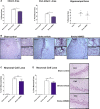
Homing and anti-inflammatory effects of bone marrow stromal cells (hBMSCs) survive better in the spleen than the brain. A, Representative merged images showing colocalization of HuNu+ and Hoeschst+ expression from grafted hBMSCs in spleen and brain (ie, striatum) in transplanted stroke rats as opposed to vehicle-infused stroke rats. Top right, Z-stack reconstruction of HuNu+ hBMSCs (green, human-specific antigen) within the spleen colocalized with Hoechst+ (blue, nuclei marker) in hBMSC-transplanted stroke animals. Bottom right, Z-stack reconstruction of HuNu+ hBMSCs (green) within the peri-infarct area of the striatum colocalized with Hoechst+ (blue) in hBMSC-transplanted stroke animals. Quantitative analyses of the estimated number of HuNu+ hBMSCs in the spleen (B) and in the brain (C) of hBMSC-transplanted stroke animals revealed 0.03% of hBMSCs survived in the spleen compared with 0.0007% survival in the brain, indicating that the mean graft survival, and likely preferential migration, of hBMSCs in the spleen was significantly higher than that found in the brain. Scale bar=50 μm. Data are expressed as mean±SEM.
Figure 6.
Figure 6.
Homing and anti-inflammatory effects of bone marrow stromal cells (hBMSCs) reduced tumor necrosis factor-α (TNF-α) density in the spleen of stroke animals. A, Quantitative analysis of TNF-α expression in the spleen revealed a significant upregulation of TNF-α density in vehicle-infused stroke animals. In contrast, hBMSC treatment promoted a 40% downregulation of TNF-α density in the spleen (*P<0.05). B, Confocal photomicrographs of positive expression of TNF-α (green) and Hoechst (blue) in the spleen of vehicle-infused (top) and transplanted stroke animals (bottom), indicating decreased expression of TNF-α in the spleen of hBMSC-transplanted stroke animals relative to vehicle-infused stroke animals. C, Correlations found between reduction in striatal infarct and peri-infarct areas, downregulation of MHCII+ cells in brain, and decreased density of TNF-α in the spleen, and the number of hBMSCs that migrated to the spleen (% infarct area: Pearson r=−0.8678, R2=−0.7531, P<0.01; % peri-infarct area: Pearson r=−0.8282, R2=−0.6859, P<0.05; the volume of MHCII+ activated cells in the striatum: Pearson r=−0.8656, R2=−0.7492, P<0.05; the density of TNF-α expression in the spleen: Pearson r=−0.8381, R2=−0.7025, P<0.05). Scale bar=50 μm. *P<0.05. Data are expressed as mean±SEM.

References

    1. Adamson J, Beswick A, Ebrahim S. Is stroke the most common cause of disability? J Stroke Cerebrovasc Dis. 2004;13:171–177. doi: 10.1016/j.jstrokecerebrovasdis.2004.06.003. - PubMed
    1. Go AS, Mozaffarian D, Roger VL, Benjamin EJ, Berry JD, Borden WB, et al. Heart disease and stroke statistics-2013 update: a report from the American Heart Association. Circulation. 2013;127:6–245. - PMC - PubMed
    1. Lo EH, Dalkara T, Moskowitz MA. Mechanisms, challenges and opportunities in stroke. Nat Rev Neurosci. 2003;4:399–415. doi: 10.1038/nrn1106. - PubMed
    1. Hacke W, Kaste M, Bluhmki E, Brozman M, Dávalos A, Guidetti D, et al. ECASS Investigators. Thrombolysis with alteplase 3 to 4.5 hours after acute ischemic stroke. N Engl J Med. 2008;359:1317–1329. doi: 10.1056/NEJMoa0804656. - PubMed
    1. The NINDS rt-PA Stroke Study Group. Intracerebral hemorrhage after intravenous tPA therapy for ischemic stroke. Stroke. 1997;28:2109–2118. - PubMed

Publication types