

Biomimetic scaffolds for regeneration of volumetric muscle loss in skeletal muscle injuries
- PMID: 26219862
- PMCID: PMC4562809
- DOI: 10.1016/j.actbio.2015.07.038
Biomimetic scaffolds for regeneration of volumetric muscle loss in skeletal muscle injuries
Abstract
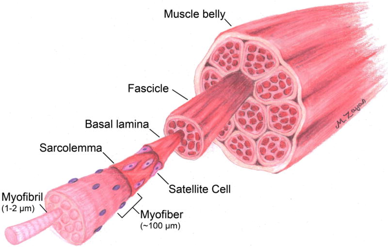
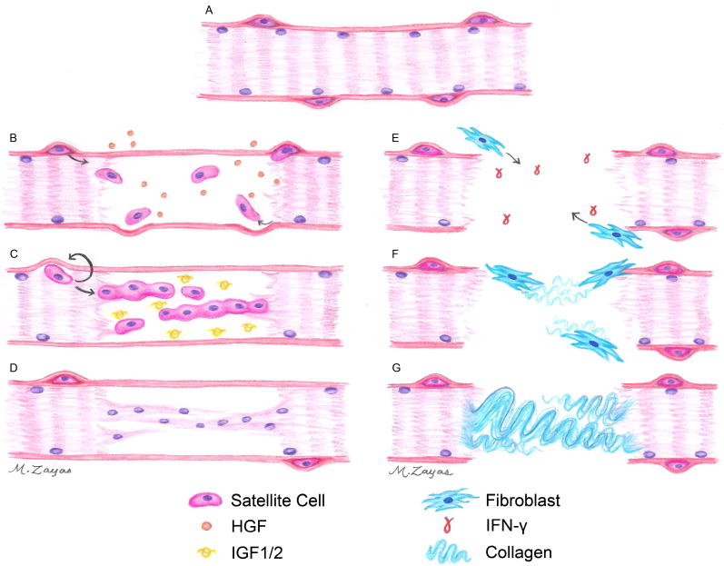
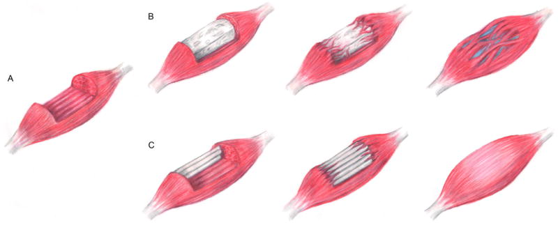
Skeletal muscle injuries typically result from traumatic incidents such as combat injuries where soft-tissue extremity injuries are present in one of four cases. Further, about 4.5 million reconstructive surgical procedures are performed annually as a result of car accidents, cancer ablation, or cosmetic procedures. These combat- and trauma-induced skeletal muscle injuries are characterized by volumetric muscle loss (VML), which significantly reduces the functionality of the injured muscle. While skeletal muscle has an innate repair mechanism, it is unable to compensate for VML injuries because large amounts of tissue including connective tissue and basement membrane are removed or destroyed. This results in a significant need to develop off-the-shelf biomimetic scaffolds to direct skeletal muscle regeneration. Here, the structure and organization of native skeletal muscle tissue is described in order to reveal clear design parameters that are necessary for scaffolds to mimic in order to successfully regenerate muscular tissue. We review the literature with respect to the materials and methodologies used to develop scaffolds for skeletal muscle tissue regeneration as well as the limitations of these materials. We further discuss the variety of cell sources and different injury models to provide some context for the multiple approaches used to evaluate these scaffold materials. Recent findings are highlighted to address the state of the field and directions are outlined for future strategies, both in scaffold design and in the use of different injury models to evaluate these materials, for regenerating functional skeletal muscle.
Statement of significance: Volumetric muscle loss (VML) injuries result from traumatic incidents such as those presented from combat missions, where soft-tissue extremity injuries are represented in one of four cases. These injuries remove or destroy large amounts of skeletal muscle including the basement membrane and connective tissue, removing the structural, mechanical, and biochemical cues that usually direct its repair. This results in a significant need to develop off-the-shelf biomimetic scaffolds to direct skeletal muscle regeneration. In this review, we examine current strategies for the development of scaffold materials designed for skeletal muscle regeneration, highlighting advances and limitations associated with these methodologies. Finally, we identify future approaches to enhance skeletal muscle regeneration.
Keywords: Biomaterials; Fibrin; Microthreads; Skeletal muscle regeneration; Tissue engineering.
Copyright © 2015 Acta Materialia Inc. Published by Elsevier Ltd. All rights reserved.
Figures
References
-
- Grogan BF, Hsu JR. Volumetric muscle loss. The Journal of the American Academy of Orthopaedic Surgeons. 2011;19(Suppl 1):S35–7. - PubMed
-
- Charge SB, Rudnicki MA. Cellular and molecular regulation of muscle regeneration. Physiol Rev. 2004;84:209–38. - PubMed
-
- Counsel P, Breidahl W. Muscle injuries of the lower leg. Semin Musculoskelet Radiol. 2010;14:162–75. - PubMed
Publication types
MeSH terms
Grants and funding
LinkOut - more resources
Full Text Sources
Other Literature Sources
Research Materials
