. 2015 Jul 29:15:254.
doi: 10.1186/s12906-015-0765-6.

Retino-protective effect of Bucida buceras against oxidative stress induced by H2O2 in human retinal pigment epithelial cells line

Affiliations

Retino-protective effect of Bucida buceras against oxidative stress induced by H2O2 in human retinal pigment epithelial cells line

Simon Bernard Iloki-Assanga et al. BMC Complement Altern Med. .

Abstract

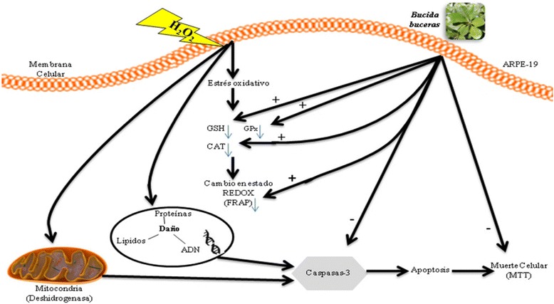
Background: Reactive Oxygen Species (ROS) impair the physiological functions of Retinal Pigment Epithelial (RPE) cells, which are known as one major cause of age-related macular degeneration and retinopathy diseases. The purpose of this study is to explore the cytoprotective effects of the antioxidant Bucida buceras extract in co-treatment with hydrogen peroxide (H2O2) delivery as a single addition or with continuous generation using glucose oxidase (GOx) in ARPE-19 cell cultures. The mechanism of Bucida buceras extract is believed to be associated with their antioxidant capacity to protect cells against oxidative stress.

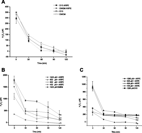
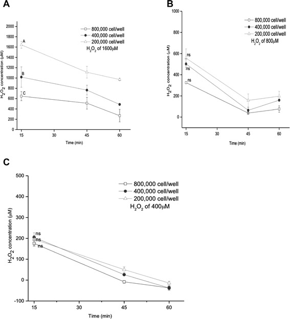
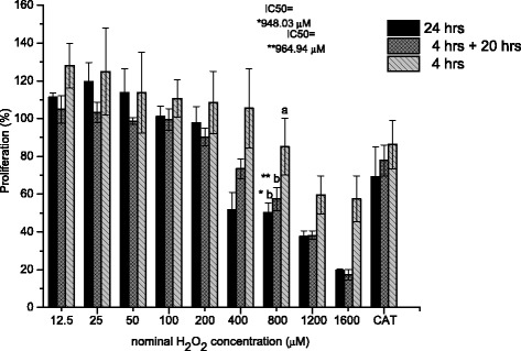
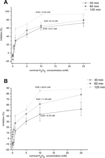
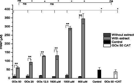
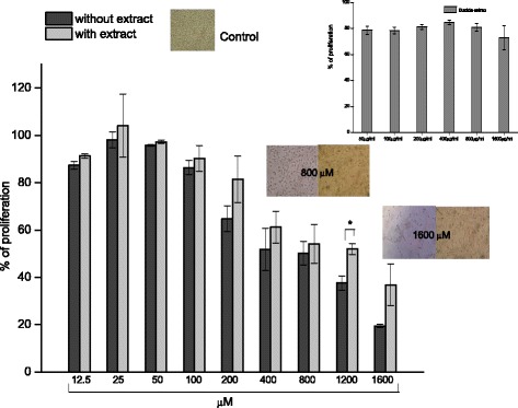
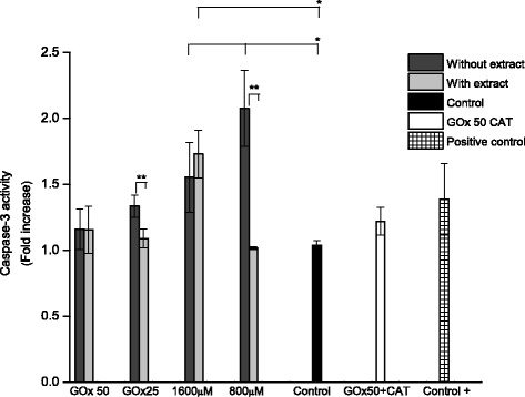
Methods: A comparative oxidative stress H2O2-induced was performed by addition and enzymatic generation using glucose oxidase on human retinal pigment epithelial cells line. H2O2-induced injury was measured by toxic effects (cell death and apoptotic pathway) and intracellular redox status: glutathione (GSH), antioxidant enzymes (catalase and glutathione peroxidase) and reducing power (FRAP). The retino-protective effect of co-treatment with Bucida buceras extract on H2O2-induced human RPE cell injury was investigated by cell death (MTT assay) and oxidative stress biomarkers (H2O2, GSH, CAT, GPx and FRAP).

Results: Bucida buceras L. extract is believed to be associated with the ability to prevent cellular oxidative stress. When added as a pulse, H2O2 is rapidly depleted and the cytotoxicity analyses show that cells can tolerate short exposure to high peroxide doses delivered as a pulse but are susceptible to lower chronic doses. Co-treatment with Bucida buceras was able to protect the cells against H2O2-induced injury. In addition to preventing cell death treatment with antioxidant plant could also reverse the significant decrease in GSH level, catalase activity and reducing power caused by H2O2.

Conclusion: These findings suggest that Bucida buceras could protect RPE against ocular pathogenesis associated with oxidative stress induced by H2O2-delivered by addition and enzymatic generation.


Figures

Fig. 1
Fig. 1
Calibration curves for H2O2 levels in the range 10–200 μM using ferrous-xylenol orange with 10 % FBS (D10; linear regression y = 0.0022x + 0.0149, R2 = 0.98616 and without FBS (DMEM linear egression: y = 0.0025x + 0.0157, R2 = 0.98643. Therefore H2O2determination was measured in PBS. Data are from three independent experiments and are the means ± SD of three culture wells per group in each experiment
Fig. 2
Fig. 2
Degradation of 250 μM H2O2 added as single pulse to medium without serum (DMEM) and with 10 % FBS (D10) in the absence of ARPE-19 cells. Exponential regression of 250 μM H2O2 in D10 y = 245.54e-0.029x, R2 = 0.96447. Therefore H2O2 determination was measured in PBS. Addition of catalase 200 U/ml to the 500 μM H2O2 with serum confirms the specificity of the determinations for H2O2. Data are from three independent experiments and are the means ± SD of three culture wells per group in each experiment
Fig. 3
Fig. 3
Dissipation of 400 μM nominal concentration of H2O2 (a), different nominal concentrations 400, 600, 800 and 1200 μM with serum-free medium (b) and with 10 % FBS (c) from the culture medium in presence o absence of ARPE-19 cells (50, 000 cell/well) with o without serum. Decay rate constants were kd = −0.065, −0.041, −0.037, −0.026 min−1 for D10 + ARPE-19 cells, D10, DMEM + ARPE-19 cells and DMEM respectively. Data represent means of triplicate measurements of 3 independent experiments. 1200 μM of H2O2 in absence of ARPE-19 cells was determined in parallel culture
Fig. 4
Fig. 4
Influence of cells number on the elimination of hydrogen peroxide in ARPE-19 cells. Cells were incubated with various concentrations of H2O2 1600 μM (a), 800 μM (b) and 400 μM (c) for the indicated times. Data are means ± SD of three independent experiments
Fig. 5
Fig. 5
Cytotoxic action of hydrogen peroxide in ARPE-19 cells. A range of concentrations of H2O2 from 12.5 to 1600 μM was added to serum-containing DMEM. Cytotoxicity was estimated 24 h after addition by the MTT assay in cultures in which the medium was not replaced (solid bars) or was replaced at 4 h with fresh H2O2-free DMEM (hatched bars) or was measured at 4 h (gray-hatched bars). Addition of catalase 200 U/ml to the 1600 μM H2O2 as CAT control cells. * IC50 in 24 h ** IC50 4 h with replace (4 h + 20 h). Data expressed as a percentage of the untreated control, are from three independent experiments and are the means ± SD
Fig. 6
Fig. 6
Influence of incubation time on the cytotoxic action of hydrogen peroxide in ARPE-19 cells. 50,000 cells were incubated for the indicated time (30, 60 and 120 min) with various concentrations of the peroxide (0.01, 0.05, 0.1, 0.5, 1, 5, 10, 25 mM) in free serum culture medium (a) and with serum (b). Data are means of at least three independent experiments
Fig. 7
Fig. 7
H2O2 concentration in culture medium generated from D-glucose after 1 h post addition of 0–10 mU/ml glucose oxidase (GOx). GOx was added to the medium of culture well without or with confluent cultures of ARPE-19 cells to illustrate the initial linear accumulation of H2O2 as a function of enzyme concentration. Data are from three independent experiments and are the means ± SD of three culture wells per group in each experiment
Fig. 8
Fig. 8
H2O2 concentration in culture medium generated from D-glucose after addition of 5, 8, 10 and 25-mU/ml glucose oxidase (GOx) during 48 h. GOx was added to the medium of culture wells (a) without or (b) with confluent cultures of ARPE-19 cells. Because higher medium concentrations are achieved in the absence of cells, different y-axis scales are used. Data are from three independent experiments and are the means ± SD of three culture wells per group in each experiment
Fig. 9
Fig. 9
Influence of glucose oxidase concentrations on the cytotoxic action of hydrogen peroxide in confluent cultures of ARPE-19 cells. The viability was determined as a percentage of the untreated controls using the MTT assay for the indicated times. IC50 values have 31.1 mU/ml and 19.5 mU/ml at 24 h y 48 h respectively. Data are from at least three independent experiments and are the means ± SD of three culture wells per group in each experiment
Fig. 10
Fig. 10
Hydrogen peroxide scavenging of ethanol Bucida buceras extract (400–1600 μg/ml) at 1 h in (a) 1600 μM H2O2 pulse y = 0.0415x - 3.2327, R2 = 0.99336 (IC50 = 1356.29 μg/ml) and (b) 50 mU/ml of GOx y = 0.0277x - 3.8591, R2 = 0.98872 (IC50 = 2050.09 μg/ml). (c) DPPH-scavenging activity of Bucida buceras (5 a 50 μg/ml), y = 1.6486x +6.8934, R2 = 0.976 IC50 = 26.14 μg/ml. (d) The ferric ion reducing antioxidant power (FRAP) in concentrations of 100 to 800 μg/ml. Values above the bars indicate % of inhibition. Each value is expressed as mean ± SD (n = 3)
Fig. 11
Fig. 11
Effects of GOx and H2O2 in addition on glutathione reduced (GSH) at 24 h (a). ARPE-19 cells treated with 12.5, 25 and 50 mU/ml of GOx and pulse of H2O2 800, 1000, 1600 μM and ARPE-19 cells treated with 1600 ug/ml of ethanolic extract of Bucida buceras, control cells (untreated with H2O2) or GOx (50 mU/ml) + 200 U/ml of catalase as CAT control; (b) The levels of H2O2 in GOx at 24 h in 12.5, 25 and 50 mU/ml, the medium was analyzed using FOX reagent. Values were expressed in means ± SD in μM (n = 3 independent experiments). *p < 0.05 significantly different from value of cells control vs. H2O2-treated **p < 0.05 significantly different from value of antioxidant treatment vs. H2O2-treated, n.s not significant differences
Fig. 12
Fig. 12
The effect of hydrogen peroxide by enzymatic generation (GOx 50, 25 and 12.5 mU/ml) and pulse (1600, 1000, 800 μM) in presence or absence of co-treatment with Bucida buceras ethanol extract (1600 μg/ml) on catalase activity (a) and GSH-peroxidase activity assay (b) H2O2 (initially, 200 μM in PBS was incubated for 1 h, at 37 °C with different cell lysates, control GOx (50 mU/ml + catalase 200 U/ml) or control cells (untreated with H2O2). Results are expressed as the means ± SD of three independent studies (each analyzed in triplicate). *p < 0.05 significantly different from value of cells control (untreated) vs. H2O2-treated **p < 0.05 significantly different from value of antioxidant treatment vs. H2O2-treated, n.s not significant differences
Fig. 13
Fig. 13
Effect of co-tretment with Bucida buceras (1600 μg/ml) in glucose oxidase (GOx) or H2O2 addition treatment on antioxidant capacity measured by FRAP assay. Control GOx + CAT (50 mU/ml + catalase 200 U/ml) or control cells (untreated with H2O2). *p < 0.05 significantly different from value of cells control vs. H2O2-treated **p < 0.05 significantly different from value of antioxidant treatment vs. H2O2-treated, n.s not significant differences
Fig. 14
Fig. 14
Effects of Bucida buceras on cell protein concentration in ARPE-19 cells H2O2-induced oxidative damage. H2O2 generated by GOx (50, 25, 12.5 mU/ml) o in addition (1600, 1000 y 800 μM) (a). Relationship between values for cell proliferation (line) and the protein concentration (bars) (b). Values are presented as means ± SD of at least 3 independent experiments. Control GOx + CAT (50 mU/ml + catalase 200 U/ml) or control cells (untreated with H2O2). *p < 0.05 significantly different from value of cells control vs. H2O2-treated **p < 0.05 significantly different from value of antioxidant treatment vs. H2O2-treated, n.s not significant differences
Fig. 15
Fig. 15
Protective effects of Bucida buceras on H2O2-treated ARPE-19 cells. Cells were co-treated with 1600 μg/ml in addition with various concentrations of H2O2-treatment by 24 h. Cell viability was measured by MTT assay: values are expressed as the percentage of cell survival relative to the medium control cells. Values are presented as means ± SD of 5–7 independent experiments. The insets show the low cytotoxicity of Bucida buceras (50 to 1600 μg/ml) on ARPE-19 cells. Cell morphology is also presented (original magnification: 200x). *p < 0.05 significantly different from value of antioxidant treatment vs. H2O2-treated alone
Fig. 16
Fig. 16
Caspase-3 activity induced by H2O2 addition (800 or 1600 μM) and GOx (25 or 50 mU/ml) in ARPE-19 cells for 24 h. Control, C+ cells were untreated with H2O, C+ (the specific caspase-3 control). Cells were co-treated with 1600 μg/ml of Bucida buceras extract. Control GOx + CAT (50 mU/ml + catalase 200 U/ml) or control cells (untreated with H2O2). Values are presented as means ± SD of at least 3 independent experiments. *p < 0.05 significantly different from value of control vs. Control GOx + CAT (50 mU/ml + catalase 200 U/ml) or control cells (untreated with H2O2) Bucida buceras, or H2O2-treated only; **p < 0.05 significantly different from value of antioxidant treatment vs. H2O2-treated alone
Fig. 17
Fig. 17
Schematic overview of the protective effects of the Bucida buceras on ARPE-19 cells. Co-treatment of ARPE-19 cells with antioxidant extract for 24 h protected cells against the toxicity of peroxide. H2O2 can cause oxidative stress in ARPE-19 cells as indicated by a depletion of GSH, GPx, Reducing power; Bucida buceras could restore GSH depletion, by enhancing the activity of glutathione peroxidase and catalase. In addition, it could also reduce H2O2-induce DNA damage by increase mitochondrial activity, and FRAP. The “+” and“-” signs indicate an increase or decrease, respectively in enzymatic activity or biochemical molecule content

Similar articles

Cited by

References

    1. Miranda M, Arnal E, Ahuja A, Alvarez R, López R, Ekstrom P, Romero F. Antioxidants rescue photoreceptors in rd1 mice: Relationship with thiol metabolism. Free Radic. Biol. Med. 2010; 48:216–22. - PubMed
    1. Yu CC, Nandrot EF, Dun Y, Finnemann SC. Dietary antioxidants prevent age-related retinal pigment epithelium actin damage and blindness in mice lacking αvβ5 integrin. Free Radical Biology and Medicine 2012;52:660–70. - PMC - PubMed
    1. Bian Q, Gao S, Zhou J, Qin J, Taylor A, Johnso EJ, et al. Lutein and zeaxanthin supplementation reduces photooxidative damage and modulates the expression of inflammation-related genes in retinal pigment epithelial cells. Free Radical Biology and Medicine, 2012;53:1298–307. - PMC - PubMed
    1. Lewis LLM, Iloki ASB, Fernández AD, Gil SAA, Lara ECL, Rubio PJL. Oxidative stress implications for the pathogenesis of ocular pathology. International Journal of Development Research 2015;5(2)3275–288, February.
    1. Panieri E, Gogvadze V, Norberg E, Venkatesh R, Orrenius S, Zhivotovky B. Reactive oxygen species generated in different compartments induce cell death, survival, or senescence. Free Radical Biology and Medicine, 2013;57:176–87. - PubMed

Publication types