. 2015 Jul 29;6(1):139.
doi: 10.1186/s13287-015-0128-8.

Hypoxia accelerates vascular repair of endothelial colony-forming cells on ischemic injury via STAT3-BCL3 axis

Affiliations

Hypoxia accelerates vascular repair of endothelial colony-forming cells on ischemic injury via STAT3-BCL3 axis

Sang Hun Lee et al. Stem Cell Res Ther. .

Abstract

Introduction: Endothelial colony-forming cells (ECFCs) significantly improve tissue repair by providing regeneration potential within injured cardiovascular tissue. However, ECFC transplantation into ischemic tissue exhibits limited therapeutic efficacy due to poor engraftment in vivo. We established an adequate ex vivo expansion protocol and identified novel modulators that enhance functional bioactivities of ECFCs.

Methods: To augment the regenerative potential of ECFCs, functional bioactivities of hypoxia-preconditioned ECFCs (hypo-ECFCs) were examined.

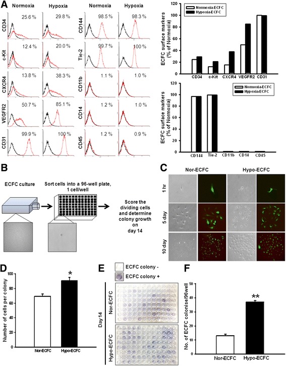
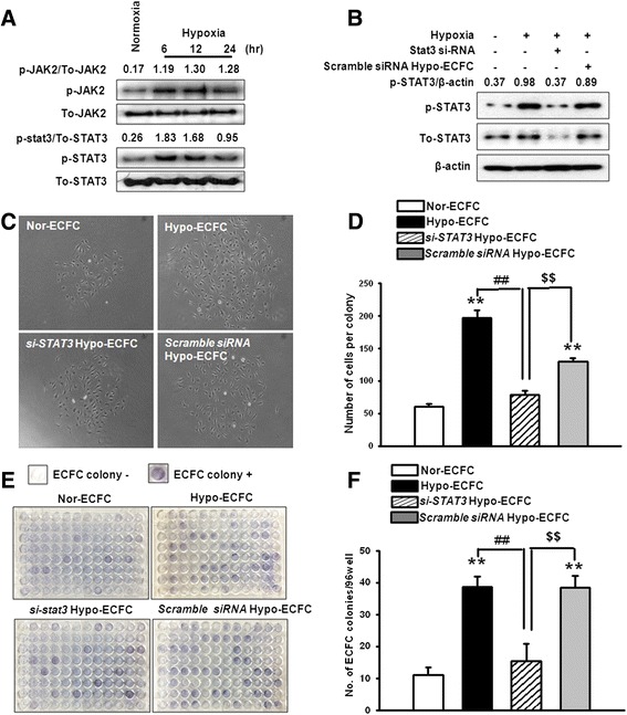
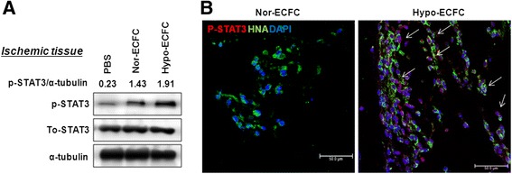
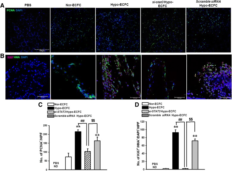
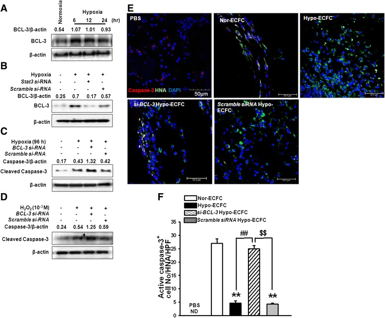
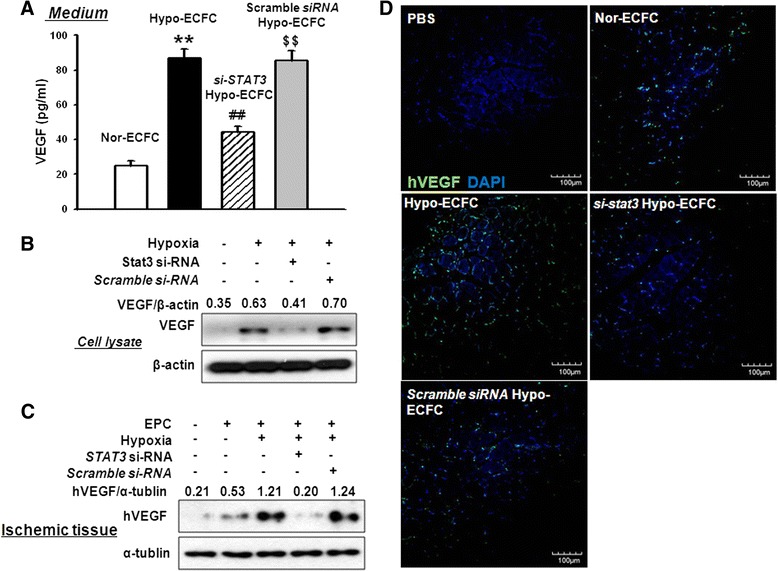
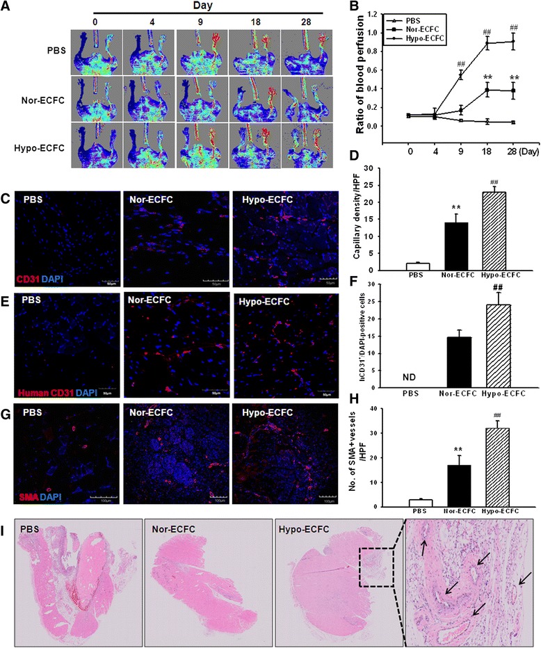
Results: Phosphorylations of the JAK2/STAT3 pathway and clonogenic proliferation were enhanced by short-term ECFC culturing under hypoxia, whereas siRNA-targeting of STAT3 significantly reduced these activities. Expression of BCL3, a target molecule of STAT3, was increased in hypo-ECFCs. Moreover, siRNA inhibition of BCL3 markedly reduced survival of ECFCs during hypoxic stress in vitro and ischemic stress in vivo. In a hindlimb ischemia model of ischemia, hypo-ECFC transplantation enhanced blood flow ratio, capillary density, transplanted cell proliferation and survival, and angiogenic cytokine secretion at ischemic sites.

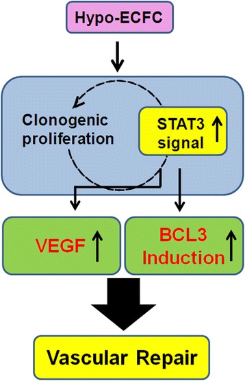
Conclusions: Hypoxia preconditioning facilitates functional bioactivities of ECFCs by mediating regulation of the STAT3-BCL3 axis. Thus, a hypoxic preconditioned ex vivo expansion protocol triggers expansion and functional bioactivities of ECFCs via modulation of the hypoxia-induced STAT3-BCL3 axis, suggesting that hypo-ECFCs offer a therapeutic strategy for accelerated neovasculogenesis in ischemic diseases.


Figures

Fig. 1
Fig. 1
Enhanced proliferative and clonogenic potential of endothelial colony-forming cells (ECFCs) after hypoxic preconditioning. a Characterization of ECFC surface markers after hypoxic preconditioning for 24 h. FACS analysis was performed using various surface markers, including progenitor markers (CD34, c-Kit, and CXCR4), endothelial lineage markers (VEGFR2, CD31, CD144, and Tie-2), and hematopoietic markers (CD11b, CD14, and CD45). b Schematic of single-cell assays using ECFCs. c Representative photomicrograph of the cell clusters derived from a single ECFC. d The number of cells per nor-ECFC or hypo-ECFC colony that underwent at least one cell division after 10 days of culture (*P < 0.05 vs. nor-ECFCs). e Image of hematoxylin-stained plate with a representative ECFC colony, and f a graph of the number of ECFC colonies per 96-well plate. The example shown is representative of four independent experiments. **P < 0.01 vs. nor-ECFCs
Fig. 2
Fig. 2
Activation of the JAK2/STAT3 signaling pathway in hypo-ECFCs and the regulation of proliferative, clonogenic potential. a Endothelial colony-forming cells (ECFCs) were exposed to hypoxia for 0–24 h, and JAK2, p-JAK2, STAT3, and p-STAT3 were detected by western blot analysis. b ECFCs were transfected with STAT3-siRNA and scramble siRNA for 48 h prior to hypoxia exposure for 24 h. p-STAT3 was detected by western blot analysis. c Representative photomicrograph of the cell clusters derived from single nor-ECFCs, hypo-ECFCs, STAT3-siRNA-transfected hypo-ECFCs (si-STAT3/hypo-ECFCs), and scramble siRNA-transfected hypo-ECFCs, as well as d a graph of the number of these cells per colony. e Images of hematoxylin-stained plates with representative nor-ECFC, hypo-ECFC, si-STAT3/hypo-ECFC, and scramble siRNA-transfected hypo-ECFCs colonies, as well as f graphs of the number of these colonies per 96-well plate. The example shown is representative of four independent experiments. **P < 0.01 vs. nor-ECFCs; ## P < 0.01 vs. hypo-ECFCs; $$ P < 0.01 vs. si-STAT3/hypo-ECFC
Fig. 3
Fig. 3
STAT3 is activated in hypo-ECFCs under ischemia. a Western blot analysis was performed to determine the levels of p-STAT3 in hindlimb ischemia injury after transplantation of hypo-ECFCs. b Co-immunofluorescence staining was used to detect p-STAT3 and hypo-ECFCs (human nuclear antigen (HNA)-positive cells, red). DAPI (blue) was used for nuclear staining. White color indicates merged color with green and red colors. Arrows indicate p-STAT3+/HNA+/DAPI+ cells. ECFC endothelial colony-forming cell, PBS phosphate-buffered saline
Fig. 4
Fig. 4
STAT3-mediated transplanted hypo-ECFC proliferation in ischemic tissues. a Immunofluorescence staining for proliferating cell nuclear antigen (PCNA, green) in ischemic hindlimb after transplantation of nor-ECFCs, hypo-ECFCs, si-STAT3/hypo-ECFCs, or scramble siRNA/hypo-ECFCs (scale bar: 100 μm). b Co-immunofluorescence staining to detect Ki-67 (a proliferation marker, red) and nor-ECFCs, hypo-ECFCs, si-STAT3/hypo-ECFCs, or scramble siRNA/hypo-ECFCs (human nuclear antigen (HNA)-positive cells, green). DAPI (blue) was used for nuclear staining (scale bar: 50 μm). c Bar graph shows the results of number of PCNA+ cells 3 days after transplantation. d Quantitative analysis of Ki-67/HNA/DAPI triple-positive cells 3 days after transplantation of nor-ECFCs, hypo-ECFCs, si-STAT3/hypo-ECFCs, or scramble siRNA/hypo-ECFCs. **P < 0.01 vs. nor-ECFCs; ## P < 0.01 vs. hypo-ECFCs; $$ P < 0.01 vs. si-STAT3/hypo-ECFC. ECFC endothelial colony-forming cell, PBS phosphate-buffered saline
Fig. 5
Fig. 5
Modulation of the STAT3-BCL3 axis enhances survival induced by ischemic stress. a Endothelial colony-forming cells (ECFCs) were exposed to hypoxia for various times (0–24 h). BCL3 expression was detected by western blot analysis. b ECFCs were transfected with STAT3-siRNA for 48 h prior to exposure to hypoxic conditions for 24 h. BCL3 was detected by western blot analysis. c ECFCs were transfected with BCL3-siRNA for 48 h prior to exposure to hypoxic conditions for 96 h. Cleaved caspase-3 was detected by western blot analysis. d ECFCs were transfected with BCL3-siRNA for 48 h prior to treatment with H2O2 (10−3 M) for 8 h followed by western blot analysis for cleaved caspase-3. e Co-immunofluorescence staining was used to detect apoptosis (caspase-3, an apoptosis marker, red) and nor-ECFCs, hypo-ECFCs, si-BCL3/hypo-ECFCs, or scramble siRNA/hypo-ECFCs (human nuclear antigen (HNA)-positive cells, green). DAPI (blue) was used for nuclear staining. Arrows indicate caspase-3+/HNA+/DAPI+ cells (scale bar: 50 μm). f Quantitative analysis of caspase-3/HNA/DAPI triple-positive cells conducted 3 days after transplantation of nor-ECFCs, hypo-ECFCs, si-BCL3/hypo-ECFCs, or scramble siRNA/hypo-ECFCs. **P < 0.01 vs. nor-ECFCs; ## P < 0.01 vs. hypo-ECFCs; $$ P < 0.01 vs. si-STAT3/hypo-ECFC; n = 10
Fig. 6
Fig. 6
Transplanted hypo-ECFCs enhance secretion of vascular endothelial growth factor (VEGF) in ischemic limb muscle via STAT3 signaling. a, b ECFCs, si-STAT3-ECFCs, and scramble siRNA hypo-ECFCs were cultured in normoxic or hypoxic conditions for 12 h, and VEGF levels were determined by using ELISA and western blot analysis. The results are expressed as the mean ± SD. c Western blotting analyses of VEGF conducted 3 days after transplantation of nor-ECFCs, hypo-ECFCs, si-STAT3/hypo-ECFCs, or scramble siRNA/hypo-ECFCs. d Immunofluorescence staining for VEGF in ischemic tissues conducted 3 days after transplantation of nor-ECFCs, hypo-ECFCs, si-STAT3/hypo-ECFCs, or scramble siRNA/hypo-ECFCs (n = 7). **P < 0.01 vs. nor-ECFCs; ## P < 0.01 vs. hypo-ECFCs; $$ P < 0.01 vs. si-STAT3/hypo-ECFC. ECFC endothelial colony-forming cell
Fig. 7
Fig. 7
Hypoxic preconditioning of endothelial colony-forming cells (ECFCs) enhances functional recovery after limb ischemia. a Improvements in blood flow recovery were evaluated using LDPI analysis in the ischemic limbs of 8-week-old Balb/C nude mice injected with phosphate-buffered saline (PBS), nor-ECFCs, and hypo-ECFCs at 0, 4, 9, 18, and 28 days post-surgery. b The ratio of blood perfusion (blood flow of the left ischemic limb/blood flow of the right non-ischemic limb) was measured in the three groups. Values represent means ± SD. **P < 0.01 vs. PBS group; ## P < 0.01 vs. nor-ECFCs. c, e Representative images of mouse-specific CD31+ and human-specific CD31+ tissue at 28 days after transplantation of nor-ECFC or hypo-ECFC into hindlimb ischemia (scale bar: 50 μm; n = 10). g Representative images of arteriole structures (α-SMA staining for arterioles, red fluorescence)-positive tissue at 28 days after transplantation of nor-ECFC or hypo-ECFC into hindlimb ischemia (scale bar: 50 μm; n = 10). Quantification (d, f, h) represents cell numbers analyzed per high-power field (HPF). Values represent means ± SD. **P < 0.01 vs. nor-ECFCs; ## P < 0.01 vs. hypo-ECFCs. i H&E staining was used to produce histological images of tissue 28 days after transplantation of nor-ECFCs or hypo-ECFCs into hindlimb ischemia
Fig. 8
Fig. 8
Schematic representation of proposed mechanisms by which hypoxia increases clonogenic and proliferative potential, and enhances hypo-ECFC-mediated neovasculogenesis. Hypoxic preconditioning increases the clonogenic and proliferative potential of endothelial colony-forming cells (ECFCs) via the STAT3 pathway and augments the survival of ECFCs via STAT3-mediated BCL3 and vascular endothelial growth factor (VEGF) expression. These effects enhance ECFC-mediated neovasculogenesis in ischemic diseases

Similar articles

Cited by

References

    1. Asahara T, Murohara T, Sullivan A, Silver M, van der Zee R, Li T, et al. Isolation of putative progenitor endothelial cells for angiogenesis. Science. 1997;275:964–7. doi: 10.1126/science.275.5302.964. - DOI - PubMed
    1. Yoo SY, Kwon SM. Angiogenesis and its therapeutic opportunities. Mediators Inflamm. 2013;2013:127170. doi: 10.1155/2013/127170. - DOI - PMC - PubMed
    1. Kalka C, Masuda H, Takahashi T, Kalka-Moll WM, Silver M, Kearney M, et al. Transplantation of ex vivo expanded endothelial progenitor cells for therapeutic neovascularization. Proc Natl Acad Sci U S A. 2000;97:3422–7. doi: 10.1073/pnas.97.7.3422. - DOI - PMC - PubMed
    1. Asahara T, Masuda H, Takahashi T, Kalka C, Pastore C, Silver M, et al. Bone marrow origin of endothelial progenitor cells responsible for postnatal vasculogenesis in physiological and pathological neovascularization. Circ Res. 1999;85:221–8. doi: 10.1161/01.RES.85.3.221. - DOI - PubMed
    1. Taguchi A, Soma T, Tanaka H, Kanda T, Nishimura H, Yoshikawa H, et al. Administration of CD34+ cells after stroke enhances neurogenesis via angiogenesis in a mouse model. J Clin Invest. 2004;114:330–8. doi: 10.1172/JCI200420622. - DOI - PMC - PubMed

Publication types

MeSH terms

LinkOut - more resources