

Loss of long-chain acyl-CoA synthetase isoform 1 impairs cardiac autophagy and mitochondrial structure through mechanistic target of rapamycin complex 1 activation
- PMID: 26220174
- PMCID: PMC4608904
- DOI: 10.1096/fj.15-272732
Loss of long-chain acyl-CoA synthetase isoform 1 impairs cardiac autophagy and mitochondrial structure through mechanistic target of rapamycin complex 1 activation
Abstract
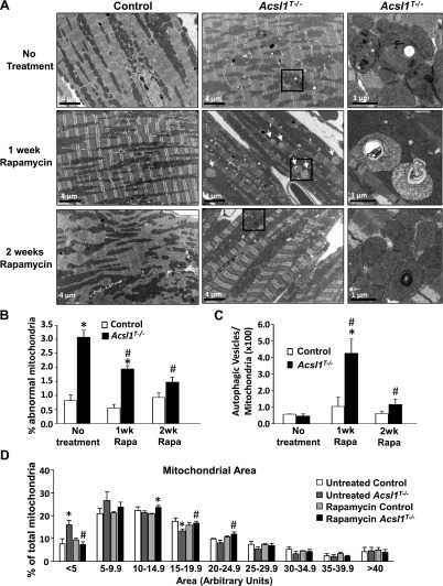
Because hearts with a temporally induced knockout of acyl-CoA synthetase 1 (Acsl1(T-/-)) are virtually unable to oxidize fatty acids, glucose use increases 8-fold to compensate. This metabolic switch activates mechanistic target of rapamycin complex 1 (mTORC1), which initiates growth by increasing protein and RNA synthesis and fatty acid metabolism, while decreasing autophagy. Compared with controls, Acsl1(T-/-) hearts contained 3 times more mitochondria with abnormal structure and displayed a 35-43% lower respiratory function. To study the effects of mTORC1 activation on mitochondrial structure and function, mTORC1 was inhibited by treating Acsl1(T-/-) and littermate control mice with rapamycin or vehicle alone for 2 wk. Rapamycin treatment normalized mitochondrial structure, number, and the maximal respiration rate in Acsl1(T-/-) hearts, but did not improve ADP-stimulated oxygen consumption, which was likely caused by the 33-51% lower ATP synthase activity present in both vehicle- and rapamycin-treated Acsl1(T-/-) hearts. The turnover of microtubule associated protein light chain 3b in Acsl1(T-/-) hearts was 88% lower than controls, indicating a diminished rate of autophagy. Rapamycin treatment increased autophagy to a rate that was 3.1-fold higher than in controls, allowing the formation of autophagolysosomes and the clearance of damaged mitochondria. Thus, long-chain acyl-CoA synthetase isoform 1 (ACSL1) deficiency in the heart activated mTORC1, thereby inhibiting autophagy and increasing the number of damaged mitochondria.
Keywords: ATP synthase; glucose metabolism; heart metabolism; lipid metabolism; β-oxidation.
© FASEB.
Figures


Similar articles
-
Mouse cardiac acyl coenzyme a synthetase 1 deficiency impairs Fatty Acid oxidation and induces cardiac hypertrophy.Mol Cell Biol. 2011 Mar;31(6):1252-62. doi: 10.1128/MCB.01085-10. Epub 2011 Jan 18. Mol Cell Biol. 2011. PMID: 21245374 Free PMC article.
-
Deficiency of cardiac Acyl-CoA synthetase-1 induces diastolic dysfunction, but pathologic hypertrophy is reversed by rapamycin.Biochim Biophys Acta. 2014 Jun;1841(6):880-7. doi: 10.1016/j.bbalip.2014.03.001. Epub 2014 Mar 12. Biochim Biophys Acta. 2014. PMID: 24631848 Free PMC article.
-
Cardiac energy dependence on glucose increases metabolites related to glutathione and activates metabolic genes controlled by mechanistic target of rapamycin.J Am Heart Assoc. 2015 Feb 24;4(2):e001136. doi: 10.1161/JAHA.114.001136. J Am Heart Assoc. 2015. PMID: 25713290 Free PMC article.
-
Nutrient-sensing mTORC1: Integration of metabolic and autophagic signals.J Mol Cell Cardiol. 2016 Jun;95:31-41. doi: 10.1016/j.yjmcc.2016.01.005. Epub 2016 Jan 7. J Mol Cell Cardiol. 2016. PMID: 26773603 Free PMC article. Review.
-
The multifaceted role of mTORC1 in the control of lipid metabolism.EMBO Rep. 2013 Mar 1;14(3):242-51. doi: 10.1038/embor.2013.5. Epub 2012 Feb 12. EMBO Rep. 2013. PMID: 23399656 Free PMC article. Review.
Cited by
-
Molecular mechanism of Cuscutae semen-radix rehmanniae praeparata in relieving reproductive injury of male rats induced with tripterygium wilfordii multiglycosides: A tandem mass tag-based proteomics analysis.Front Pharmacol. 2023 Feb 17;14:1050907. doi: 10.3389/fphar.2023.1050907. eCollection 2023. Front Pharmacol. 2023. PMID: 36874004 Free PMC article.
-
It takes a village: channeling fatty acid metabolism and triacylglycerol formation via protein interactomes.J Lipid Res. 2019 Mar;60(3):490-497. doi: 10.1194/jlr.S091843. Epub 2019 Jan 25. J Lipid Res. 2019. PMID: 30683668 Free PMC article. Review.
-
Myeloid-Specific Deficiency of Long-Chain Acyl CoA Synthetase 4 Reduces Inflammation by Remodeling Phospholipids and Reducing Production of Arachidonic Acid-Derived Proinflammatory Lipid Mediators.J Immunol. 2021 Dec 1;207(11):2744-2753. doi: 10.4049/jimmunol.2100393. Epub 2021 Nov 1. J Immunol. 2021. PMID: 34725110 Free PMC article.
-
Autophagy modulation: a potential therapeutic approach in cardiac hypertrophy.Am J Physiol Heart Circ Physiol. 2017 Aug 1;313(2):H304-H319. doi: 10.1152/ajpheart.00145.2017. Epub 2017 Jun 2. Am J Physiol Heart Circ Physiol. 2017. PMID: 28576834 Free PMC article. Review.
-
Access and utilization of long chain fatty acyl-CoA by zDHHC protein acyltransferases.Curr Opin Struct Biol. 2022 Dec;77:102463. doi: 10.1016/j.sbi.2022.102463. Epub 2022 Sep 29. Curr Opin Struct Biol. 2022. PMID: 36183446 Free PMC article. Review.
References
-
- Duncan J. G., Bharadwaj K. G., Fong J. L., Mitra R., Sambandam N., Courtois M. R., Lavine K. J., Goldberg I. J., Kelly D. P. (2010) Rescue of cardiomyopathy in peroxisome proliferator-activated receptor-alpha transgenic mice by deletion of lipoprotein lipase identifies sources of cardiac lipids and peroxisome proliferator-activated receptor-alpha activators. Circulation 121, 426–435 - PMC - PubMed
-
- Belke D. D., Larsen T. S., Gibbs E. M., Severson D. L. (2000) Altered metabolism causes cardiac dysfunction in perfused hearts from diabetic (db/db) mice. Am. J. Physiol. Endocrinol. Metab. 279, E1104–E1113 - PubMed
-
- Mazumder P. K., O’Neill B. T., Roberts M. W., Buchanan J., Yun U. J., Cooksey R. C., Boudina S., Abel E. D. (2004) Impaired cardiac efficiency and increased fatty acid oxidation in insulin-resistant ob/ob mouse hearts. Diabetes 53, 2366–2374 - PubMed
-
- Allard M. F., Henning S. L., Wambolt R. B., Granleese S. R., English D. R., Lopaschuk G. D. (1997) Glycogen metabolism in the aerobic hypertrophied rat heart. Circulation 96, 676–682 - PubMed
Publication types
MeSH terms
Substances
Grants and funding
LinkOut - more resources
Full Text Sources
Other Literature Sources
Molecular Biology Databases
Miscellaneous
