

Changes in central corneal thickness and refractive error after thin-flap laser in situ keratomileusis in Chinese eyes
- PMID: 26220189
- PMCID: PMC4517627
- DOI: 10.1186/s12886-015-0083-2
Changes in central corneal thickness and refractive error after thin-flap laser in situ keratomileusis in Chinese eyes
Abstract
Background: Refractive stability is influenced by alterations in corneal curvature and corneal thickness after laser in situ keratomileusis (LASIK). The aim of this study was to analyze the changes of central corneal thickness (CCT) and refractive error following thin-flap LASIK surgery in Chinese eyes.
Methods: One hundred and fifty-eight myopic patients (302 eyes) who underwent thin-flap LASIK surgery were prospectively evaluated. CCT was measured by non-contact specular microscopy before, and 1 day, 1 week, and 1, 3, and 6 months following surgery. Age, refractive error, and optic zone diameter were also recorded.
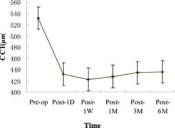
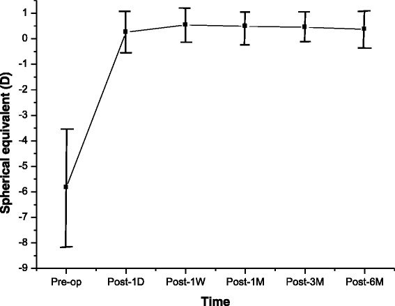
Results: Preoperatively, the mean CCT was 531.6 ± 24.3 μm. At 1 day, 1 week, and 1, 3, and 6 months after surgery, mean CCTs were 431.4 ± 38.4 μm, 422.6 ± 3 7.8 μm, 427.2 ± 38.0 μm, 434.4 ± 38.2 μm, and 435.6 ± 38.0 μm, respectively. Significant changes were detected in CCT values at each time point after thin-flap LASIK treatment (P < 0.05). The mean preoperative spherical equivalent (SE) was -5.73 ± 2.30 diopters (D). At 1 day, 1 week, and 1, 3, and 6 months after surgery, it was 0.26 ± 0.58 D, 0.54 ± 0.52 D, 0.49 ± 0.53 D, 0.45 ± 0.49 D, and 0.37 ± 0.42 D, respectively. The spherical equivalent refraction at 6 months postsurgery was close to the predicted value (0.34 ± 0.30 D). The changes in CCT within 6 months (4.06 ± 9.99 μm) were negatively correlated with age, preoperative refractive error, and optical zone diameter, respectively (r = -0.180, P < 0.05; r = -0.187, P < 0.001; r = -0.171, P < 0.05, respectively). No significant correlation was found between CCT changes and SE changes at different time points, postoperatively.
Conclusions: CCTs decreased significantly at 1 day after surgery, and continued to decline at 1 week after surgery, then increased over time. From postoperative 1 week, SE over time continually shifted to the myopic side.
Figures
References
-
- Duffey RJ, Leaming D. US trends in refractive surgery: 2003 ISRS ⁄AAO survey. J Refract Surg. 2005;21:87–91. - PubMed
-
- Azar DT, Ghanem RC, de la Cruz J, Hallak JA, Kojima T, Al-Tobaigy FM, et al. Thin-flap (sub-Bowman keratomileusis) versus thick-flap laser in situ keratomilusis for moderate to high myopia: case–control analysis. J Cataract Refract Surg. 2008;34:2073–78. doi: 10.1016/j.jcrs.2008.08.019. - DOI - PMC - PubMed
-
- Zhao MH, Zou J, Wang WQ, Li J. Comparison of central corneal thickness as measured by non-contact specular microscopy and ultrasound pachymetry before and post-LASIK. Clin Experiment. 2007;35(9):818–23. - PubMed
-
- Thomas J, Wang J, Rollins AM, Sturm J. Comparison of corneal thickness measured with optical coherence tomography, ultrasonic pachymetry, and scanning slit method. J Refract Surg. 2006;22:671–78. - PubMed
MeSH terms
LinkOut - more resources
Full Text Sources
Other Literature Sources
