. 2016 Feb;15(1):9-28.
doi: 10.1007/s10237-015-0704-y. Epub 2015 Jul 30.

Poromicromechanics reveals that physiological bone strains induce osteocyte-stimulating lacunar pressure

Affiliations

Poromicromechanics reveals that physiological bone strains induce osteocyte-stimulating lacunar pressure

Stefan Scheiner et al. Biomech Model Mechanobiol. 2016 Feb.

Abstract

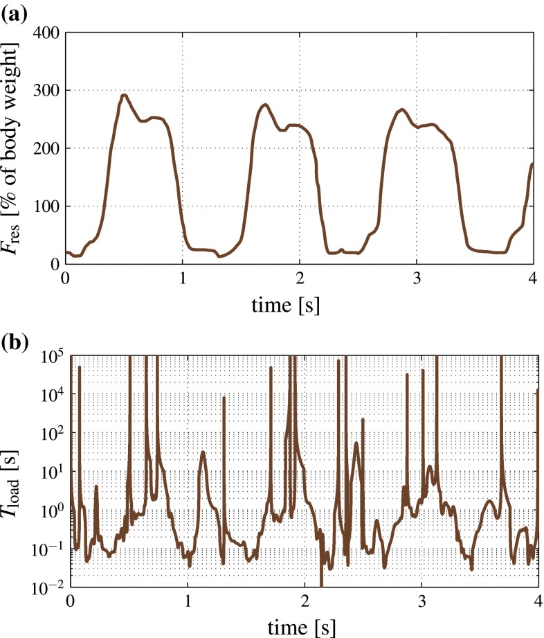
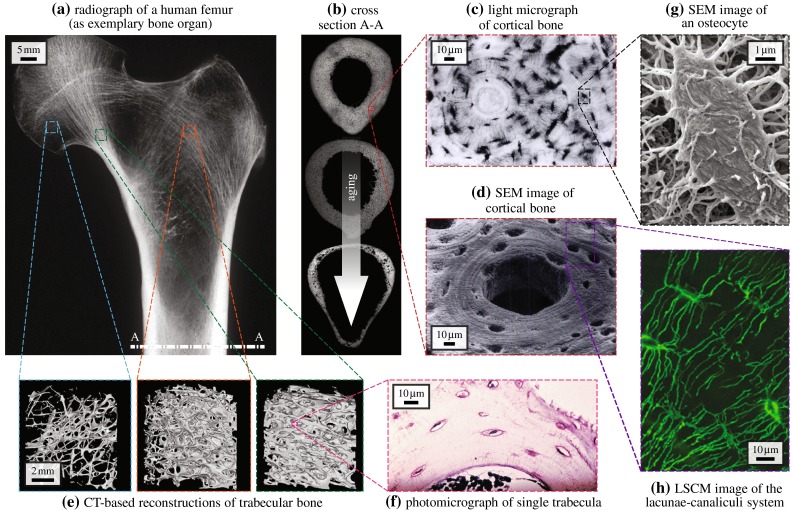
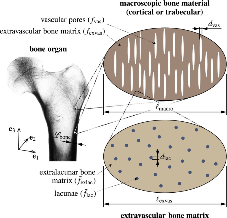
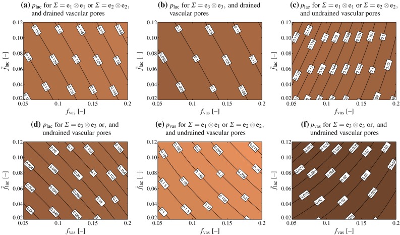
Mechanical loads which are macroscopically acting onto bony organs, are known to influence the activities of biological cells located in the pore spaces of bone, in particular so the signaling and production processes mediated by osteocytes. The exact mechanisms by which osteocytes are actually able to "feel" the mechanical loading and changes thereof, has been the subject of numerous studies, and, while several hypotheses have been brought forth over time, this topic has remained a matter of debate. Relaxation times reported in a recent experimental study of Gardinier et al. (Bone 46(4):1075-1081, 2010) strongly suggest that the lacunar pores are likely to experience, during typical physiological load cycles, not only fluid transport, but also undrained conditions. The latter entail the buildup of lacunar pore pressures, which we here quantify by means of a thorough multiscale modeling approach. In particular, the proposed model is based on classical poroelasticity theory, and able to account for multiple pore spaces. First, the model reveals distinct nonlinear dependencies of the resulting lacunar (and vascular) pore pressures on the underlying bone composition, highlighting the importance of a rigorous multiscale approach for appropriate computation of the aforementioned pore pressures. Then, the derived equations are evaluated for macroscopic (uniaxial as well as hydrostatic) mechanical loading of physiological magnitude. The resulting model-predicted pore pressures agree very well with the pressures that have been revealed, by means of in vitro studies, to be of adequate magnitude for modulating the responses of biological cells, including osteocytes. This underlines that osteocytes may respond to many types of loading stimuli at the same time, in particular so to fluid flow and hydrostatic pressure.

Keywords: Bone remodeling; Hydrostatic pressure; Mechanosensing; Micromechanics; Osteocytes; Poroelasticity.


Figures

Fig. 1
Fig. 1
Bone loading experienced in the hip joint during walking on a treadmill at 2 km/h: a resultant force history as recorded by Bergmann et al. (1993), and b corresponding loading times Tload according to Eq. (1)
Fig. 2
Fig. 2
Hierarchical organization of bone relevant for bone remodeling–modulating pore pressures, presented by the example of the human femur: a X-ray image of the proximal part of a human femur, reproduced from Sinclair et al. (2013), with permission from Elsevier B.V.; b midshaft cross section A-A, illustrated through corresponding microradiographs of femur cross sections, by courtesy of John G. Clement and David Thomas (taken from the Melbourne Femur Collection), shows deteriorating integrity with increasing age; cortical bone microstructure and its main constituents acquired by means of c light microscopy, reprinted from Buckwalter and Cooper (1987), with permission from the American Academy of Orthopaedic Surgeons (AAOS), or d scanning electron microscopy (SEM), reprinted from Kessel and Kardon (1979), by courtesy of Randy H. Kardon; e shows computed tomography (CT) images of trabecular bone acquired at different locations showing different porosities, reproduced from Padilla et al. (2008), with permission from Elsevier B.V.; f a photomicrograph of a single trabecula shows the composition of trabecular bone, reproduced from Sinclair et al. (2013), with permission from Elsevier B.V.; g SEM allows to visualize the osteocytes residing in the lacunar pores detectable in cortical bone and trabecular bone, reprinted from Pajevic (2009), by permission from Macmillan Publishers Ltd. on behalf of Cancer Research UK: IBMS BoneKey,  2009; h laser scanning confocal microscopy (LSCM) shows the canaliculi connecting the lacunae and the therein residing osteocytes, forming a dense network embedded in the extracellular bone matrix, reproduced from Ebacher et al. (2012), with permission from Elsevier B.V.
Fig. 3
Fig. 3
Micromechanical representation of cortical bone, based on which the poromicromechanical model is developed: Cortical bone microstructure is composed of extravascular bone matrix, with volume fraction fexvas, and vascular pore space, with volume fraction fvas, fexvas+fvas=1, Lbonemacrodvas, whereas extravascular bone matrix is composed of extracellular bone matrix, with volume fraction f¯exlac, and lacunar pores, with volume fraction f¯lac, f¯exlac+f¯lac=1, exvasdlac; the X-ray image of the bone organ was reproduced from Sinclair et al. (2013), with permission of Elsevier B.V.
Fig. 4
Fig. 4
Lacunar (ad) and vascular (e, f) pore pressures building up in response to uniaxial macroscopic unit stresses; the pore pressures shown in a and b follow from evaluation of Eq. (10), whereas the pore pressures shown in cf follow from evaluation of Eqs. (18) and (19)
Fig. 5
Fig. 5
Lacunar (ad) and vascular (e, f) pore pressures, in kPa, building up in response to uniaxial macroscopic unit microstrains; the pore pressures shown in a and b follow from evaluation of Eq. (6), whereas the pore pressures shown in cf follow from evaluation of Eqs. (13) and (14)
Fig. 6
Fig. 6
Lacunar and vascular pore pressures building up in correspondence to physiological macroscopic (uniaxial and hydrostatic) strains; strain magnitudes are chosen according the requirement of strain energy density equivalence
Fig. 7
Fig. 7
Lacunar pore pressure evolution during bone aging: a the effect of decreasing lacunar porosity, b the effect of increasing vascular porosity, and c combination of a and b; all computations are performed for undrained and drained vascular pores

References

    1. Abdalrahman T, Scheiner S, Hellmich C. Is trabecular bone permeability governed by molecular ordering-induced fluid viscosity gain? Arguments from re-evaluation of experimental data in the framework of homogenization theory. J Theor Biol. 2015;365:433–444. doi: 10.1016/j.jtbi.2014.10.011. - DOI - PubMed
    1. Abousleiman Y, Cheng A-D, Cui L, Detournay E, Roegiers J-C. Mandel’s problem revisited. Géotechnique. 1996;46(2):187–195. doi: 10.1680/geot.1996.46.2.187. - DOI
    1. Ahn A, Grodzinsky A. Relevance of collagen piezoelectricity to “Wolff’s law”: a critical review. Med Eng Phys. 2009;31(7):733–741. doi: 10.1016/j.medengphy.2009.02.006. - DOI - PMC - PubMed
    1. Ashman R, Cowin S, Van Buskirk W, Rice J. A continuous wave technique for the measurement of the elastic properties of cortical bone. J Biomech. 1984;17(5):349–361. doi: 10.1016/0021-9290(84)90029-0. - DOI - PubMed
    1. Auriault J-L, Boutin C, Geindreau C (2009) Homogenization of coupled phenomena in heterogeneous media. Wiley-Iste

Publication types

LinkOut - more resources