. 2015 Jul 29:5:12564.
doi: 10.1038/srep12564.

LK6/Mnk2a is a new kinase of alpha synuclein phosphorylation mediating neurodegeneration

Affiliations

LK6/Mnk2a is a new kinase of alpha synuclein phosphorylation mediating neurodegeneration

Shiqing Zhang et al. Sci Rep. .

Abstract

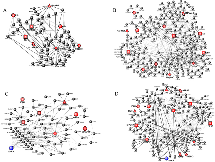
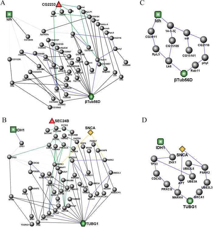
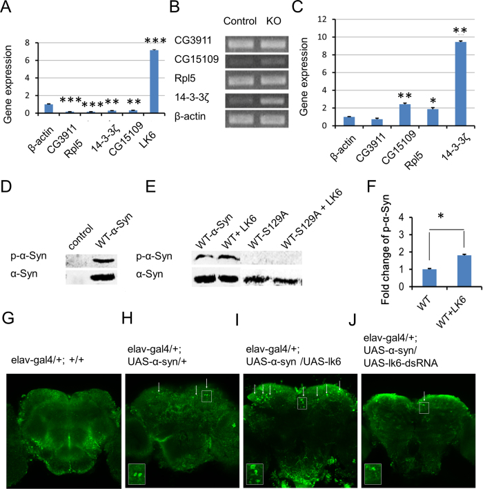
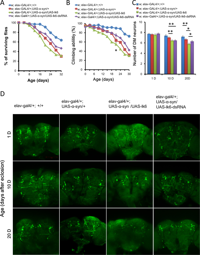
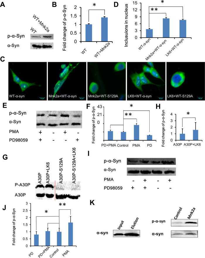
Parkinson's disease (PD) is a movement disorder due to the loss of dopaminergic (DA) neurons in the substantia nigra. Alpha-synuclein phosphorylation and α-synuclein inclusion (Lewy body) become a main contributor, but little is known about their formation mechanism. Here we used protein expression profiling of PD to construct a model of their signalling network from drsophila to human and nominate major nodes that regulate PD development. We found in this network that LK6, a serine/threonine protein kinase, plays a key role in promoting α-synuclein Ser129 phosphorylation by identification of LK6 knockout and overexpression. In vivo test was further confirmed that LK6 indeed enhances α-synuclein phosphorylation, accelerates the death of dopaminergic neurons, reduces the climbing ability and shortens the the life span of drosophila. Further, MAP kinase-interacting kinase 2a (Mnk2a), a human homolog of LK6, also been shown to make α-synuclein phosphorylation and leads to α-synuclein inclusion formation. On the mechanism, the phosphorylation mediated by LK6 and Mnk2a is controlled through ERK signal pathway by phorbolmyristate acetate (PMA) avtivation and PD98059 inhibition. Our findings establish pivotal role of Lk6 and Mnk2a in unprecedented signalling networks, may lead to new therapies preventing α-synuclein inclusion formation and neurodegeneration.


Conflict of interest statement

The authors declare no competing financial interests.

Figures

Figure 1
Figure 1. α-synuclein A53T/A30P PIN.
Drosophila expressing α-synuclein A53T/A30P related PIN was constructed with our experimental data and the DIP database. (A) The network of Drosophila expressing α-synuclein A53T shows 60 proteins and 100 interactions in 20720 interactions from DIP database of Drosophila. The 8 biggest nodes identified by mass spectrometry with red color stand for the corresponding altered proteins in human. (B) The network of Drosophila expressing α-synuclein A30P shows 217 proteins and 330 interactions in 20720 interactions from DIP database of Drosophila. The 13 biggest nodes with red color stand for the corresponding altered proteins in human. Nodes highlighted in redare from our experimental data. Broad lines represent the proteins interaction both exist in Drosophila and Human networks. Red nodes with the same shapes represent the same functions both in networks of Drosophila and Human (C), The human network in relationship to Drosophila expressing α-synuclein A53T. It shows 83 proteins of 6340 proteins and 146 interactions in 23591 interactions from HPRD database of human. (D) The human network in relationship to Drosophila expressing α-synuclein A30P. It shows 161 proteins of 6340 proteins and 316 interactions in 23591 interactions from HPRD data base of human. The 13 biggest nodes with red color stand for the corresponding altered proteins in the Drosophila expressing α-synuclein A30P. The big green node (SNCA) stands for α-synuclein. Broad lines represent that the protein interactions exist in both Drosophila and Human networks. Each node and its counterpart in Drosophila and Human networks are shown with the same color and same shape in these two figures.
Figure 2
Figure 2. The common interaction networks between β-tub56D and Idh.
(A) The network between β-tub56D and Idh included 56 proteins and 104 interactions in 20720 interactions from DIP database of Drosophila. (B) The network between β-tub56D and Idh included 61 proteins of 6340 proteins and 84 interactions of 23591 interactions from HPRD database of human. Blue lines represent the proteins interaction also exists in Drosophila network and human network. (C) Representative graph of two main pathways in Drosophila network. (D) Representative graph of two main pathways in Human network. The two main pathways in (C,D) are extremely similar.
Figure 3
Figure 3. LK6 phosphorylates α-synuclein.
(A) Quantitative analysis of the expression changes of CG3911, Rpl5, 14-3-3ζ, LK6, CG15109 in the PIN were detected with real-time PCR. (B) RT-PCR analysis showing expression of CG3911, Rpl5, 14-3-3ζ, CG15109 between LK6 (-/-) drosophila and wild type (+/+). (C) Quantitative analysis of the expression changes of mRNAs in the LK6 (-/-) drosophila. Results are shown as means ± SE of triplicate in three independent experiments. The statistical significance of differences was calculated by using P value (*<0.01; **<0.001; ***<0.0001). (D) The expression of endogenous α-synuclein in HEK293T cells. Western blots of protein lysates from cells were transient transfected with WT-α-synuclein and the parental HEK293T cells. (E) LK6 phosphorylates WT-α-synuclein and A30P-α-synuclein at Ser129. Phosphorylation was not detected in mutants S129A and S129A-LK6. (F) Quantitative results from (E) between α-synuclein and α-synuclein–LK6 show a significant increase (n = 3, *p<0.05). Immuno histochemical staining in drosophila brains with antibody indicate a siginificant increase in expressing α-synuclein-LK6(I) than α-synuclein line (H) and blank control (G). Silence of LK6 by RNAi α-synuclein phosphorylation was significantly decreased (Fig. 3J).
Figure 4
Figure 4. α-synuclein phosphorylation by LK6 shortened drosophila lifespan and caused loss of dopaminergic neurons.
(A) Quantitative analysis of survival rate in transgenic flies. Data were analyzed by Log-rank (Mantel-Cox) test with *P<0.05, compared to PD flies. Genotypes: control flies are w; elav-gal4/+;+/+. α-synuclein flies are w; elav-gal4/+; UAS-synuclein/+. lk6 overexpression α-synuclein flies are elav-gal4/+; UAS-synuclein/UAS-lk6. lk6 silencing α-synuclein flies are elav-gal4/+; UAS-synuclein/UAS-lk6-dsRNA. (B) Overexpression Lk6 accelerates loss of climbing ability in α-synuclein transgenic flies throughout the nervous system (elav-GAL4 driver). Quantitative analysis of climbing ability in transgenic flies. Values represent mean ± SEM. (***P<0.001, compared to wild type flies, #P<0.05, ##P<0.01, compared to PD flies, multivariant ANOVA with Bonferroni’s Multiple Comparison Test). Genotypes: control flies are w; elav-gal4/+;+/+. α-synuclein flies are w; elav-gal4/+; UAS-synuclein/+. lk6 overexpression α-synuclein flies are elav-gal4/+; UAS-synuclein/UAS-lk6. lk6 silencing α-synuclein flies are elav-gal4/+; UAS-synuclein/UAS-lk6-dsRNA. (C) Confocal images of dopaminergic neurons regions immune stained with anti-TH antibody on 1-d, 10-d, and 20-d fly brains, respectively, with box for DM clusters. A normal number of dorsomedial neurons was identified in 1-d flies in a pan-neural pattern, whereas loss of dopaminergic neurons was observed in 10-d and 20-d α-synuclein transgenic flies with lk6 overexpression. Silence of lk6 could rescue the defect of dopaminergic neurons derived from α-synuclein. Quantitative analysis of TH immunoreactive dorsomedial cluster dopamine neurons in transgenic flies. Values represent mean ± SEM. (*P<0.05, multivariant ANOVA with Bonferroni’s Multiple Comparison Test) .(D) Quantitative analysis of dopaminergic neuron numbers over time in the DM clusters ofdifferent genotypes of α-synuclein flies.
Figure 5
Figure 5. LK6/Mnk2a mediate α-synuclein phosphorylation signal mechanism.
(A) Mnk2a phosphorylates WT-α-synucleinat Ser129. Quantitative results show a significant increase of α-synuclein phosphorylation level by Mnk2a (B). (C) Overexpression of Mnk2a and WT-α-synuclein enhanced α-synuclein inclusion formation in nucleus. Cells were transfected with WT-α-synuclein and co-transfected with WT-α-synuclein and Mnk2a or WT-α-synuclein-S129A and Mnk2a. (D) Statistical analysis of α-synuclein inclusion formation in nucleus. (E) LK6 phosphorylates WT-α-synuclein Ser129 by ERK signaling. Western blots of protein lysates from cells were co-transfected with WT-α-synuclein and LK6. (F) Quantitative results of the comparison from (I) showing a 1.83-fold greater capacity for PMA (n = 3, **p<0.01) and a 0.61-fold lower capacity for PD98059 (n = 3, *p<0.05). (G) LK6 α-synuclein phosphorylation detection indicates enhanced status in mutant A30P-LK6, whereas phosphorylation disappear in mutants A30P-S129A and A30P-S129A-LK6. Quantitative results confirmed the significant difference between A30P and A30P-LK6 (H). (I) Mnk2a phosphorylates WT-α-synuclein Ser129 by ERK signaling. (J) Quantitative results of the comparison from (I) showing a 1.65-fold greater capacity for PMA (n = 3, **p<0.01) and a 0.80-fold lower capacity for PD98059 (n = 3, *p<0.05). (K) Purified α-synuclein was incubated with cell lysate including overexpressed Mnk2a at concentration of 1 ng/μl with 200 μM ATP for 1 hour. The protein was analyzed by western blotting probed with anti-phospho-α-synuclein and anti α-synuclein antibodies. Cell lysate from 293T overexpressed Mnk2a showed more α-synuclein phosphorylation than control group.

References

    1. Karuppagounder S. S. et al. The c-Abl inhibitor, nilotinib, protects dopaminergic neurons in a preclinical animal model of Parkinson’s disease. Sci Rep 4, 4874 (2014). - PMC - PubMed
    1. Jiang P. et al. Adenosine monophosphate-activated protein kinase overactivation leads to accumulation of alpha-synuclein oligomers and decrease of neurites. Neurobiol Aging 34, 1504–1515 (2013). - PMC - PubMed
    1. Wolters E. Variability in the clinical expression of Parkinson’s disease. J Neurol Sci 266, 197–203 (2008). - PubMed
    1. Polymeropoulos M. H. et al. Mutation in the alpha-synuclein gene identified in families with Parkinson’s disease. Science 276, 2045–2047 (1997). - PubMed
    1. Sandal M. et al. Conformational equilibria in monomeric alpha-synuclein at the single-molecule level. PLoS Biol 6, e6 (2008). - PMC - PubMed

Publication types

MeSH terms