. 2015 Nov 15:122:262-71.
doi: 10.1016/j.neuroimage.2015.07.051. Epub 2015 Jul 26.

Medial prefrontal pathways for the contextual regulation of extinguished fear in humans

Affiliations

Medial prefrontal pathways for the contextual regulation of extinguished fear in humans

Fredrik Åhs et al. Neuroimage. .

Abstract

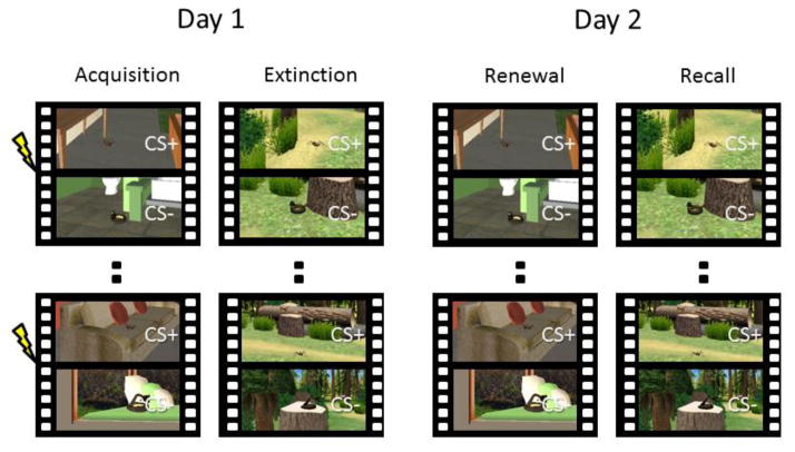
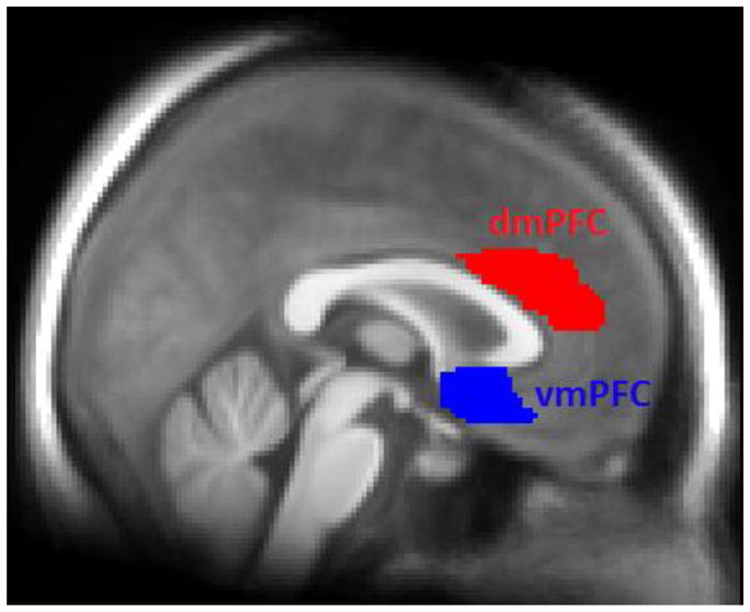
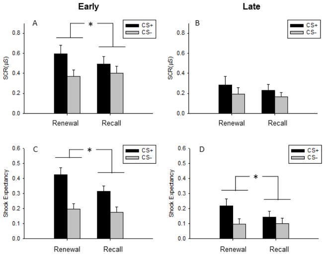
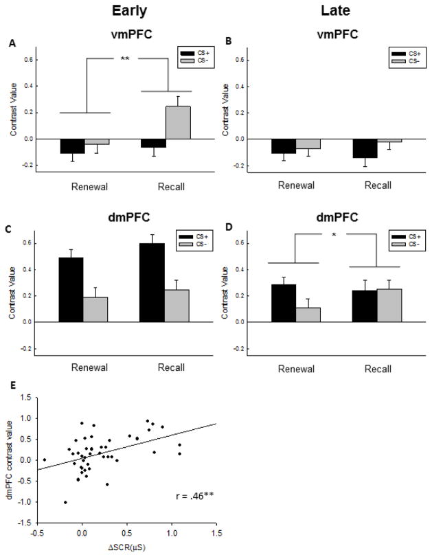
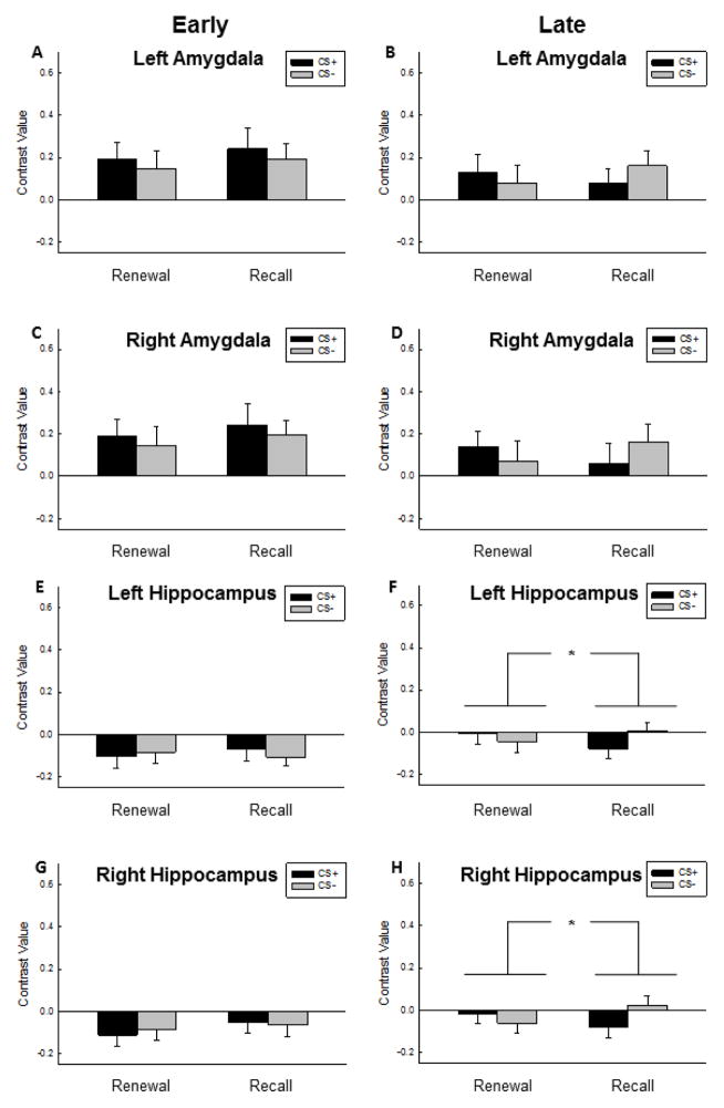
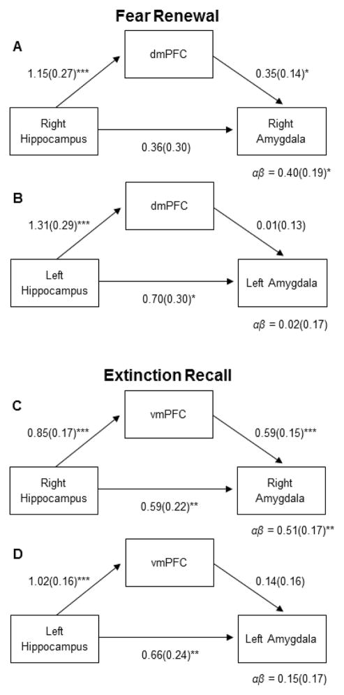
The maintenance of anxiety disorders is thought to depend, in part, on deficits in extinction memory, possibly due to reduced contextual control of extinction that leads to fear renewal. Animal studies suggest that the neural circuitry responsible fear renewal includes the hippocampus, amygdala, and dorsomedial (dmPFC) and ventromedial (vmPFC) prefrontal cortex. However, the neural mechanisms of context-dependent fear renewal in humans remain poorly understood. We used functional magnetic resonance imaging (fMRI), combined with psychophysiology and immersive virtual reality, to elucidate how the hippocampus, amygdala, and dmPFC and vmPFC interact to drive the context-dependent renewal of extinguished fear. Healthy human participants encountered dynamic fear-relevant conditioned stimuli (CSs) while navigating through 3-D virtual reality environments in the MRI scanner. Conditioning and extinction were performed in two different virtual contexts. Twenty-four hours later, participants were exposed to the CSs without reinforcement while navigating through both contexts in the MRI scanner. Participants showed enhanced skin conductance responses (SCRs) to the previously-reinforced CS+ in the acquisition context on Day 2, consistent with fear renewal, and sustained responses in the dmPFC. In contrast, participants showed low SCRs to the CSs in the extinction context on Day 2, consistent with extinction recall, and enhanced vmPFC activation to the non-reinforced CS-. Structural equation modeling revealed that the dmPFC fully mediated the effect of the hippocampus on right amygdala activity during fear renewal, whereas the vmPFC partially mediated the effect of the hippocampus on right amygdala activity during extinction recall. These results indicate dissociable contextual influences of the hippocampus on prefrontal pathways, which, in turn, determine the level of reactivation of fear associations.

Keywords: Amygdala; Anterior cingulate cortex; Extinction; Fear conditioning; Functional magnetic resonance imaging; Hippocampus; Ventromedial prefrontal cortex; Virtual reality.


Conflict of interest statement

Conflict of interest

The authors declare no competing financial interests.

Figures

Figure 1
Figure 1
Experimental design. Participants were conditioned to dynamic cues (spiders or snakes) while travelling through a virtual reality environment (indoor or outdoor) presented in 3-D goggles. The fear cue was paired with shock on 5 out of the 16 trials (CS+) whereas the safe cue (CS−) was never paired with shock. Participants were not informed about which cue would serve as CS+ and CS−. Fear was extinguished in another environment following conditioning. Twenty-four hours later, responses to the fear cue and the safe cue were tested in the original acquisition context (Fear Renewal context) and the extinction context (Extinction Recall context), with presentation order counterbalanced between subjects. Trials were divided into Early (trials 1–8) and Late (9–16) phases for analysis purposes to map gross temporal dynamics in responses over time (not shown). CS, conditioned stimulus.
Figure 2
Figure 2
Prefrontal anatomical regions of interest overlaid on participants’ average T1-weighted MR image. These regions were used for extraction of fMRI data. dmPFC, dorsomedial prefrontal cortex; vmPFC, ventromedial prefrontal cortex.
Figure 3
Figure 3
Behavioral test of fear renewal. A) During Early trials (first half of the trial blocks), the difference in skin conductance response (SCR) between the reinforced cue (CS+) and the control cue (CS−) was greater in the Fear Renewal context than the Extinction Recall context. B) During late trials, SCRs to the conditioned cues were similar, suggesting re-extinction of the conditioned response. C) The difference in shock expectancy between the CS+ and CS− was greater in the Fear Renewal context than the Extinction Recall context during early trials. D) The CS-difference in shock expectancy was also greater in the Fear Renewal context during late trials. Bars represent mean and standard error of the mean. *P < 0.05.
Figure 4
Figure 4
Functional MRI responses in the medial prefrontal cortex (mPFC) during Early and Late trials in the Fear Renewal and Extinction Recall contexts. A) During Early trials, ventromedial PFC (vmPFC) safety signaling (response to the CS− > CS+) was pronounced in the Extinction Recall context relative the Renewal context. B) Early trials were associated with an increased dorsomedial PFC (dmPFC) response to the CS+ in both contexts. C) During Late trials, the vmPFC response was low and non-differential across cues or contexts. D) During Late trials, the dmPFC signaling of fear responses (CS+ > CS−) was significant only in the Fear Renewal context, suggesting faster habituation in the Extinction Recall context. E) Fear expression is correlated with dmPFC reactivity during Early trials in the Fear Renewal context. Contrast values from Early trials are derived from the comparison CS+ > CS− and are plotted against the same comparison in skin conductance response (ΔSCR), averaged across subjects. A–D) Bars represent mean and standard error. * p < .05; ** p < .01..
Figure 5
Figure 5
Functional MRI responses in the amygdala and hippocampus during Early and Late trials in the Fear Renewal and Extinction Recall contexts. During Early trials, similar mean levels of responding to the CS+ relative the CS− in the Extinction Recall context and the Fear Renewal context was observed in the A) left and C) right amygdala. Also during Late trials, the B) left and D) right amygdala response was low and non-differential across cues or contexts. A lack of differential responding across cues or contexts was further noted in the E) left and G) right hippocampus during Early trials. During Late trials however, hippocampal signaling of fear responses was attenuated relative safety responses (CS− > CS+) in the Extinction Recall context compared to the Fear Renewal context when considering both the F) left and H) right hippocampus signal. Bars represent mean and standard error.
Figure 6
Figure 6
Results from the statistical mediation analysis investigating hippocampal influences on the amygdala via direct projections and through indirect dorsal and ventral medial prefrontal pathways during Fear Renewal and Extinction Recall. This analysis targeted Early trials, for which the psychophysiological effects were maximal (see Fig. 4). A) During Fear Renewal, the dmPFC fully mediated the effect of the right hippocampus on the right amygdala. B) In the left hemisphere, the effect of the hippocampus on the amygdala was not mediated by the dmPFC. C) During Extinction Recall, the vmPFC partially mediated the effect of the right hippocampus on the right amygdala. D) In the left hemisphere, we did not observe mediation of the effect of the hippocampus on the amygdala by the vmPFC. * p < .05; ** p < .01; *** p < .001.

References

    1. Aguirre GK, Zarahn E, D’Esposito M. The variability of human, BOLD hemodynamic responses. Neuroimage. 1998;8:360–369. - PubMed
    1. Ahs F, Dunsmoor JE, Zielinski D, LaBar KS. Spatial proximity amplifies valence in emotional memory and defensive approach-avoidance. Neuropsychologia. 2015;70:476–485. - PMC - PubMed
    1. Apergis-Schoute AM, Schiller D, LeDoux JE, Phelps EA. Extinction resistant changes in the human auditory association cortex following threat learning. Neurobiology of Learning and Memory. 2014;113:109–114. - PMC - PubMed
    1. Baronchelli A, Ferrer-i-Cancho R, Pastor-Satorras R, Chater N, Christiansen MH. Networks in cognitive science. Trends in Cognitive Sciences. 2013;17:348–360. - PubMed
    1. Beckmann M, Johansen-Berg H, Rushworth MF. Connectivity-based parcellation of human cingulate cortex and its relation to functional specialization. J Neurosci. 2009;29:1175–1190. - PMC - PubMed

Publication types