

Hepatopulmonary Fistula: a life threatening complication of hydatid disease
- PMID: 26220789
- PMCID: PMC4518634
- DOI: 10.1186/s13019-015-0311-0
Hepatopulmonary Fistula: a life threatening complication of hydatid disease
Abstract
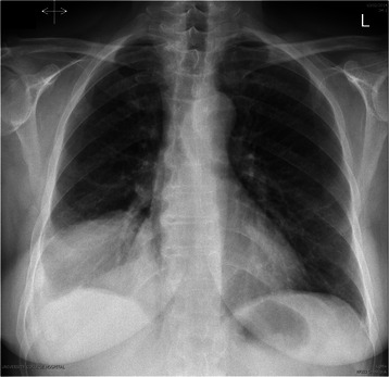
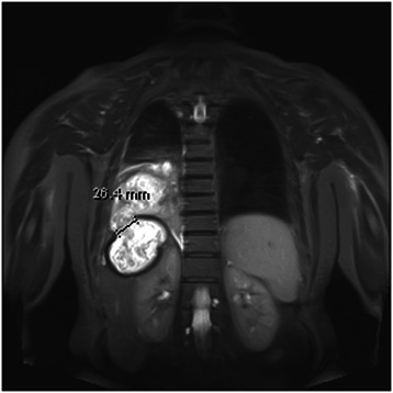
Despite extensive infection control measures against parasitic diseases, hydatid disease, caused by Echinococcus granulosus, still occurs in a minor group of our population. If the infection is not treated adequately, it goes on to developing life-threatening complications, one of which is hepatopulmonary fistula. These complications usually warrant early surgical intervention, or else may lead to extensive sepsis and ultimately death. We discuss the case of an elderly female suffering from pulmonary hydatid disease, further complicated by a hepatopulmonary fistula and underwent surgical treatment. This case emphasises the importance of early recognition of pulmonary hydatid disease given its atypical nature of presentation before the disease is further exacerbated by this aggressive complication. Furthermore, it is imperative to incorporate radical surgery as the first-line treatment in established hepatopulmonary fistula, in order to prevent further clinical deterioration and curative outcome.
Figures

References
-
- Kourias B. Second World Congress of Gastroenterology, Karger AG Publisher, Basel, Switzerland: Munich, 1963; 556.
Publication types
MeSH terms
LinkOut - more resources
Full Text Sources
Other Literature Sources
Medical
