. 2016 Sep:150:44-61.
doi: 10.1016/j.exer.2015.07.016. Epub 2015 Jul 26.

Idiopathic preretinal glia in aging and age-related macular degeneration

Affiliations

Idiopathic preretinal glia in aging and age-related macular degeneration

Malia M Edwards et al. Exp Eye Res. 2016 Sep.

Abstract

During analysis of glia in wholemount aged human retinas, frequent projections onto the vitreal surface of the inner limiting membrane (ILM) were noted. The present study characterized these preretinal glial structures. The amount of glial cells on the vitreal side of the ILM was compared between eyes with age-related macular degeneration (AMD) and age-matched control eyes. Retinal wholemounts were stained for markers of retinal astrocytes and activated Müller cells (glial fibrillary acidic protein, GFAP), Müller cells (vimentin, glutamine synthetase) and microglia/hyalocytes (IBA-1). Retinal vessels were labeled with UEA lectin. Images were collected using a Zeiss LSM 710 confocal microscope. Retinas were then cryopreserved. Laminin labeling of cryosections determined the location of glial structures in relation to the ILM. All retinas investigated herein had varied amounts of preretinal glia. These glial structures were classified into three groups based on size: sprouts, blooms, and membranes. The simplest of the glial structures observed were focal sprouts of singular GFAP-positive cells or processes on the vitreal surface of the ILM. The intermediate structures observed, glial blooms, were created by multiple cells/processes exiting from a single point and extending along the vitreoretinal surface. The most extensive structures, glial membranes, consisted of compact networks of cells and processes. Preretinal glia were observed in all areas of the retina but they were most prominent over large vessels. While all glial blooms and membranes contained vimentin and GFAP-positive cells, these proteins did not always co-localize. Many areas had no preretinal GFAP but had numerous vimentin only glial sprouts. In double labeled glial sprouts, vimentin staining extended beyond that of GFAP. Hyalocytes and microglia were detected along with glial sprouts, blooms, and membranes. They did not, however, concentrate in the retina below these structures. Cross sectional analysis identified small breaks in the ILM above large retinal vessels through which glial cells exited the retina. Preretinal glial structures of varied sizes are a common occurrence in aged retinas and, in most cases, are subclinical. While all retinal glia are found in blooms, vimentin labeling suggests that Müller cells form the leading edge. All retinas investigated from eyes with active choroidal neovascularization (CNV) had extensive glial membranes on the vitreal surface of the ILM. Although these structures may be benign, they may exert traction on the retina as they spread along the vitreoretinal interface. In cases with CNV, glial cells in the vitreous could bind intravitreally injected anti-vascular endothelial growth factor. These preretinal glial structures indicate the remodeling of both astrocytes and Müller cells in aged retinas, in particular those with advanced AMD.

Keywords: Age-related macular degeneration; Astrocytes; Epiretinal membranes; Glia; Muller cells.


Figures

Figure 1
Figure 1. Quantification of GFAP-positive processes/cells on the vitreoretinal surface
(A) Retinas were labelled with GFAP and tiled 5x5 confocal Z stacks were collected at 10x to generate a map of each retinal region. (B) A new image was created after selecting GFAP+ processes/cells on the vitreoretinal surface. Scale bar indicates: 500 μm.
Figure 2
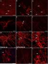
Figure 2. Glial sprouts were the simplest glial structures observed on the vitreal surface of the ILM
Retinas were labelled with anti-GFAP. (A) A low magnification tiled image of a posterior pole containing numerous focal glial sprouts (arrows), the fovea is shown with an asterisk. (B) At higher magnification, a Z stack slice demonstrates that glial sprouts lie in a focal plane above retinal astrocytes. Numerous branching processes from these cells extend into the retina below (arrows). (C) In the retina below, the astrocyte pattern is normal with the exception of tangled stalks where cells have exited the retina (arrows). (D) Closer investigation of a glial sprout demonstrates the length of processes. (E) Connections to cells within the retina are visible in the focal plane below “D” (arrows). (F) The overall astrocyte pattern below this sprout is normal. (G) A 3-D rendering of the same sprout demonstrates the main stalk (asterisk). Processes on the vitreal surface extend radially along the retinal surface. (H) When the opposing view of this 3-D image is examined, with the normal astrocyte pattern facing up and the sprout below, numerous connections are observed between the retinal astrocytes and the sprout (arrows). (I) Closer investigation provides better visualization of these connections (arrows) as well as the primary stalk (asterisk). (J) Some sprouts had a more stellate morphology extending numerous processes. Sprouts often joined together (arrow). (K) In the focal plane below, astrocytes within the retina were normal in density and pattern. (L) 3-D rendering of this glial sprout better demonstrates the connections between two sprouts by a fine process (arrow) as well as to astrocytes within the retina. Scale bars indicate: A: 500 μm, B-F, J, K: 50 μm and G-I, L: 20 μm.
Figure 3
Figure 3. Multiple GFAP-positive cells exited the retina through one point to create glial blooms
Retinas stained with anti-GFAP are shown. (A, B) Low magnification tiled images showing posterior pole regions containing glial blooms (arrows). An asterisk indicates the fovea. (C) Higher magnification of two glial blooms joined by fine processes (arrow). (D) A glial bloom at high magnification shows multiple stalks exiting from one site in the retina (arrow). (E) In some cases, thick processes terminated as arborizations, creating a net-like appearance. (F) In others, stalks branched into multiple long fine processes (arrow). (G) The astrocytes in the retina below have a normal pattern with only a focal disruption where cells exited the retina (arrow). (H) Three dimensional rendering further demonstrates that this bloom lies above the normal astrocyte layer (asterisk). (I) The exit of glial processes from the retina (arrow) is evident in the cross section rendering. (J) Another example of a glial bloom is shown along with the cross sectional view demonstrating the bright GFAP labeling above the retina. An asterisk indicates an area without glial cells on the vitreoretinal surface where retinal astrocytes are visible. (K) The arborizations of the thick glial processes shown by the arrow in “J” are shown at higher magnification. (L) Three dimensional rendering of a bloom shows the layer of astrocytes below (asterisk). Scale bars indicate: A, B: 500 μm; C, D, G, H, J, L: 50 μm; E, F, I, K: 20 μm.
Figure 4
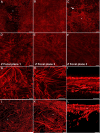
Figure 4. Multiple glial blooms coalesced to form continuous glial membranes
GFAP labeling of wholemount retinas demonstrates large glial membranes. (A) This tiled image of a posterior pole shows two large membranes merging above a normal astrocyte pattern (asterisk). (B) A continuous glial membrane covers the entire area of this tiled imaged. (C) Connections between blooms (arrow) create this larger glial membrane (asterisk). (D) Higher magnification demonstrates the density of these structures. (E) In the focal plane below “D”, numerous GFAP+ processes are present. (F) Astrocytes in the retina below are tortuous with misaligned processes. (G) In another membrane, thick, GFAP+ processes arborize into smaller processes which connect with one another. (H) Many smaller structures within this membrane appear to be individual cells. Long processes extend between these glial cells and into the retina. (I) A digital cross sectional view demonstrates the multiple layers of GFAP+ cells. The connections between the top layer (which lies in the vitreous) and astrocytes below can be observed. (J) The complexity of membranes is also shown in this 3D rendering. The astrocytes in an area lacking membrane (asterisk) are visible. (K) A side view of this membrane shows the astrocyte layer below (asterisks). (L) A cross sectional view demonstrates the astrocytes (asterisk) below the glial membrane. Scale bars indicate: A-C: 500 μm; D-H: 50 μm; I: 10 μm, and J-L: 20 μm.
Figure 5
Figure 5. Preretinal glial structures were associated with retinal vessels
Retinal wholemounts shown were stained with anti-GFAP (red, glia) as well as UEA lectin (grey, blood vessels). (A-C) A glial sprout is shown extending onto the vitreal surface above a large vessel branch point. (D-F) Tiling of multiple images shows glial sprouts and blooms above retinal capillaries (arrow) as well as a larger vessel and branch point. (G) A 3D rendering of multiple glial blooms (asterisks) joined by a glial sprout (arrow) above a large retinal vessel. (H) The same 3D image viewed from the blood vessels side. Scale bars indicate: A-C: 50 μm; D-H: 100 μm.
Figure 6
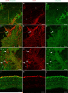
Figure 6. Glial cells exited the retina through breaks in the ILM
(A) Retinal wholemounts labeled for GFAP (red) were cryopreserved and sections stained for laminin (green) to label the ILM. GFAP+ cells are observed in the vitreous adjacent to a break (arrow) in the ILM. (B) Breaks in the ILM (arrow) are evident in JB4-embedded regions of a retina stained with PAS and hematoxylin. (C) Nuclei (arrow) are visible on the vitreoretinal surface in sections stained with toluidine blue. Scale bars indicate: A: 40 μm and B, C: 20 μm.
Figure 7
Figure 7. Retinal wholemounts stained for GFAP (red) and vimentin (green) demonstrate both Müller cells and astrocytes in preretinal glial structures
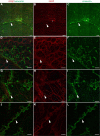
(A-C) GFAP+ and vimentin+ cells are observed in glial sprouts above a normal astrocyte template and Müller cell endfeet (asterisks). (D-I) In larger glial blooms and membranes, most cells are double-positive. Some cells/processes (arrow) have only GFAP while others (asterisk) have only vimentin. (J-L) A wholemount cross sectioned demonstrates staining of Müller cells with vimentin (green) and astrocytes with GFAP (red). Some lighter GFAP staining is seen in some Müller cells. The membrane above the Müller cell endfeet is double-positive. Scale bars indicate 40 μm.
Figure 8
Figure 8. Retinal wholemounts labelled with GFAP (red) and GS (green) confirm two cell populations in preretinal glial structures
(A-C) Above a retinal vessel branch point (asterisk), glial sprouts contain GFAP and GS+ long processes that extend into the vitreous (arrows). GFAP+, GS tangles are also observed (asterisk). In addition, intense GS staining (arrowhead) could indicate either swelling or aggregation of Müller cell endfeet at the ILM. (D-F) The glial bloom shown contains primarily GFAP+, GS processes/cells (arrow) with a few GFAP-, GS+ processes (arrowhead). (G-I) A glial sprout along a large retinal vessel (asterisk) contains double labelled processes/cells (arrowhead) as well as cells expressing only GFAP (arrows), which appears blebbed. (J-L) A dense area of GS+ cells (arrowhead) lies above a GFAP+ glial bloom (arrow). Scale bars represent 40 μm.
Figure 9
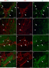
Figure 9. Müller cells exited the retina ahead of astrocytes
Retinal wholemounts stained for GFAP (red) and vimentin (green) are shown. (A-C) This area had no preretinal GFAP but contained vimentin+ sprouts (arrowhead). One small area (asterisk) had a tangle of GFAP+ processes. (D-F) Vimentin+ glial sprouts (arrowhead) are shown at higher magnification above a normal astrocyte pattern. (G-I) Under other areas of vimentin only sprouts (arrowhead), singular GFAP+ processes extended onto the vitreal side of the ILM. (J-L) Vimentin+ sprouts with a few GFAP+ processes (arrowhead) are shown in an area above a retinal vessel. Scale bar represent: A-C: 200 μm, D-L: 40 μm.
Figure 10
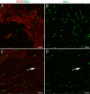
Figure 10. IBA-1 positive cells associated with glial cells on the vitreoretinal surface
Wholemount retinas labeled with anti-GFAP (red) and anti-IBA-1 (green). (A, B) IBA-1+ cells associate with glial sprouts and blooms. (C, D) These cells were not concentrated at the base where glia exited the retina (arrow). Scale bars indicate 50 μm.
Figure 11
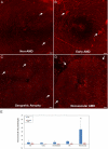
Figure 11. Large glial membranes were most prominent in neovascular AMD retinas
Retina wholemounts stained for GFAP (red) are shown. Tiled images taken at 10x are shown from representative (A) non-AMD retina, (B) early AMD, (C) geographic atrophy, and (D) neovascular AMD. In all images, arrows indicate some of the preretinal glia. Images were taken with the fovea centered. (E) Graphs show the average percentage of posterior pole tiled images covered with GFAP+ cells on the vitreal surface. (F) Graphs show the average percentage of the ILM covered by preretinal GFAP+ structures. As shown, there was a significant increase in the GFAP+ cells on the vitreal side of the ILM in neovascular AMD in the posterior pole but not outside this area. Scale bars indicate 200 μm. Asterisks indicate significant difference compared to the non-AMD group.

Similar articles

Cited by

References

    1. Blasiak J, Salminen A, Kaarniranta K. Potential of epigenetic mechanisms in AMD pathology. Front Biosci (Schol Ed) 2013;5:412–425. - PubMed
    1. Bringmann A, Wiedemann P. Involvement of Muller glial cells in epiretinal membrane formation. Graefes Arch Clin Exp Ophthalmol. 2009;247:865–883. - PubMed
    1. Charteris DG, Sethi CS, Lewis GP, Fisher SK. Proliferative vitreoretinopathy-developments in adjunctive treatment and retinal pathology. Eye. 2002;16:369–374. - PubMed
    1. Edwards MM, McLeod DS, Grebe R, Heng C, Lefebvre O, Lutty GA. Lama1 mutations lead to vitreoretinal blood vessel formation, persistence of fetal vasculature, and epiretinal membrane formation in mice. BMC Dev Biol. 2011;11:60. - PMC - PubMed
    1. Fisher SK, Lewis GP. Muller cell and neuronal remodeling in retinal detachment and reattachment and their potential consequences for visual recovery: a review and reconsideration of recent data. Vision Res. 2003;43:887–897. - PubMed

Publication types