. 2015 Jul 28;6(4):e00767.
doi: 10.1128/mBio.00767-15.

Pediatric Cystic Fibrosis Sputum Can Be Chemically Dynamic, Anoxic, and Extremely Reduced Due to Hydrogen Sulfide Formation

Affiliations

Pediatric Cystic Fibrosis Sputum Can Be Chemically Dynamic, Anoxic, and Extremely Reduced Due to Hydrogen Sulfide Formation

Elise S Cowley et al. mBio. .

Abstract

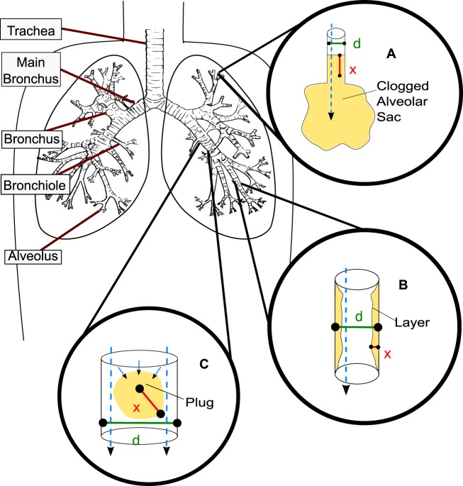
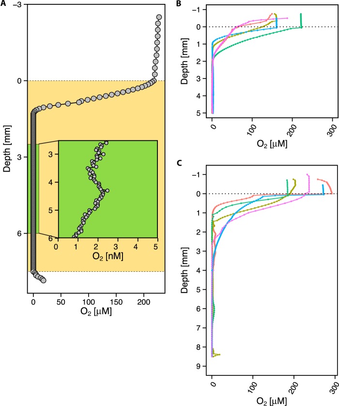
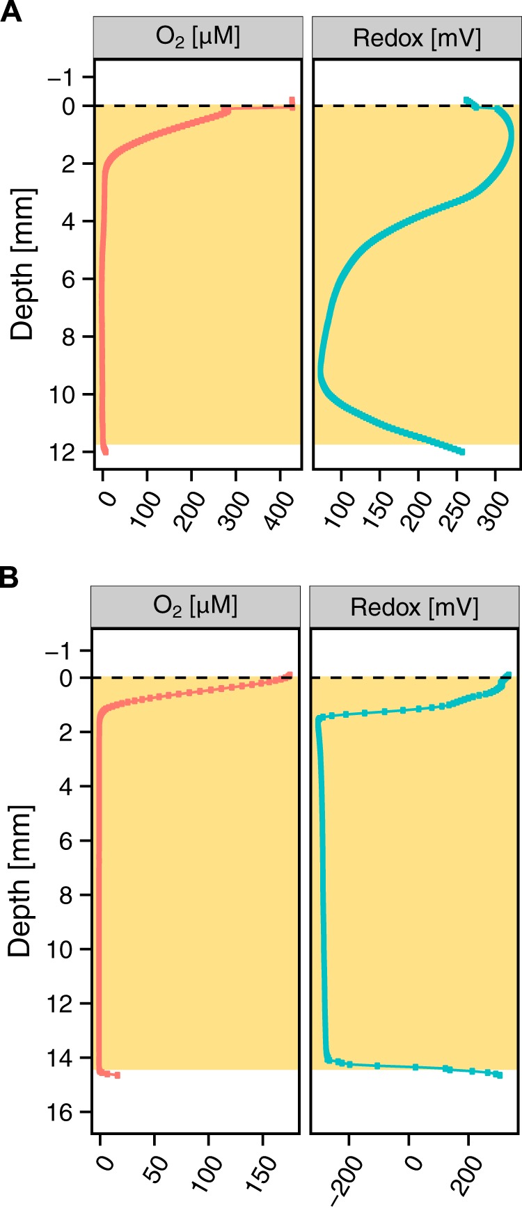
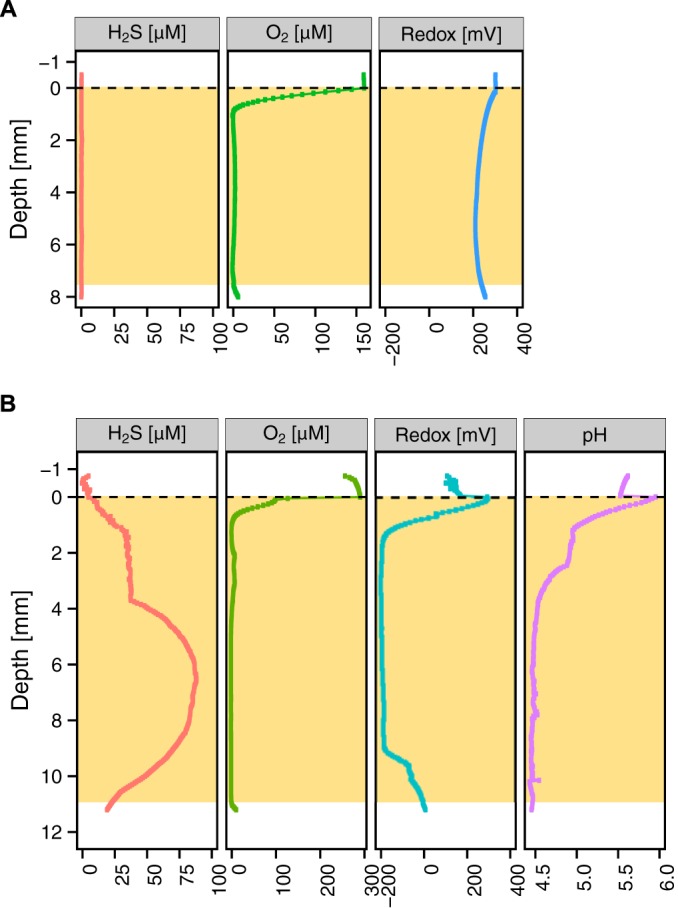
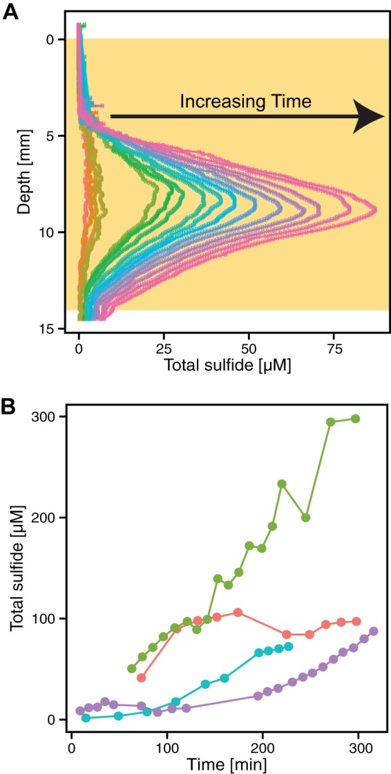
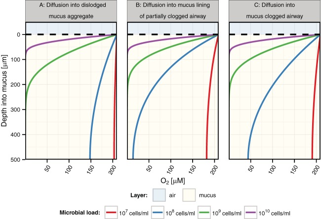
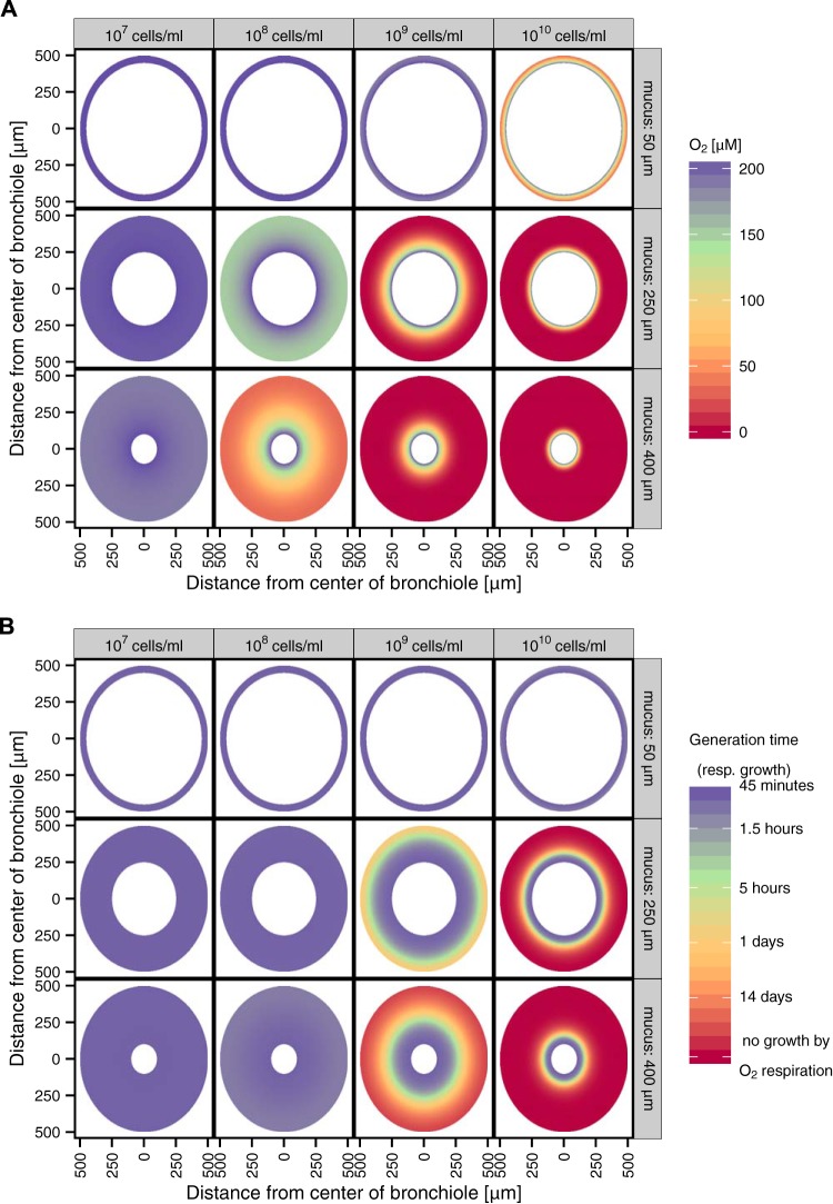
Severe and persistent bacterial lung infections characterize cystic fibrosis (CF). While several studies have documented the microbial diversity within CF lung mucus, we know much less about the inorganic chemistry that constrains microbial metabolic processes and their distribution. We hypothesized that sputum is chemically heterogeneous both within and between patients. To test this, we measured microprofiles of oxygen and sulfide concentrations as well as pH and oxidation-reduction potentials in 48 sputum samples from 22 pediatric patients with CF. Inorganic ions were measured in 20 samples from 12 patients. In all cases, oxygen was depleted within the first few millimeters below the sputum-air interface. Apart from this steep oxycline, anoxia dominated the sputum environment. Different sputum samples exhibited a broad range of redox conditions, with either oxidizing (16 mV to 355 mV) or reducing (-300 to -107 mV) potentials. The majority of reduced samples contained hydrogen sulfide and had a low pH (2.9 to 6.5). Sulfide concentrations increased at a rate of 0.30 µM H2S/min. Nitrous oxide was detected in only one sample that also contained sulfide. Microenvironmental variability was observed both within a single patient over time and between patients. Modeling oxygen dynamics within CF mucus plugs indicates that anoxic zones vary as a function of bacterial load and mucus thickness and can occupy a significant portion of the mucus volume. Thus, aerobic respiration accounts only partially for pathogen survival in CF sputum, motivating research to identify mechanisms of survival under conditions that span fluctuating redox states, including sulfidic environments.

Importance: Microbial infections are the major cause of morbidity and mortality in people living with CF, and yet microbial growth and survival in CF airways are not well understood. Insufficient information about the chemistry of the in vivo environment contributes to this knowledge gap. Our documentation of variable redox states corresponding to the presence or absence of sulfide begins to fill this void and motivates understanding of how different opportunistic pathogens adapt in these dynamic environments. Given the changing chemical state of CF sputum over time, it is important to consider a spectrum of aerobic and anaerobic lifestyles when studying CF pathogens in the laboratory. This work not only provides relevant constraints that can shape the design of laboratory experiments, it also suggests that sulfide might be a useful proxy for assessing the redox state of sputum in the clinic.


Figures

FIG 1
FIG 1
Overview of the respiratory airways. Tan coloring indicates mucus. Cyan dashed arrows indicate the direction of possible oxygen transport. In CF lungs, mucus can aggregate in different scenarios. In each scenario, “d” indicates the diameter of the airway and “x” indicates the mucus thickness. (A) Although less common in CF, the small airways, the alveolar sacs, can clog with mucus entirely (d ≤ 200 µm). (B) The surface layer of the bronchioles becomes dehydrated, and mucus thickening begins (300 µm ≤ d ≤ 1,000 µm). (C) Mucus aggregates can dislodge from lower airways and become lodged in larger, upper airways (the d value depends on the airway the aggregate is dislodged into; 300 µm ≤ d ≤ 5,000 µm).
FIG 2
FIG 2
Representative oxygen microprofiles. (A) STOX microprofile, with the sputum sample marked in tan, representative of n = 8. The green rectangle shows an expanded view of a portion of the anoxic zone in the sputum sample. The detection limit is 2 nM. (B) Oxygen microelectrode profiles of 5-mm-deep expectorated sputum samples from 5 different patients, representative of n = 12. At the air-sputum interface, a steep oxycline begins and is followed by an anoxic zone that persists for the remaining portion of the sputum. (C) Oxygen microelectrode profiles of 8-mm-deep expectorated sputum samples from 5 different patients that were larger in volume, representative of n = 27. The same trend in profile occurs, with a steep oxycline followed by anoxia.
FIG 3
FIG 3
Representative examples of high and low sputum ORPs. The tan-shaded boxes indicate the extent of the sputum sample. (A) Of 23 of the redox-profiled samples, 11 displayed a positive redox potential (16 mV to 355 mV) indicative of an oxidizing microenvironment. (B) Of 23 of the profiled samples, 17 displayed a negative redox potential (−300 mV to −107 mV) indicative of a reducing microenvironment.
FIG 4
FIG 4
Representative examples of ORPs in tandem with H2S detection. The tan-shaded boxes indicate the extent of the sputum sample. (A) A total of 14 of the 16 sputum samples that were highly oxidized samples (positive reduction potential) did not exhibit a presence of H2S. (B) A total of 5 of the 7 sputum samples that were highly reduced (negative reduction potential) exhibited the presence of H2S and were acidic.
FIG 5
FIG 5
Sulfide can build up rapidly in sputum. (A) Dynamics of total sulfide production over time in one sputum sample incubated for a total of 240 min. Time increases from left to right (orange to pink). This is the same sample represented by the purple line in panel B. The tan box indicates the depth of the sputum sample. (B) Increases in sulfide levels over time in sputum samples incubated at 37°C for 4 different patients. Each color represents a different sputum sample profiled over time. Each time point represents the maximum sulfide concentration for a specific profile. Not all of the maxima were recorded at the same depth in the samples. For the sample represented by the red line, the average maximum sulfide depth was within the range of 6.85 to 7.40 mm; for the green sample, 9.85 to 13.30 mm; for the blue sample, 15.60 to 18.30 mm; and for the purple sample, 8.15 to 11.25 mm. Five of the sulfidic samples were profiled over a period of up to 300 min and demonstrated a significant increase in the sulfide level.
FIG 6
FIG 6
Comparison of levels of oxygen diffusion into mucus based on different respiratory airway geometry constraints and bacterial densities. Data represent oxygen diffusion into different respiratory airways clogged with mucus at various bacterial densities. The described scenarios correspond to the diagram in Fig. 1 with a modeled mucus thickness (x) of 500 µm and an airway diameter of 1.5 mm for scenario B (model scenarios A and C are not affected by the airway diameter). The upper blue shaded area indicates air, and the tan lower layer indicates mucus.
FIG 7
FIG 7
Predicted oxygen concentrations and generation times due to aerobic respiration in bronchioles clogged with mucus. (A) Differential oxygen concentrations affecting the thickness of the mucus and the bacterial density. (B) A two-dimensional visualization of the bronchiole with variation in the mucus thickness and bacterial density and the resulting predicted generation time of the pathogens in the mucus during growth only via aerobic respiration (resp.). This accounts only for aerobic respiration and neglects other catabolic pathways.

References

    1. Riordan JR, Rommens JM, Kerem B, Alon N, Rozmahel R, Grzelczak Z, Zielenski J, Lok S, Plavsic N, Chou JL. 1989. Identification of the cystic fibrosis gene: cloning and characterization of complementary DNA. Science 245:1066–1073. doi: 10.1126/science.2475911. - DOI - PubMed
    1. Boucher RC. 2007. Airway surface dehydration in cystic fibrosis: pathogenesis and therapy. Annu Rev Med 58:157–170. doi: 10.1146/annurev.med.58.071905.105316. - DOI - PubMed
    1. Knowles MR, Boucher RC. 2002. Mucus clearance as a primary innate defense mechanism for mammalian airways. J Clin Invest 109:571–577. doi: 10.1172/JCI15217. - DOI - PMC - PubMed
    1. Koch C, Høiby N. 2000. Diagnosis and treatment of cystic fibrosis. Respiration 67:239–247. doi: 10.1159/000029503. - DOI - PubMed
    1. Chmiel JF, Davis PB. 2003. State of the art: why do the lungs of patients with cystic fibrosis become infected and why can’t they clear the infection? Respir Res 4:8. doi: 10.1186/1465-9921-4-8. - DOI - PMC - PubMed

Publication types