

Human SERPINB12 Is an Abundant Intracellular Serpin Expressed in Most Surface and Glandular Epithelia
- PMID: 26220980
- PMCID: PMC4812677
- DOI: 10.1369/0022155415600498
Human SERPINB12 Is an Abundant Intracellular Serpin Expressed in Most Surface and Glandular Epithelia
Abstract
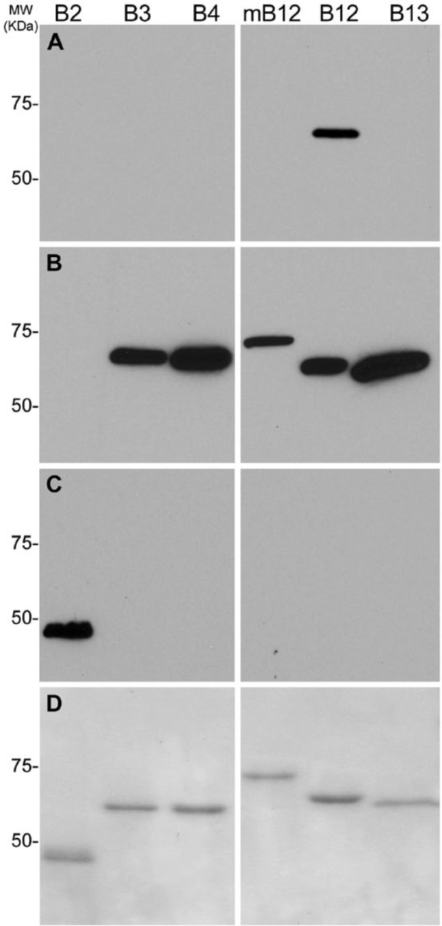
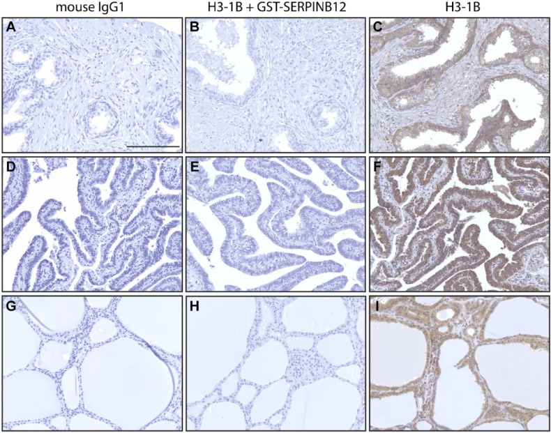
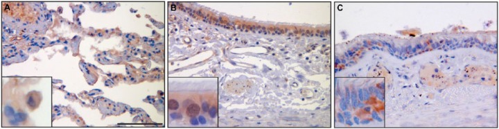
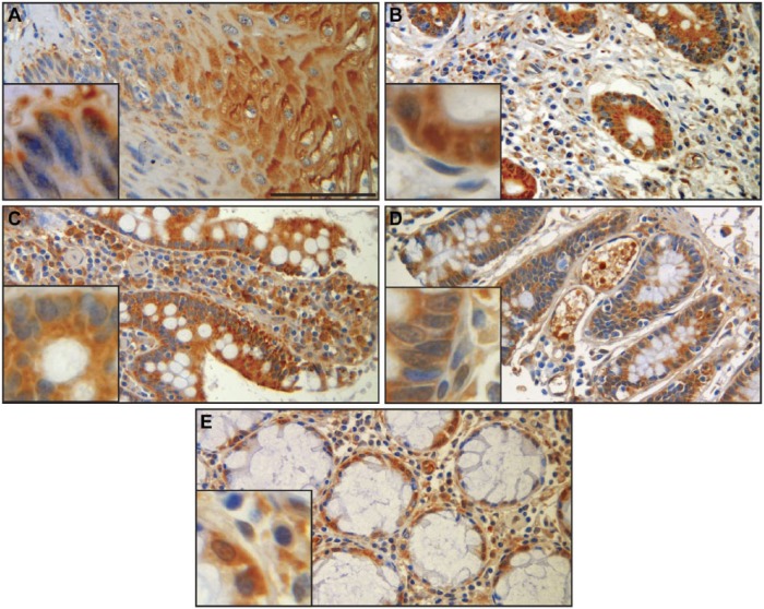
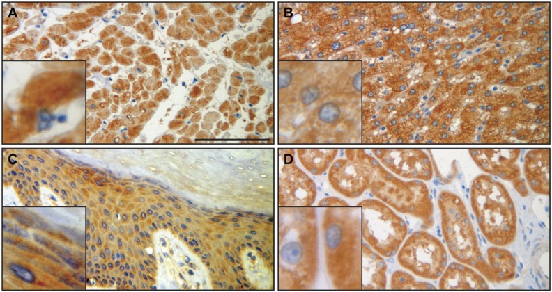
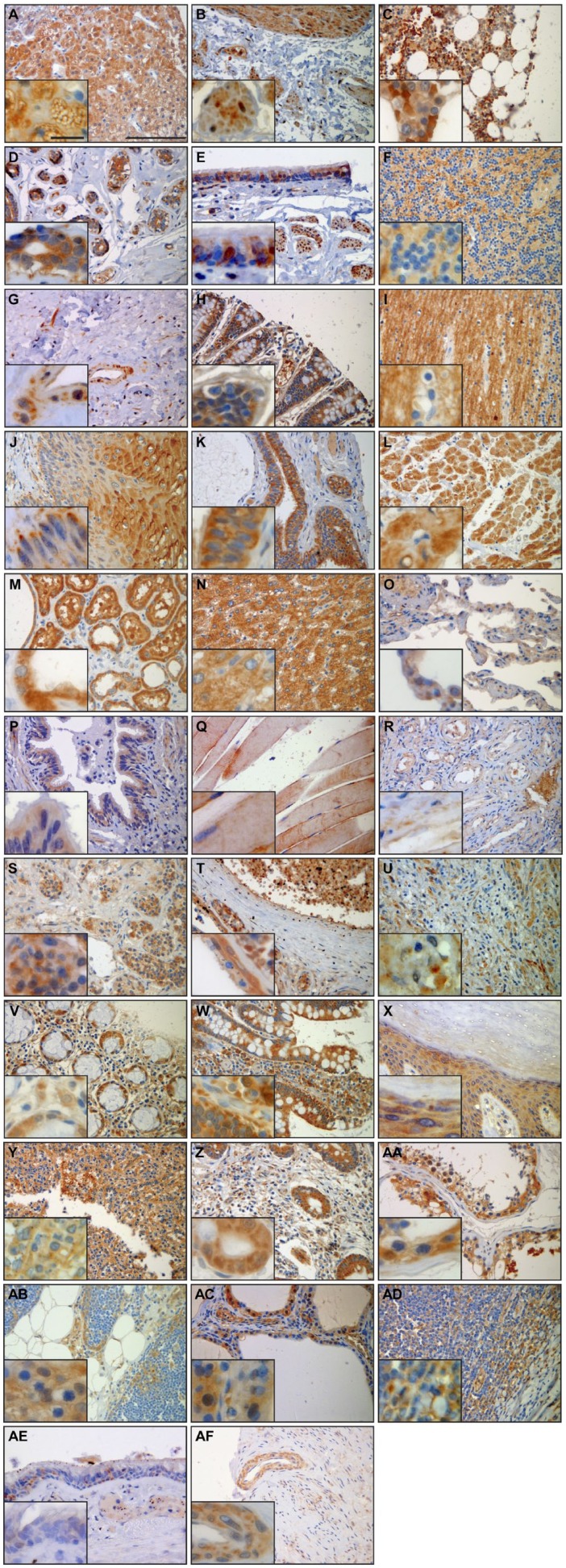
The intracellular serine protease inhibitors (serpins) are an important family of proteins that protect cells form proteinase-mediated injury. Understanding the tissue and cellular expression pattern of this protein family can provide important insights into their physiologic roles. For example, high expression in epithelial tissues, such as lung, may suggest a biologic function in cellular defense, secretion, or selective absorption. Although the expression pattern of many of the intracellular serpins has been well described, one member of this class, SERPINB12, has not been carefully examined. We generated a mouse monoclonal antibody directed against human SERPINB12 and delineated its specificity and tissue and cell type distribution pattern through immunoblotting and immunohistochemistry, respectively. This monoclonal antibody was human specific and did not cross-react with other human intracellular serpins or mouse Serpinb12. SERPINB12 was found in nearly all the tissues investigated. In addition, this serpin was found in multiple cell types within individual tissues but primarily the epithelium. These data suggest that SERPINB12, like some other intracellular serpins, may play a vital role in barrier function by providing protection of epithelial cells.
Keywords: epithelium; human; immunohistochemistry; protease; serpin; tissue array.
© The Author(s) 2015.
Conflict of interest statement
Figures
Similar articles
-
SERPINB12 is a novel member of the human ov-serpin family that is widely expressed and inhibits trypsin-like serine proteinases.J Biol Chem. 2001 Dec 28;276(52):49320-30. doi: 10.1074/jbc.M108879200. Epub 2001 Oct 16. J Biol Chem. 2001. PMID: 11604408
-
SERPINB12 Is a Slow-Binding Inhibitor of Granzyme A and Hepsin.Biochemistry. 2015 Nov 17;54(45):6756-9. doi: 10.1021/acs.biochem.5b01042. Epub 2015 Nov 5. Biochemistry. 2015. PMID: 26497600 Free PMC article.
-
Distribution of the human intracellular serpin protease inhibitor 8 in human tissues.J Histochem Cytochem. 2002 Nov;50(11):1443-54. doi: 10.1177/002215540205001103. J Histochem Cytochem. 2002. PMID: 12417609
-
Production, characterization, and use of serpin antibodies.Methods. 2004 Feb;32(2):141-9. doi: 10.1016/s1046-2023(03)00205-6. Methods. 2004. PMID: 14698626 Review.
-
Serpin protease inhibitors in plant biology.Physiol Plant. 2012 May;145(1):95-102. doi: 10.1111/j.1399-3054.2011.01540.x. Epub 2011 Dec 16. Physiol Plant. 2012. PMID: 22085334 Review.
Cited by
-
Exosome Liberation by Human Neutrophils under L-Amino Acid Oxidase of Calloselasma rhodostoma Venom Action.Toxins (Basel). 2023 Oct 25;15(11):625. doi: 10.3390/toxins15110625. Toxins (Basel). 2023. PMID: 37999488 Free PMC article.
-
A role for whey acidic protein four-disulfide-core 12 (WFDC12) in the pathogenesis and development of psoriasis disease.Front Immunol. 2022 Sep 6;13:873720. doi: 10.3389/fimmu.2022.873720. eCollection 2022. Front Immunol. 2022. PMID: 36148224 Free PMC article.
-
A Proteomic Approach to Elucidate the Changes in Saliva and Serum Proteins of Pigs with Septic and Non-Septic Inflammation.Int J Mol Sci. 2022 Jun 16;23(12):6738. doi: 10.3390/ijms23126738. Int J Mol Sci. 2022. PMID: 35743177 Free PMC article.
-
Skin tape proteomics identifies pathways associated with transepidermal water loss and allergen polysensitization in atopic dermatitis.J Allergy Clin Immunol. 2020 Dec;146(6):1367-1378. doi: 10.1016/j.jaci.2020.04.022. Epub 2020 Apr 28. J Allergy Clin Immunol. 2020. PMID: 32360271 Free PMC article. Clinical Trial.
-
Regulation of epidermal barrier function and pathogenesis of psoriasis by serine protease inhibitors.Front Immunol. 2024 Dec 16;15:1498067. doi: 10.3389/fimmu.2024.1498067. eCollection 2024. Front Immunol. 2024. PMID: 39737188 Free PMC article. Review.
References
-
- Askew DJ, Askew YS, Kato Y, Luke CJ, Pak SC, Bromme D, Silverman GA. (2004). The amplified mouse squamous cell carcinoma antigen gene locus contains a serpin (serpinb3b) that inhibits both papain-like cysteine and trypsin-like serine proteinases. Genomics 84:166-175. - PubMed
-
- Askew DJ, Cataltepe S, Kumar V, Edwards C, Pace SM, Howarth RN, Pak SC, Askew YS, Bromme D, Luke CJ, Whisstock JC, Silverman GA. (2007). SERPINB11 is a new noninhibitory intracellular serpin: common single nucleotide polymorphisms in the scaffold impair conformational change. J Biol Chem 282:24948-24960. - PubMed
-
- Askew YS, Pak SC, Luke CJ, Askew DJ, Cataltepe S, Mills DR, Kato H, Lehoczky J, Dewar K, Birren B, Silverman GA. (2001). SERPINB12 is a novel member of the human ov-serpin family that is widely expressed and inhibits trypsin-like serine proteinases. J Biol Chem 276:49320-49330. - PubMed
-
- Bird CH, Sutton VR, Sun J, Hirst CE, Novak A, Kumar S, Trapani JA, Bird PI. (1998). Selective regulation of apoptosis: the cytotoxic lymphocyte serpin proteinase inhibitor 9 protects against granzyme B–mediated apoptosis without perturbing the Fas cell death pathway. Mol Cell Biol 18:6387-6398. - PMC - PubMed
-
- Cataltepe S, Gornstein ER, Schick C, Kamachi Y, Chatson K, Fries J, Silverman GA, Upton MP. (2000). Co-expression of the squamous cell carcinoma antigens 1 and 2 in normal adult human tissues and squamous cell carcinomas. J Histochem Cytochem 48:113-122. - PubMed
Publication types
MeSH terms
Substances
Grants and funding
LinkOut - more resources
Full Text Sources
Other Literature Sources
Molecular Biology Databases
