Review
. 2015 Jul 15;29(14):1463-86.
doi: 10.1101/gad.266551.115.

Fibroblast growth factor signaling in skeletal development and disease

Affiliations
Review

Fibroblast growth factor signaling in skeletal development and disease

David M Ornitz et al. Genes Dev. .

Abstract

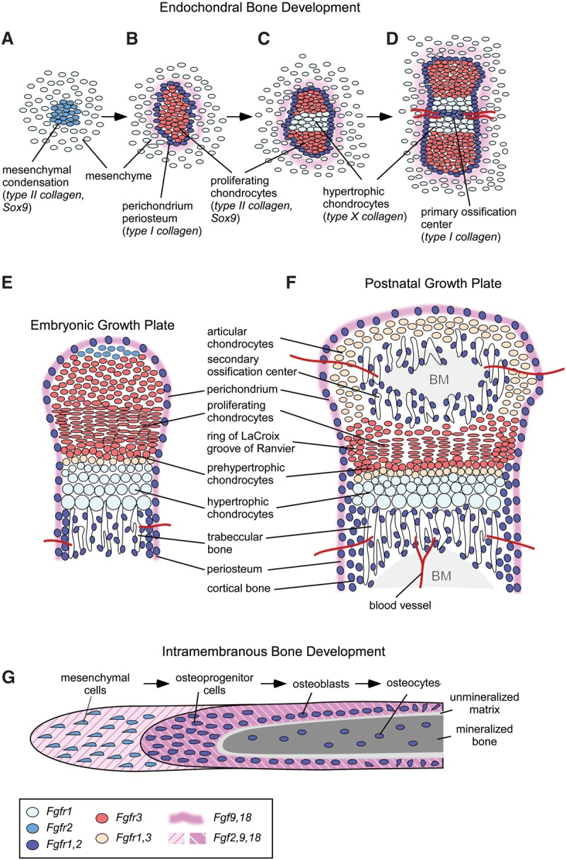
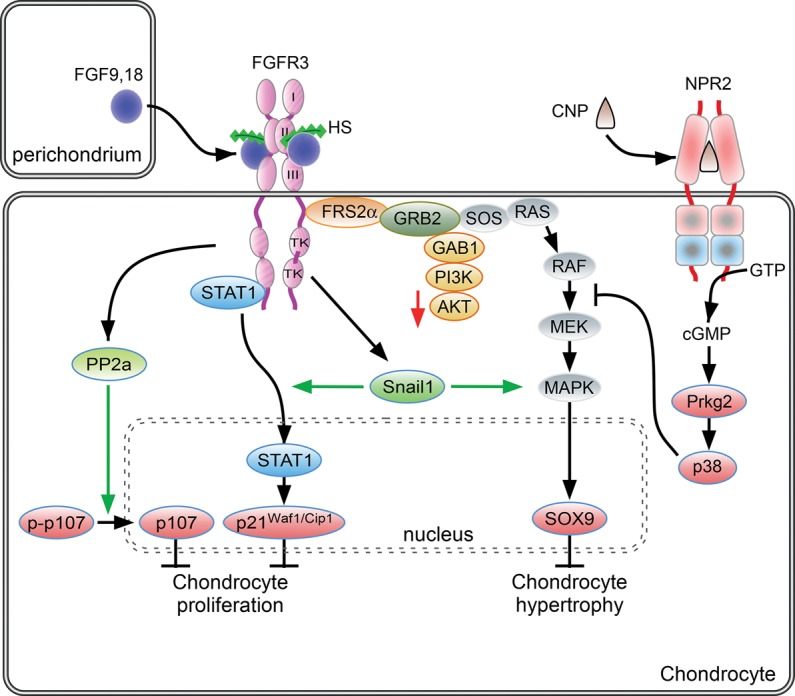
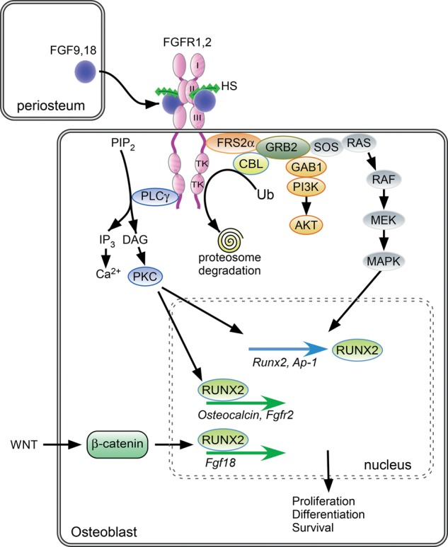
Fibroblast growth factor (FGF) signaling pathways are essential regulators of vertebrate skeletal development. FGF signaling regulates development of the limb bud and formation of the mesenchymal condensation and has key roles in regulating chondrogenesis, osteogenesis, and bone and mineral homeostasis. This review updates our review on FGFs in skeletal development published in Genes & Development in 2002, examines progress made on understanding the functions of the FGF signaling pathway during critical stages of skeletogenesis, and explores the mechanisms by which mutations in FGF signaling molecules cause skeletal malformations in humans. Links between FGF signaling pathways and other interacting pathways that are critical for skeletal development and could be exploited to treat genetic diseases and repair bone are also explored.

Keywords: FGF; FGFR; bone; cartilage; chondrocyte; osteoblast.


Figures

Figure 1.
Figure 1.
Fibroblast growth factor (FGF) and FGF receptor (FGFR) expression patterns during endochondral bone development. (AD) Progression of endochondral bone development from the mesenchymal condensation to formation of the primary ossification center. (E) Embryonic growth plate. (F) Postnatal growth plate after formation of the secondary ossification center. (G) Developmental progression of intramembranous bone development. Cells and tissues are color-coded for expression domains of FGFs and FGFRs. (BM) Bone marrow.
Figure 2.
Figure 2.
FGF signaling pathways in proliferating chondrocytes. During endochondral bone development, FGF9 and FGF18, derived from the perichondrium and surrounding tissue, signal to chondrocytes. Activity is mediated in part by regulated diffusion through the extracellular matrix through affinity for HS and potentially other sulfated glycosaminoglycans. Activation of FGFR3 in proliferating chondrocytes activates the STAT1 and MAPK signaling pathways. FGFR3 signaling results in increased expression of Snail1, which in turn is required for STAT1 and MAPK signaling. FGFR3 signaling can also activate PP2a. Activation of downstream signals, p107, p21Waf1/Cip1, and Sox9 regulates chondrocyte proliferation and differentiation to hypertrophic chondrocytes. C-type natriuretic peptide (CNP) signals through the natriuretic peptide receptor 2 (NPR2), a guanylyl cyclase. cGMP activates cyclic GMP-dependent protein kinase II (cGKII), which, through p38 MAPK activation, functionally antagonizes RAF1 activation of MEK. See the text for more details.
Figure 3.
Figure 3.
FGF/FGFR signaling in cells of the osteoblast lineage. FGF9 and FGF18, expressed in periosteal and surrounding tissue, interact with HS, FGFR1, and FGFR2. Activation of the FGFR tyrosine kinase domain leads to the recruitment of various substrates and activation of phospholipase Cγ/PKCα, ERK MAPKs, and PI3K/AKT, resulting in the modulation of transcription factors that control cell proliferation, differentiation, and survival in cells of the osteoblast lineage. Following FGFR activation, CBL is recruited and mediates FGFR ubiquitination and proteasome degradation, leading to FGFR down-regulation. Activated FGFRs may also be retained in intracellular compartments and degraded following c-Cbl-dependent ubiquitination. FGFR signaling activates expression of the transcription factor RUNX2, which regulates the expression of osteoblast-specific genes. In cooperation with Wnt/β-catenin signaling, RUNX2 activates Fgf18 gene expression. See the text for more details.

References

    1. Abad V, Meyers JL, Weise M, Gafni RI, Barnes KM, Nilsson O, Bacher JD, Baron J. 2002. The role of the resting zone in growth plate chondrogenesis. Endocrinology 143: 1851–1857. - PubMed
    1. Agas D, Marchetti L, Menghi G, Materazzi S, Materazzi G, Capacchietti M, Hurley MM, Sabbieti MG. 2008. Anti-apoptotic Bcl-2 enhancing requires FGF-2/FGF receptor 1 binding in mouse osteoblasts. J Cell Physiol 214: 145–152. - PubMed
    1. Agas D, Sabbieti MG, Marchetti L, Xiao L, Hurley MM. 2013. FGF-2 enhances Runx-2/Smads nuclear localization in BMP-2 canonical signaling in osteoblasts. J Cell Physiol 228: 2149–2158. - PubMed
    1. Agoston H, Khan S, James CG, Gillespie JR, Serra R, Stanton LA, Beier F. 2007. C-type natriuretic peptide regulates endochondral bone growth through p38 MAP kinase-dependent and -independent pathways. BMC Dev Biol 7: 18. - PMC - PubMed
    1. Aikawa T, Segre GV, Lee K. 2001. Fibroblast growth factor inhibits chondrocytic growth through induction of p21 and subsequent inactivation of cyclin E–Cdk2. J Biol Chem 276: 29347–29352. - PubMed

Publication types