. 2015 Jul 15;29(14):1507-23.
doi: 10.1101/gad.267583.115.

The oncogenic BRD4-NUT chromatin regulator drives aberrant transcription within large topological domains

Affiliations

The oncogenic BRD4-NUT chromatin regulator drives aberrant transcription within large topological domains

Artyom A Alekseyenko et al. Genes Dev. .

Abstract

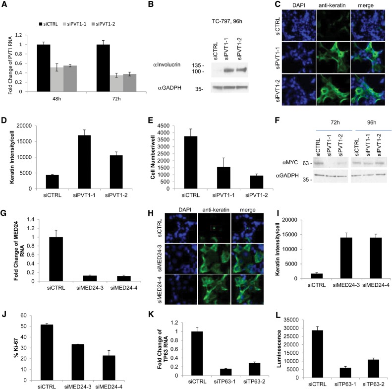
NUT midline carcinoma (NMC), a subtype of squamous cell cancer, is one of the most aggressive human solid malignancies known. NMC is driven by the creation of a translocation oncoprotein, BRD4-NUT, which blocks differentiation and drives growth of NMC cells. BRD4-NUT forms distinctive nuclear foci in patient tumors, which we found correlate with ∼100 unprecedented, hyperacetylated expanses of chromatin that reach up to 2 Mb in size. These "megadomains" appear to be the result of aberrant, feed-forward loops of acetylation and binding of acetylated histones that drive transcription of underlying DNA in NMC patient cells and naïve cells induced to express BRD4-NUT. Megadomain locations are typically cell lineage-specific; however, the cMYC and TP63 regions are targeted in all NMCs tested and play functional roles in tumor growth. Megadomains appear to originate from select pre-existing enhancers that progressively broaden but are ultimately delimited by topologically associating domain (TAD) boundaries. Therefore, our findings establish a basis for understanding the powerful role played by large-scale chromatin organization in normal and aberrant lineage-specific gene transcription.

Keywords: BRD4; chromatin hyperacetylation; topological domains.


Figures

Figure 1.
Figure 1.
The BRD4-NUT complex forms chromosomal megadomains driving ectopic transcription. (A) Endogenous BRD4-NUT localization in interphase and metaphase nuclei, detected with rabbit monoclonal anti-NUT antibody clone C52. (Left) Immunofluorescence confocal microscopy of endogenous BRD4-NUT expressed in TC-797 cells. (Middle) A human NMC biopsy immunohistochemically stained with anti-NUT. (Right) Immunofluorescence of a metaphase preparation of TC-797 NMC cells stained with anti-NUT. (Inset) Magnified view of chromosomes from the same image. (B) Immunofluorescence of BioTAP-tagged BRD4-NUT. 797TRex and 293TRex Flp-in cells were induced to express C-terminal-tagged or N-terminal-tagged BRD4-NUT. (C) Example of a BRD4-NUT megadomain in 293T cells. An ∼900-kb region of chromosome 20 is shown, illustrating extensive enrichment for BioTAP-tagged BRD4-NUT (16 h), N-Flag-C-HA-BRD4-NUT (7 h), and H3K27ac (7 h) following induction as indicated. The calculated extent of the megadomain is shown as a bar below the H3K27ac track. Transcriptional changes ([red] + strand; [blue] − strand) accompanying induction of BRD4-NUT in 293T cells are illustrated by the 0-h (before induction) and 7-h (following induction) nascent RNA sequencing (RNA-seq) reads. (D) The extent of BRD4-NUT megadomains is an order of magnitude larger than superenhancer regions. The size distribution of H3K27ac regions is shown for 293T cells before (blue) and after (red) induction of BRD4-NUT. (E) Average log2 enrichment profiles of H3K27ac are shown for enhancer regions, superenhancers, and BRD4-NUT megadomains detected in 293T cells. The profiles are scaled to their corresponding median sizes (8 kb, 28 kb, and 177 kb, respectively). The gray area around the average enrichment curve shows the 99.9% confidence interval. (F) Example of a BRD4-NUT megadomain in patient-derived TC-797 cells. A representative ∼900-kb region of chromosome 10q21.1 is shown. The megadomains and transcripts within them are sensitive to JQ1 treatment as measured by nascent RNA-seq. The “IP BRD4-NUT (4 h JQ1 vs. no JQ1)” track shows log2 fold change in BRD4-NUT enrichment with and without JQ1 treatment ([blue] depleted at 4 h; [brown] enriched), illustrating significant reduction of BRD4-NUT enrichment 4 h after JQ1 treatment. Most transcripts within the domain are also attenuated at 4 h (see also Supplemental Fig. S1).
Figure 2.
Figure 2.
Transcriptionally active, BET inhibitor-sensitive BRD4-NUT megadomains arise from seed regions. (A) BRD4-NUT megadomains are enriched for histone marks associated with active transcription. Average enrichment profiles within the megadomains (scaled to the same size) and 100-kb flanking regions are shown for TC-797 cells. (B) Average BRD4-NUT enrichment within the megadomains is reduced following JQ1 treatment of TC-797 cells. Scaled average profiles of BRD4-NUT enrichment are shown for TC-797 cells without JQ1 and 4 h following JQ1 treatment. (C) The average level of transcriptional activity is reduced within the megadomains and their flanking regions following JQ1 treatment of TC-797 cells. Similar to Figure 1F, the scaled average profile of nascent RNA-seq is shown for cells without JQ1 treatment and for cells 1 h, 2.5 h, and 4 h following JQ1 treatment. (D) Expression levels (log2[FPKM]) of all genes within the megadomain regions in TC-797 cells show broad reduction of transcriptional activity following JQ1 treatment. Each column corresponds to a gene within the TC-797 megadomain. Each row indicates JQ1 treatment condition: no JQ1 or 1 h, 2.5 h, or 4 h following treatment. (FPKM) Fragments per kilobase of exon per million reads mapped. (E) BRD4-NUT and H3K27ac enrichment in a 293T BRD4-NUT induction time course. The resulting domain calls (based on H3K27ac profiles) are shown as bars below the enrichment profiles. The domain border expansion is traced using green (expanding to the left) and red (expanding to the right) arrows. Seed regions are indicated by brown dotted circles, and an interseed region that will later “fill in” with BRD4-NUT and H3K27ac is indicated with a dotted green circle. For illustrative purposes, dotted arrows from seed and interseed regions point to corresponding regions in the violin plot (F) of all seed and interseed regions. (F) BRD4-NUT megadomain seed regions are enriched with H3K27ac. The violin plots show H3K27ac enrichment in 293T cells prior to BRD4-NUT induction for the seed regions that will show BRD4-NUT enrichment within 2 h following induction (early domains; brown) and the interseed regions that will be bound by BRD4-NUT only after 2 h (late domains; green). Colored dotted arrows pointing from the corresponding examples of these regions in Figure 1E are shown for illustrative purposes. The H3K27ac enrichment level found at regular enhancers within 293T cells is shown as a reference (gray). Genome-wide average enrichment of H3K27ac is shown with the dashed brown line. (G) Examples of the three classes of BRD4-NUT domain formation. Domains of the first class are formed by expanding from a single pre-existing H3K27ac region. Class 2 domains form by expanding from multiple pre-existing H3K27ac regions. Class 3 domains start with one or more pre-existing H3K27ac regions and activate additional enhancer-like regions during formation (see also Supplemental Fig. S2).
Figure 3.
Figure 3.
BRD4-NUT domain placement in different cells reveals a limited number of common regions. (A) Overview of BRD4-NUT megadomain positions within the genome. Positions of the BRD4-NUT megadomains detected in the five examined NMC cell types and derivatives (TC-797, 797TRex, Per403, 1015 tissue, and 1015 cell line) are shown as black rectangles. Loci at which domains were observed in three or more cell types are highlighted with red. (B) Related cell types show higher similarity of megadomain positions. A clustered matrix shows pair-wise similarity of BRD4-NUT megadomain positions detected in different cell types. The number of megadomains in each cell type (row) overlapped by another cell type (column) is shown by the numbers in each cell, and the percentage overlap is shown with color. (C) An example of BRD4-NUT megadomains within the MYC locus in different cell types. Tracks shown in brown represent enrichment and the calculated megadomain of BRD3-NUT in this region. The plot also shows the effect of JQ1 treatment on the transcriptional activity (as measured by nascent RNA-seq) over time in TC-797 cells as described above (see also Supplemental Fig. S3).
Figure 4.
Figure 4.
BRD4-NUT megadomains expand from a select set of pre-existing enhancers to fill topological domains. (A) The extent of the BRD4-NUT domains tends to correspond to topological domains. Hi-C observed/expected signal (from GM12878 cells) is shown for the c-MYC region. While the extent of BRD4-NUT megadomains (gray bars on the margins) varies between the cells, their boundaries tend to correspond to the boundaries of different TADs. (B) BRD4-NUT megadomains tend to coincide with the topological domains. An average observed/expected Hi-C interaction signal is shown for the domains identified in all of the examined cell lines. For the regions where domains were observed in multiple cell types, a union of all domain regions was used. The profile reveals strong interactions between domain ends and within the domain and a lack of interaction of the domain with the neighboring regions. (C) Most of the BRD4-NUT megadomain boundaries coincide with topological domain boundaries. A Hi-C topological domain boundary score (see the Materials and Methods) is shown for all of the detected megadomains (heat map at the bottom), with the average profile shown on the top. (D) BRD4-NUT megadomain boundaries tend to coincide with enhancer positions. The average frequency of occurrence of annotated enhancer positions (across 86 cell and tissue types) (Hnisz et al. 2013) is shown. The orange curve and light-orange area show the mean and standard error of the occurrence frequency of megadomain-sized blocks randomly positioned within the genome. (E) BRD4-NUT megadomain boundaries tend to coincide with CTCF-binding sites. The orange curve and light-orange area show the mean and standard error of the occurrence frequency of randomly placed megadomain-sized blocks (see also Supplemental Fig. S4).
Figure 5.
Figure 5.
Evidence that the BRD4-NUT megadomain-associated genes PVT1, MED24, and TP63 are oncogenic drivers in NMC. (A) qRT–PCR of PVT1 levels following transfection of control or PVT1 siRNAs into TC-797 cells. qRT–PCR was performed in triplicate. Error bars indicate the mean ± SD of the triplicate wells. (B) Immunoblot of involucrin levels (Sigma, catalog no. I9018) in TC-797 cells subjected to siRNA knockdown of PVT1. (C) High-throughput 384-well plate immunofluorescent assay of keratin expression in TC-797 cells 96 h following siRNA knockdown of PVT1. TC-797 cells were stained with anti-keratin (Dako), and nuclei were counterstained with 4′,6-diamidino-2-phenylindole (DAPI). Representative photographs use identical magnification (×400). (D,E) Quantitative analysis of keratin intensity and cell numbers from the high-throughput assay of PVT-1 knockdown in C. Average results from three biological replicates, each performed in triplicate, are shown. Error bars indicate the mean ± SD of the triplicate experiments. (F) Immunoblot of MYC levels (Cell Signaling, catalog no. 9402) in TC-797 cells subjected to siRNA knockdown of PVT1. Protein levels are decreased after 72 h but appear to recover at 96 h. However, at both time points, differentiation has already commenced, beginning at 48 h. (G) qRT–PCR of MED24 mRNA levels following transfection of control or MED24 siRNAs into TC-797 cells. qRT–PCR was performed in triplicate. Error bars indicate the mean ± SD of the triplicate wells. (H) Immunofluorescent assay of keratin expression in TC-797 cells 96 h following MED24 knockdown as in C. Representative photographs use identical magnification (×400). (I) Quantitative analysis of keratin intensity after MED24 knockdown in TC-797 cells in H. Results are from triplicate experiments, as in D. (J) Ki-67 fraction (marker of cell division) quantified from MED24 knockdown in TC-797 cells in H. Results are from triplicate experiments, as in D. (K) qRT–PCR of TP63 mRNA levels following transfection of control or TP63 siRNAs into TC-797 cells. qRT–PCR was performed in triplicate. Error bars indicate the mean ± SD of the triplicate wells. (L) Cell viability assay (CellTiter-Glo) of TC-797 cells 96 h following siRNA knockdown of TP63 versus control. Results are from three biological replicates, each performed in triplicate. Error bars indicate the mean ± SD of the triplicate wells (see also Supplemental Fig. S5).
Figure 6.
Figure 6.
Feed-forward model of megadomain formation. BRD4-NUT is tethered to acetylated chromatin by BRD4 bromodomains. The NUT portion of the fusion protein recruits p300, leading to increased local histone acetylation and thus a self-perpetuating recruitment of BRD4-NUT. The ultimate result is uncontrolled spreading of BRD4-NUT across chromatin, forming megadomains that typically are limited only by TAD boundaries.

Comment in

References

    1. Alekseyenko AA, Gorchakov AA, Kharchenko PV, Kuroda MI. 2014a. Reciprocal interactions of human C10orf12 and C17orf96 with PRC2 revealed by BioTAP-XL cross-linking and affinity purification. Proc Natl Acad Sci 111: 2488–2493. - PMC - PubMed
    1. Alekseyenko AA, Gorchakov AA, Zee BM, Fuchs SM, Kharchenko PV, Kuroda MI. 2014b. Heterochromatin-associated interactions of Drosophila HP1a with dADD1, HIPP1, and repetitive RNAs. Genes Dev 28: 1445–1460. - PMC - PubMed
    1. Alekseyenko AA, McElroy KA, Kang H, Zee BM, Kharchenko PV, Kuroda MI. 2015. BioTAP-XL: cross-linking/tandem affinity purification to study DNA targets, RNA, and protein components of chromatin-associated complexes. Curr Protoc Mol Biol 109: 21.30.21–21.30.32. - PMC - PubMed
    1. Alsarraj J, Hunter KW. 2012. Bromodomain-containing protein 4: a dynamic regulator of breast cancer metastasis through modulation of the extracellular matrix. Int J Breast Cancer 2012: 670632. - PMC - PubMed
    1. Anders S, Huber W. 2010. Differential expression analysis for sequence count data. Genome Biol 11: R106. - PMC - PubMed

Publication types

MeSH terms

Associated data