. 2015 Sep 11;290(37):22602-11.
doi: 10.1074/jbc.M115.663666. Epub 2015 Jul 28.

FANCD2-associated nuclease 1, but not exonuclease 1 or flap endonuclease 1, is able to unhook DNA interstrand cross-links in vitro

Affiliations

FANCD2-associated nuclease 1, but not exonuclease 1 or flap endonuclease 1, is able to unhook DNA interstrand cross-links in vitro

Julia Pizzolato et al. J Biol Chem. .

Abstract

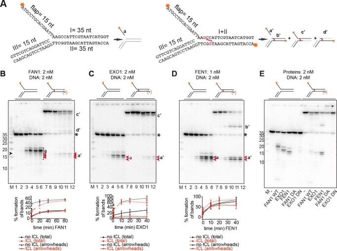
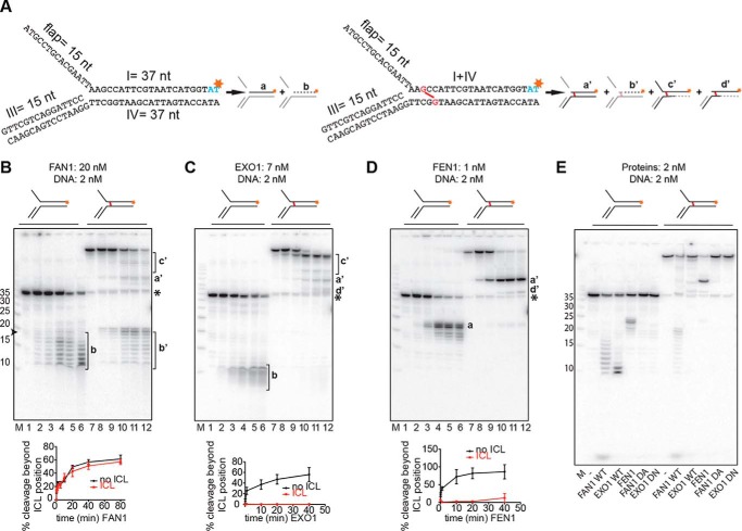
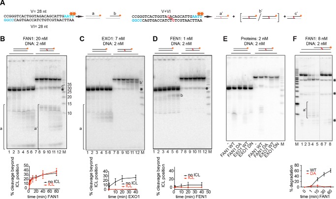
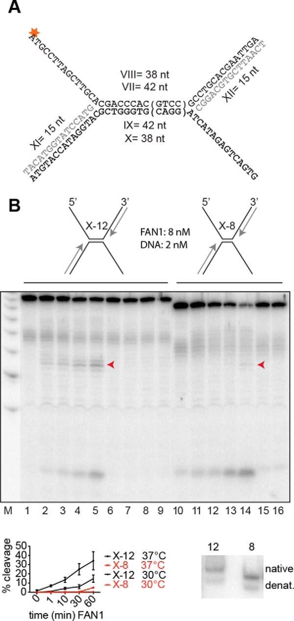
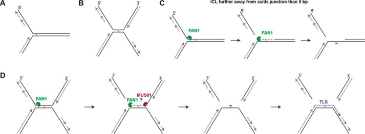
Cisplatin and its derivatives, nitrogen mustards and mitomycin C, are used widely in cancer chemotherapy. Their efficacy is linked primarily to their ability to generate DNA interstrand cross-links (ICLs), which effectively block the progression of transcription and replication machineries. Release of this block, referred to as unhooking, has been postulated to require endonucleases that incise one strand of the duplex on either side of the ICL. Here we investigated how the 5' flap nucleases FANCD2-associated nuclease 1 (FAN1), exonuclease 1 (EXO1), and flap endonuclease 1 (FEN1) process a substrate reminiscent of a replication fork arrested at an ICL. We now show that EXO1 and FEN1 cleaved the substrate at the boundary between the single-stranded 5' flap and the duplex, whereas FAN1 incised it three to four nucleotides in the double-stranded region. This affected the outcome of processing of a substrate containing a nitrogen mustard-like ICL two nucleotides in the duplex region because FAN1, unlike EXO1 and FEN1, incised the substrate predominantly beyond the ICL and, therefore, failed to release the 5' flap. We also show that FAN1 was able to degrade a linear ICL substrate. This ability of FAN1 to traverse ICLs in DNA could help to elucidate its biological function, which is currently unknown.

Keywords: DNA damage; DNA endonuclease; DNA repair; DNA-protein interaction; enzyme mechanism; exonuclease; interstrand cross-links.


Figures

FIGURE 1.
FIGURE 1.
Comparison of 5′ flap endonuclease activities and the specificities of FAN1, EXO1, and FEN1. A, schematic representation of the DNA substrates used in this study. The unmodified flap substrates (left panel) were generated as described under “Experimental Procedures,” and the labeling of the flap substrates with a nitrogen mustard-like interstrand cross-link (right panel, cross-link shown in red) resulted primarily, but not exclusively, in the labeling of the I strand. The likely products generated by the above enzymes from these substrates are also shown. Red asterisks indicate the positions of the 32P-labeled 5′ phosphates. Fragments invisible on the autoradiograph are shown in gray. Dashed lines represent regions of exonucleolytic degradation. nt, nucleotides. B–D, product generated upon incubation of the substrates shown above the panels with FAN1 (B), EXO1 (C), and FEN1 (D). Aliquots were withdrawn at 0, 10 s, 2 min, 20 min, 40 min, and 80 min (B, lanes 1–6 and 7–12, respectively) or at 0, 10 s, 1 min, 10 min, 20 min, and 40 min (C and D, lanes 1–6 and 7–12, respectively). The protein-to-DNA ratios were 1:1 for FAN1 and EXO1 and 1:2 for FEN1. M, low molecular weight marker (Affymetrix). The oligonucleotide sizes are indicated on the left, and the position of the cross-link is indicated by a black arrowhead. The lowercase letters on the right correspond to the products in A. (Products b'd' are seen solely in the reactions using the cross-linked substrate, in which both strands were labeled.) The black asterisk indicates the position of the non-cross-linked 35-mer oligonucleotide that was present in small amounts in the ICL substrate. The graphs below the autoradiographs of 20% denaturing polyacrylamide gels represent the quantification of either all product bands (total) or of only the bands indicated in red (arrowheads). The most prominent product bands in the reaction of FAN1 with the unmodified substrate resulted from incisions beyond the position of the ICL and are therefore not detectable in digestions of the cross-linked substrate. Error bars show mean ± S.D. (n = 3). E, comparative analysis of digestions of the indicated 5′ flap substrates by the three structure-specific endonucleases, including the nuclease-dead mutants of FAN1 (D960A) and EXO1 (D173N). DNA and proteins were all in equimolar ratios, and the reactions were carried out for 30 min at 37 °C.
FIGURE 2.
FIGURE 2.
Comparison of 5′ to 3′ exonuclease activities and the specificities of FAN1, EXO1, and FEN1. A, schematic of the DNA substrates used in this study. The unmodified flap substrates (left panel) and the substrates with a nitrogen mustard-like interstrand cross-link (right panel, cross-link shown in red) were produced as described under “Experimental Procedures.” Schematics of products generated by the above enzymes are shown next to the corresponding substrates. Red asterisks indicate the positions of the 32P-labeled nucleotides. Fragments invisible on the autoradiograph are shown in gray. B–D, incubation of the indicated substrates with FAN1 (B), EXO1 (C), or FEN1 (D). Aliquots were withdrawn after 0, 10 s, 2 min, 20 min, 40 min, and 80 min (B, lanes 1–6 and 7–12, respectively) or 0, 10 s, 1 min, 20 min, and 40 min (C and D, lanes 1–6 and 7–12, respectively). Lowercase letters on the right correspond to the products in A. The protein-to-DNA ratios were FAN1 (10:1), EXO1 (3.5:1), and FEN1 (1:2). M, low molecular weight marker (Affymetrix). The oligonucleotide sizes are indicated on the left. The black asterisk indicates the position of the non-cross-linked 37-mer oligonucleotide that was present in small amounts in the ICL substrate. The graphs below the autoradiographs of 20% denaturing polyacrylamide gels show the quantification of bands a or b, respectively. b' was generated by incisions in the top strand. Error bars show mean ± S.D. (n = 3). E, comparative analysis of cleavage products generated on the 3′ side of the cross-link by the three nucleases as well as by the nuclease-dead mutants of FAN1 (D960A) and EXO1 (D173N). The reaction conditions were as in Fig. 1E.
FIGURE 3.
FIGURE 3.
FAN1 can also traverse a cross-link on a linear DNA substrate. A, schematic of the linear DNA substrates labeled at the 3′ terminus of the upper strand by fill-in reactions with [α-32P]dTTP. A schematic of the expected products is shown next to the corresponding substrates (a, b, and a'c'). Red asterisks indicate the positions of the 32P-labeled nucleotides. Fragments invisible on the autoradiograph are shown in gray. B–D, 5′ to 3′ exonuclease activities of FAN1 (B), EXO1 (C), and FEN1 (D) on the indicated substrates. Reaction conditions and protein concentrations were as in Fig. 2. Lowercase letters correspond to the products in A. M, low molecular weight marker (Affymetrix). The oligonucleotide sizes are indicated. The black asterisk indicates the position of the non-cross-linked 28-mer oligonucleotide that was present in small amounts in the ICL substrate. Bottom panels, quantifications of the degradation fragments. Error bars show mean ± S.D. (n = 3). E, comparative analysis of cleavage products generated on the 3′ side of the cross-link by the three nucleases as well as by the nuclease-dead mutants of FAN1 (D960A) and EXO1 (D173N). The reaction conditions were as in Fig. 1E. F, FAN1 WT and D960A activity on a recessed linear DNA substrate shown schematically above. Concentrations were as indicated, and samples were withdrawn after 1, 10, 30, or 60 min (WT, lanes 2–5) or after 1, 30, and 60 min (D960A, lanes 6–8). For the substrate sequence, see “Experimental Procedures.” Quantifications are as indicated above. Black asterisks indicate the non-cross-linked oligonucleotides present in the substrate preparation. The dashed line represents missing lanes.
FIGURE 4.
FIGURE 4.
FAN1 cleavage requires ∼10 base pairs of dsDNA on both sides of the flap. A, schematic of the X-12 and X-8 substrates. The tetranucleotide sequence in parentheses is absent from the X-8 substrate. The asterisk indicates the 32P-labeled phosphate. B, the substrates were incubated with the enzymes at the indicated enzyme:substrate ratio at 37 °C for 1, 10, 30, and 60 min (FAN1-WT, lanes 1–5 and 10–14; FAN1 D960A, lanes 6–9) or 1 and 60 min (lanes 15 and 16). The X-8 substrate was extremely inefficiently processed even at 30 °C (bottom left panel), which indicates that the lack of flap cleavage was not caused by denaturation (denat) of the X structure. (Only the bands indicated by the arrowheads were quantified.) Bottom right panel, 10% native polyacrylamide gel of the two substrates, incubated at 37 °C for 30 min. This additional control experiment shows that both substrates were predominantly annealed under the conditions of the reaction.
FIGURE 5.
FIGURE 5.
FAN1- and/or MUS81-dependent DSB induction upon MMC treatment of human cells. A, top panel, FAN1 and/or MUS81 siRNA-mediated knockdown efficiencies assessed by Western blotting of total cell extracts of untreated (−) or MMC-treated (+) U2OS cells. TFIIH was used as a loading control. Quantification of the knockdown efficiencies shown in the bottom panel was carried out using ImageJ, and the graph was produced by GraphPad Prism (n = 3). The same extracts were probed for the markers of DSB metabolism RPA and CtIP. Center panel, Western blot analysis of the chromatin-enriched fraction of the above extracts probed for RPA, phospho-RPA, and γH2AX. Lamin was used as the loading control. B, time course of DSB formation assessed by pulsed field gel electrophoresis after MMC treatment (3 μg/ml) of U2OS cells, in which FAN1 or MUS81 were knocked down by siRNA. C, representative pulsed field gel electrophoresis image of DSBs induced by 24 h MMC (3 μg/ml) treatment of U2OS cells in which FAN1 and/or MUS81 were knocked down. The left panel shows a quantification of three independent experiments. siLUC was used as a control, and the ratio of DSBs of the MMC-treated samples divided by the untreated samples is shown for each siRNA condition. Error bars show mean ± S.E. D, quantifications of a FACS analysis of EdU-labeled cells pretreated with the indicated siRNAs and subsequently treated for 24 h with 3 μg/ml MMC. The knockdown did not affect cell viability during the course of the experiment. Error bars show mean ± S.E. (n = 3). E, clonogenic survival assay of U2OS cells treated with the indicated siRNAs and drugs. Colonies were counted 8 days after treatment, and MMC was washed out 24 h after treatment.
FIGURE 6.
FIGURE 6.
The putative mechanism of FAN1-dependent ICL unhooking. A, schematic of a single replication fork arrested at an ICL. B, the X-shaped structure arising through the convergence of two replication forks at an ICL. C, a single fork arrested at an ICL that is more than 5 bp from the ss/ds boundary would be incised by FAN1 5′ from the ICL, which would release the lagging strand. The enzyme would then degrade the nicked strand in a 5′-to-3′ direction to generate a substrate for translesion polymerases and subsequent repair by nucleotide excision repair. D, an X-shaped structure that contains a duplex longer than 10 bp where the lagging and leading strand flaps could be released by the action of FAN1 and, e.g., MUS81.

References

    1. Schärer O. D. (2005) DNA interstrand crosslinks: natural and drug-induced DNA adducts that induce unique cellular responses. ChemBioChem 6, 27–32 - PubMed
    1. Hinz J. M. (2010) Role of homologous recombination in DNA interstrand crosslink repair. Environ. Mol. Mutagen. 51, 582–603 - PubMed
    1. Auerbach A. D. (2009) Fanconi anemia and its diagnosis. Mutat. Res. 668, 4–10 - PMC - PubMed
    1. Crossan G. P., Patel K. J. (2012) The Fanconi anaemia pathway orchestrates incisions at sites of crosslinked DNA. J. Pathol. 226, 326–337 - PubMed
    1. Zhang J., Walter J. C. (2014) Mechanism and regulation of incisions during DNA interstrand cross-link repair. DNA Repair 19, 135–142 - PMC - PubMed

Publication types

LinkOut - more resources