

The effect of PDIA3 gene knockout on the mucosal immune function in IBS rats
- PMID: 26221224
- PMCID: PMC4509169
The effect of PDIA3 gene knockout on the mucosal immune function in IBS rats
Abstract
Objective: To observe the changes of intestinal inflammation on PDIA3 gene knockout IBS rats and its effect on immune function.
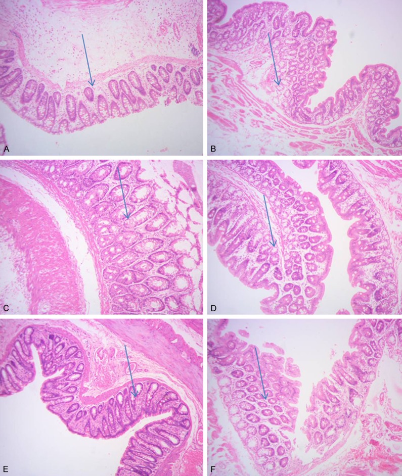
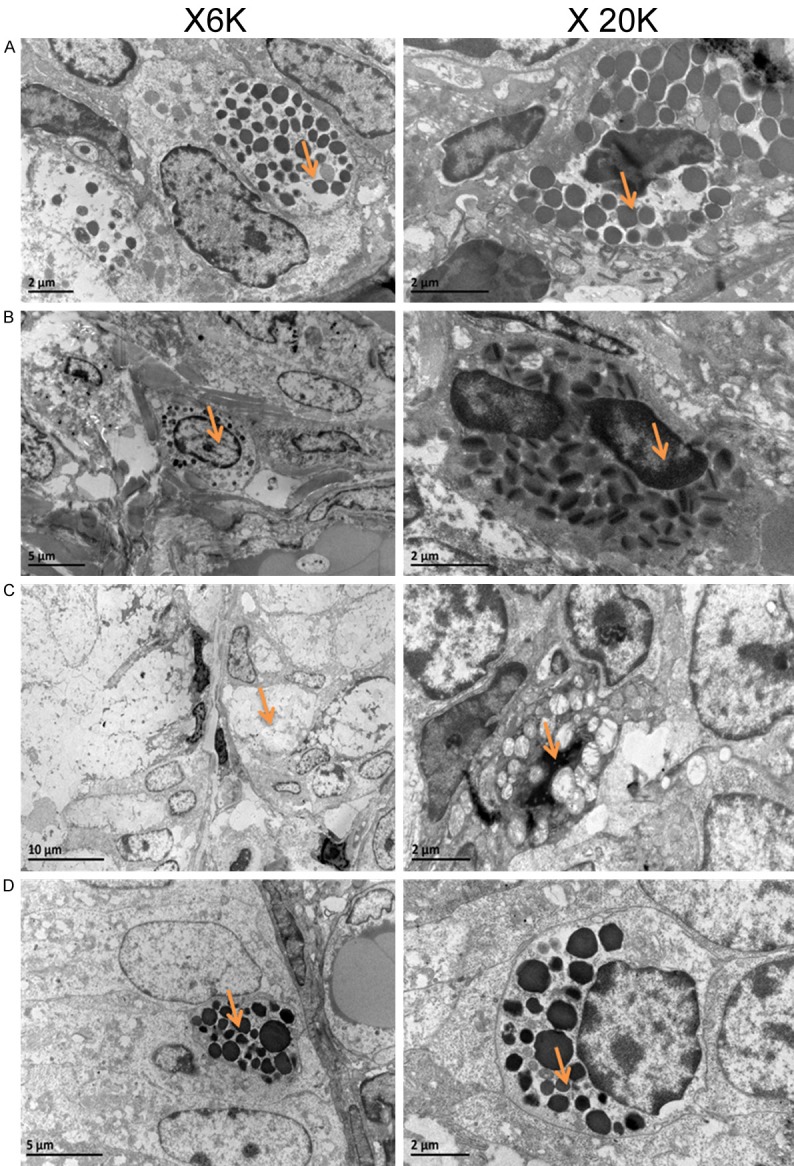
Methods: 36 SD rats were randomly divided into four groups: the control group (n = 8); IBS- empty virus group (IBS-GFP, which); IBS-PDIA3 knockout group (n = 12); IBS- the control group (n = 12). After modeling, colon and ileocecal tissue pathology in each group were observed separately. Changes of immune and inflammatory markers were measured. At the same time, ultrastructural changes in each group were observed by electron microscopy.
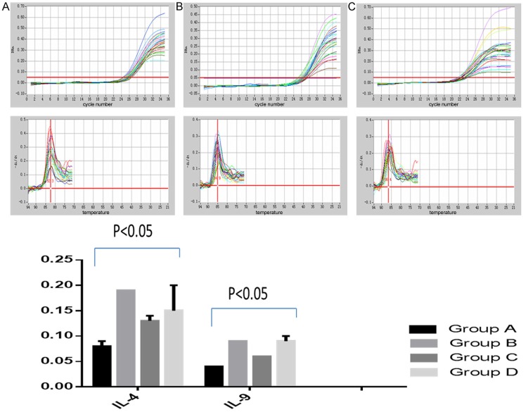
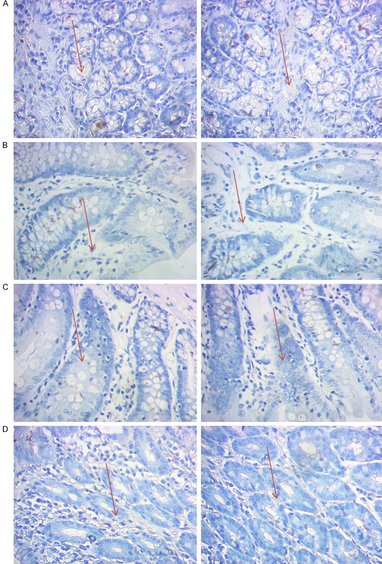
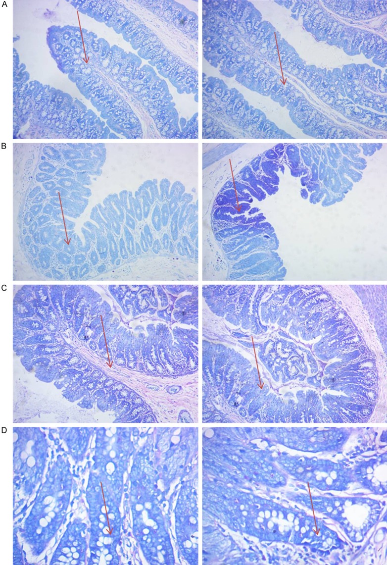
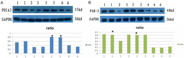
Results: Compared with the IBS control group, inflammation was reduced significantly in IBS-PDIA3 knockout group. IgE, IL-4 and IL-9 and the level of intestinal trypsin type were decreased significantly. Furthermore, mast cell degranulation and PAR 2 receptor reduced significantly.
Conclusion: PDIA3 may play an important role in the development of IBS by mediating through immune responses of mucosal abnormalities. However, the mechanism needs to be confirmed in further study.
Keywords: PDIA3; colon; immune response; irritable bowel syndrome; knockout.
Figures
References
-
- Spinelli A. Irritable bowel syndrome. Clin Drug Investig. 2007;27:15–33. - PubMed
-
- Spiegel BM. The burden of IBS: looking at metrics. Curr Gastroenterol Rep. 2009;11:265–269. - PubMed
-
- Ghaith O, El-Halabi M, Hashash JG, Sharara AI. Investigational agents for the irritable bowel syndrome. Expert Opin Investig Drugs. 2010;19:1161–1178. - PubMed
-
- Cremon C, Gargano L, Morselli-Labate AM, Santini D, Cogliandro RF, De Giorgio R, Stanghellini V, Corinaldesi R, Barbara G. Mucosal immune activation in irritable bowel syndrome: gender-dependence and association with digestive symptoms. Am J Gastroenterol. 2009;104:392–400. - PubMed
LinkOut - more resources
Full Text Sources
Miscellaneous