. 2015 Jul 29;7(298):298ra118.
doi: 10.1126/scitranslmed.aac5546.

Endotoxemia-mediated inflammation potentiates aminoglycoside-induced ototoxicity

Affiliations

Endotoxemia-mediated inflammation potentiates aminoglycoside-induced ototoxicity

Ja-Won Koo et al. Sci Transl Med. .

Abstract

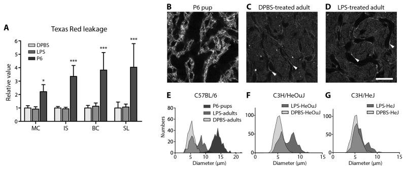
The ototoxic aminoglycoside antibiotics are essential to treat severe bacterial infections, particularly in neonatal intensive care units. Using a bacterial lipopolysaccharide (LPS) experimental model of sepsis, we tested whether LPS-mediated inflammation potentiates cochlear uptake of aminoglycosides and permanent hearing loss in mice. Using confocal microscopy and enzyme-linked immunosorbent assays, we found that low-dose LPS (endotoxemia) greatly increased cochlear concentrations of aminoglycosides and resulted in vasodilation of cochlear capillaries without inducing paracellular flux across the blood-labyrinth barrier (BLB) or elevating serum concentrations of the drug. Additionally, endotoxemia increased expression of both serum and cochlear inflammatory markers. These LPS-induced changes, classically mediated by Toll-like receptor 4 (TLR4), were attenuated in TLR4-hyporesponsive mice. Multiday dosing with aminoglycosides during chronic endotoxemia induced greater hearing threshold shifts and sensory cell loss compared to mice without endotoxemia. Thus, endotoxemia-mediated inflammation enhanced aminoglycoside trafficking across the BLB and potentiated aminoglycoside-induced ototoxicity. These data indicate that patients with severe infections are at greater risk of aminoglycoside-induced hearing loss than previously recognized.


Conflict of interest statement

Competing interests: All authors declare no competing interests.

Figures

Fig. 1
Fig. 1. Cochlear lateral wall uptake of GTTR is enhanced by LPS
(A) In xz planes of the cochlear lateral wall 1 hour after GTTR injection, F-actin labeling (green,) revealed tight junctions (arrowheads) between marginal cells (MC), with amorphous labeling in basal cells (BC, arrows). In DPBS-treated mice, intense GTTR fluorescence (red) distinguished strial capillaries (c), with less intense fluorescence in marginal cells, intra-strial layer (IS), and basal cells of the stria vascularis. The spiral ligament (SL) fibrocytes presented substantially less intense GTTR fluorescence compared to strial cells. LPS-treated mice displayed more intense GTTR fluorescence in the lateral wall (right panel) compared to DPBS-treated mice (left panel). (B) A focal series of xy planes through marginal cells, intra-strial tissues, basal cells, and fibrocytes at successively lower xy planes in the z-axis, 1 hour after GTTR injection. LPS-treated mice exhibited more intense GTTR fluorescence in grayscale (right panels) compared to DPBS-treated mice (left panels). Scale bar, 50 μm. (C) Mean pixel intensities of GTTR fluorescence in lateral wall ROIs (excluding capillary structures) are dose-dependently increased with increasing doses of LPS at 1 and 3 hours after GTTR injection (relative to DPBS-treated mice at 1 hour), with statistical significance in every cell type at 1 hour at 1 mg/kg or higher dose of LPS (Wilcoxon signed-rank test; *P<0.05; **P<0.01; ***P<0.001; error bars=s.e.m.; N as in Table S1).
Fig. 2
Fig. 2. Low dose LPS does not alter serum concentrations but does alter cochlear concentrations of aminoglycosides
(A) Using immunoturbidimetry, GTTR serum concentrations were significantly higher in 2.5 and 10 mg/kg LPS-treated mice than in controls at 1 or 3 hours. There was no difference between DPBS-treated mice and those dosed with 0.1 and 1 mg/kg LPS, nor between mice dosed with 2.5 and 10 mg/kg LPS. Serum concentrations of GTTR in 2.5 mg/kg LPS-treated mice were significantly higher than in 0.1 and 1 mg/kg LPS-treated mice at both time points (P<0.05). Elevated serum GTTR concentrations in 10 mg/kg LPS-treated mice showed borderline significance at 1 hour compared to 0.1 and 1 mg/kg LPS-treated mice (P=0.087, 0.053, respectively), and variable significance at 3 hours (P=0.27, 0.028, respectively; see also Table S2; Mann-Whitney U test; *P<0.05; **P<0.01; ***P<0.005; N in Table S1). (B, D) Using ELISA, serum concentrations of GTTR or gentamicin were not statistically different between DPBS-treated or 1 mg/kg LPS-treated mice at 1 or 3 hours after injection. (C, E) Cochlear concentrations of GTTR or gentamicin (GT) were significantly increased in 1 mg/kg LPS-treated mice compared to controls at 1 or 3 hours after injection (*P<0.05; N=4/group; Student’s unpaired t-test). Error bars=s.e.m; au, arbitrary units.
Fig. 3
Fig. 3. LPS does not alter BLB permeability but vasodilates basal strial capillaries
(A) The relative mean intensities of hTR fluorescence in marginal cell (MC), intra-strial tissue (IS), basal cell (BC) and spiral ligament (SL) layers from P6 pups were significantly elevated compared to the same ROIs in adult mice. There was no difference in hTR fluorescence of lateral wall ROIs between DPBS-treated and LPS-treated adult mice (*P<0.05, ***P<0.001; N=6 cochleae/group; error bars=s.d.; 2-way ANOVA with Bonferroni post-hoc tests). Absolute fluorescent intensities are shown in fig. S5A. (B, C) In P6 mice, the lumen of strial capillaries, revealed by phalloidin labeling, was larger than in adult DPBS-treated mice (endothelial cells indicated by white arrowheads). (D) Twenty-five hours after LPS treatment, a subpopulation of strial capillaries were dilated compared to DPBS-treated mice (C). Scale bar, 20 μm. (E) Strial capillary diameters in P6 mice were wider than in DPBS-treated adults (see also Table 1). LPS-treated adult mice had a subset of dilated strial capillaries, resulting in a bimodal distribution. (F) LPS also dilated a subset of strial capillaries in C3H/HeOuJ mice compared to DPBS-treated C3H/HeOuJ mice. (G) In TLR4-hyporesponsive C3H/HeJ mice, LPS dilated fewer strial capillaries compared to LPS-treated control C3H/HeOuJ mice (F), resulting in an asymmetrical bimodal distribution. A Gaussian regression curve fit was applied to obtain the bimodal peak means in Table 1.
Fig. 4
Fig. 4. Low dose LPS induced major inflammatory responses in serum and cochleae within 6 hours
(A) Significant increases in selected serum inflammatory proteins were observed 6 hours after LPS (±gentamicin) injection compared to DPBS-treated mice (±gentamicin; N=10/cohort). (B) Significant increases in cochlear inflammatory proteins were observed 6 hours after LPS (±gentamicin) injection for TNFα, IL-1α, IL-6, IL-8, MIP-1α and MIP-2α (but not IL-1β and IL-10) compared to DPBS-treated mice (±gentamicin; N=5/cohort; 4 cochleae/sample; measured in duplicate; *significant difference after 1-way ANOVA with Bonferroni multiple comparison correction and a family-wise 95% confidence level; error bars, 95% confidence intervals [CI] derived from Student’s t-tests). (C) Significant increases in cochlear mRNA for selected inflammatory markers were observed 6 hours after LPS (±gentamicin) injection when normalized to DPBS-treated mice (N=6/cohort; 2 cochleae/sample; *significant difference if the 95% CI does not overlap with 1 [i.e., DPBS-treated mice baseline]). Gentamicin did not modulate serum or cochlear expression of inflammatory proteins or mRNA for inflammatory markers.
Fig. 5
Fig. 5. TLR4-mediated cochlear inflammatory markers are attenuated in C3H/HeJ mice
(A) All selected acute phase inflammatory proteins (except for IL-10) were significantly elevated in cochleae from LPS-treated C57BL/6 and C3H/HeOuJ mice compared to DPBS-treated mice of the same strain. Several inflammatory proteins (TNFα, IL-6, IL-8, MIP-1α, MIP-2α) were more elevated in C57BL/6 compared to C3H/HeOuJ mice after LPS. In TLR4-hyporesponsive cochleae from C3H/HeJ mice, only a subset of inflammatory proteins (IL-1α, IL-6, IL-8, MIP-1α) were elevated after LPS, with small differences between the means for TNFα and IL-10. Expression of predominantly later-expressing inflammatory markers (IL-1α, IL-6, IL-8, MIP-1α) was significantly attenuated in LPS-treated C3H/HeJ mice compared to LPS-treated C3H/HeOuJ and C57BL/6 mice (N=4/cohort; 6 cochleae from 3 mice/sample) (B) In C57BL/6 and C3H/HeOuJ mice, significant increases were observed in cochlear expression of Il-1β, Il-6, Il-8, Il-10, Mip-1α and Mip-2α mRNA after LPS treatment when normalized to DPBS-treated mice. These increases were attenuated for Il-8, Mip-1α and Mip-2α in LPS-treated C3H/HeJ mice compared to LPS-treated C3H/HeOuJ mice. Il-10 mRNA expression was significantly higher in C3H/HeJ mice compared to C3H/HeOuJ and C57BL/6 mice (N=4/cohort; 2 cochleae from 1 mouse/sample). Error bars, 95% CI derived from Student’s t-tests; *significant difference compared to C3H/HeOuJ mice after 1-way ANOVA with Dunnett’s post-hoc tests and a family-wise 95% confidence level). See also fig. S6.
Fig. 6
Fig. 6. LPS-induced GTTR uptake by lateral wall cells is attenuated in TLR4-hyporesponsive C3H/HeJ mice
The fold change in GTTR intensity in LPS-treated mice over DPBS-treated mice is shown. GTTR fluorescence was significantly enhanced in strial marginal (MC), intermediate (IC) and basal (BC) cells, as well as fibrocytes (FC) of LPS-treated C3H/HeOuJ mice compared to that in DPBS-treated C3H/HeOuJ mice. LPS also significantly enhanced GTTR fluorescence intensities in strial cells (but not fibrocytes) in LPS-treated C3H/HeJ mice compared to that in DPBS-treated C3H/HeJ mice. Note that LPS-induced GTTR uptake was significantly attenuated (P<0.05) in marginal cells, intermediate cells and fibrocytes in C3H/HeJ mice compared to C3H/HeOuJ mice. (*P<0.05; N=8/bar; error bars=95% CI derived from Student’s t-tests; significance was determined if 95% CI did not overlap with 1; #P<0.05; unpaired 1-way t-test between strains; see fig. S8 for raw data).
Fig. 7
Fig. 7. Chronic endotoxemia potentiates kanamycin ototoxicity
(A) Three weeks after chronic LPS (or DPBS) exposure ±kanamycin (see fig. S13), ABR threshold shifts for LPS-only mice (N=5) were not different from DPBS-treated mice (N=4). Kanamycin alone (N=5) induced a small but significant PTS at only 32 kHz (**P<0.01) compared to DPBS-treated mice. Mice that received LPS+kanamycin (N=6) had significant PTS at 16, 24 (##P<0.01) and 32 kHz (#P<0.05) compared to mice treated with kanamycin, DPBS or LPS only (**P<0.01). Mice receiving LPS+kanamycin also had significant PTS at 12 kHz compared to mice treated with DPBS or LPS only (фP<0.05 and P<0.01, respectively), or LPS-only mice at 8 kHz (†P<0.05). Error bars=s.d. All statistical results were produced using 2-way ANOVA with Bonferroni post-hoc correction with 95% family-wise confidence intervals. (B) Cytocochleogram for mice in (A) revealed that OHC loss in the basal cochlear regions was greater and over a wider frequency range in LPS+kanamycin-treated mice compared to mice treated with LPS, DBPS, or kanamycin. Mean cochlear length = 6.84 (±0.79, s.d.) mm. Error bars, 95% CI derived from Student’s t-tests. See also figs. S9–S11, and tables S5, S7 for statistical comparisons using 2-way ANOVA Bonferroni post-hoc correction with 95% family-wise confidence intervals.

Comment in

References

    1. Kimberlin DW. Meningitis in the Neonate. Current treatment options in neurology. 2002;4:239–248. - PubMed
    1. Radigan EA, Gilchrist NA, Miller MA. Management of aminoglycosides in the intensive care unit. Journal of intensive care medicine. 2010;25:327–342. - PubMed
    1. Cheng AG, Johnston PR, Luz J, Uluer A, Fligor B, Licameli GR, Kenna MA, Jones DT. Sensorineural hearing loss in patients with cystic fibrosis. Otolaryngol Head Neck Surg. 2009;141:86–90. - PubMed
    1. Pillers DM, Schleiss MR. Gentamicin in the Clinical Setting. The Volta Review. 2005;105:205–210.
    1. Erenberg A, Lemons J, Sia C, Trunkel D, Ziring P. Newborn and infant hearing loss: detection and intervention. American Academy of Pediatrics. Task Force on Newborn and Infant Hearing, 1998–1999. Pediatrics. 1999;103:527–530. - PubMed

Publication types

MeSH terms

Supplementary concepts

LinkOut - more resources