. 2015 Oct;14(10):2814-23.
doi: 10.1074/mcp.O115.049650. Epub 2015 Jul 29.

MStern Blotting-High Throughput Polyvinylidene Fluoride (PVDF) Membrane-Based Proteomic Sample Preparation for 96-Well Plates

Affiliations

MStern Blotting-High Throughput Polyvinylidene Fluoride (PVDF) Membrane-Based Proteomic Sample Preparation for 96-Well Plates

Sebastian T Berger et al. Mol Cell Proteomics. 2015 Oct.

Abstract

We describe a 96-well plate compatible membrane-based proteomic sample processing method, which enables the complete processing of 96 samples (or multiples thereof) within a single workday. This method uses a large-pore hydrophobic PVDF membrane that efficiently adsorbs proteins, resulting in fast liquid transfer through the membrane and significantly reduced sample processing times. Low liquid transfer speeds have prevented the useful 96-well plate implementation of FASP as a widely used membrane-based proteomic sample processing method. We validated our approach on whole-cell lysate and urine and cerebrospinal fluid as clinically relevant body fluids. Without compromising peptide and protein identification, our method uses a vacuum manifold and circumvents the need for digest desalting, making our processing method compatible with standard liquid handling robots. In summary, our new method maintains the strengths of FASP and simultaneously overcomes one of the major limitations of FASP without compromising protein identification and quantification.


Figures

Fig. 1.
Fig. 1.
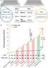
FASP versus MStern blot. (A) Comparison of the physical properties of the ultrafiltration membrane used for FASP and the membrane used for MStern blot. FASP uses physical retention while in MStern blotting proteins are adsorbed onto the hydrophobic membrane surface. (B) Time advantage of MStern blotting (blue curve) versus FASP (yellow curve) without considering potentially different digestion times. Major time savers are the fast liquid transfer steps (1 min versus 100 min; red) and the omission of any desalting (green).
Fig. 2.
Fig. 2.
Performance comparison MStern blot versus FASP. (A) Comparison of proteins identified from CSF, HeLa lysate, and urine after loading approx. 10 μg, 10 μg, and 15 μg, respectively. Each sample type was processed in quadruplicate. (B) Testing the loading capacity of the PVDF membrane used for MStern blotting based on proteins identified adsorbed to the PVDF membrane (i.e. MStern blotting, blue curve) and the respective flow through processed by FASP (red curve), in comparison to standard FASP of the same sample (yellow curve). A HeLa lysate was used. Values shown demonstrate average protein identifications. (C) Comparison of the dynamic range in three different biological samples (CSF, HeLa lysate, and urine); MaxQuant-based iBAQ intensities are marked blue (MStern blotting) and yellow (FASP).
Fig. 3.
Fig. 3.
Comparison of the properties of the identified proteins. Venn diagram of the proteins (A) and peptides (B) identified from CSF, HeLa lysate and urine. On the bottom, GO annotations (cellular compartment; C) of the method specific proteins, namely MStern blotting (blue) or FASP (yellow).
Fig. 4.
Fig. 4.
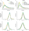
Physical-chemical properties. (A) Comparison of three different properties: molecular weight (top), isoelectric pH (middle) and GRAVY score (bottom) at protein level for MStern-blotting-specific proteins (blue trace), FASP-specific proteins (yellow trace), shared proteins (green trace), and theoretical distribution of the entire human proteome (dashed gray trace). (B) Comparison of physic-chemical property changes at peptide level: molecular weight (top), isoelectric pH (middle) and GRAVY score (bottom) for MStern-blotting-specific peptides (blue trace), FASP-specific peptides (yellow trace), shared peptides (green trace), and theoretical distribution upon tryptically digesting the entire human proteome assuming no missed cleavages (0 MC; dashed dark gray trace) or two missed cleavages (2 MC; dashed light gray trace).
Fig. 5.
Fig. 5.
Correlation of FASP- and MStern-blotting-based protein quantifications. Correlation of the ProteinPilot-derived signal intensities of the proteins identified in CSF, HeLa lysate and urine (see Fig. 2): MStern blot versus MStern blot (left), FASP versus FASP (middle), and MStern blot versus FASP (right).
Fig. 6.
Fig. 6.
Statistical analysis of serpin B3 and four iBAQ intensities in urine. (A) Box plot diagrams of the normalized iBAQ intensities of urinary serpin B3 and B4; listed p values are Bonferroni corrected. (B) Receiver operating characteristic curves for urine serpin B3 and B4 based on 10 ovarian cyst cases and 74 abdominal pain controls.

References

    1. Manza L. L., Stamer S. L., Ham A. J., Codreanu S. G., Liebler D. C. (2005) Sample preparation and digestion for proteomic analyses using spin filters. Proteomics 5, 1742–1745 - PubMed
    1. Wiśniewski J. R., Zougman A., Nagaraj N., Mann M. (2009) Universal sample preparation method for proteome analysis. Nature Meth. 6, 359–362 - PubMed
    1. Switzar L., van Angeren J., Pinkse M., Kool J., Niessen W. M. (2013) A high-throughput sample preparation method for cellular proteomics using 96-well filter plates. Proteomics 13, 2980–2983 - PubMed
    1. Yu Y., Suh M. J., Sikorski P., Kwon K., Nelson K. E., Pieper R. (2014) Urine sample preparation in 96-well filter plates for quantitative clinical proteomics. Anal. Chem. 86, 5470–5477 - PMC - PubMed
    1. Naldrett M. J., Zeidler R., Wilson K. E., Kocourek A. (2005) Concentration and desalting of peptide and protein samples with a newly developed C18 membrane in a microspin column format. J. Biomolecular Techniques 16, 423–428 - PMC - PubMed

Publication types