. 2015 Oct 1;593(19):4341-60.
doi: 10.1113/JP270849. Epub 2015 Aug 30.

The extracellular matrix molecule brevican is an integral component of the machinery mediating fast synaptic transmission at the calyx of Held

Affiliations

The extracellular matrix molecule brevican is an integral component of the machinery mediating fast synaptic transmission at the calyx of Held

Maren Blosa et al. J Physiol. .

Abstract

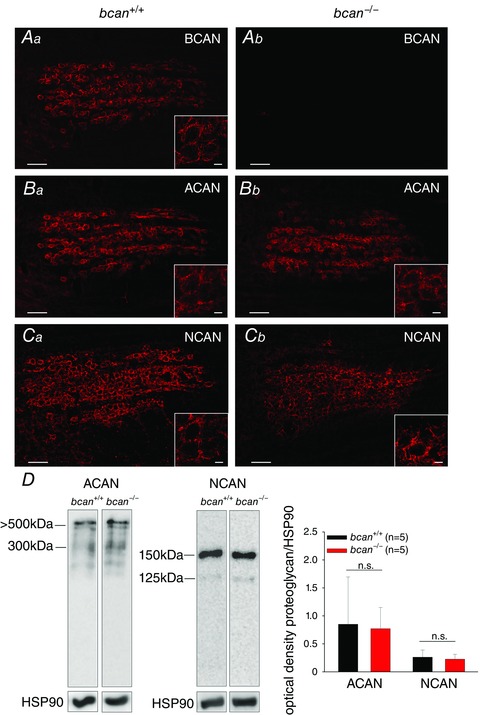
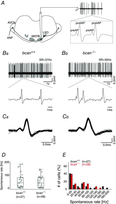
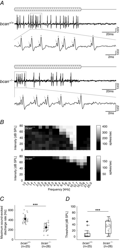
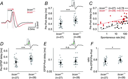
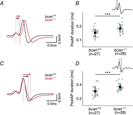
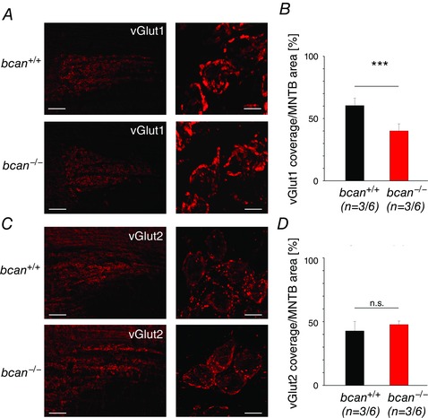
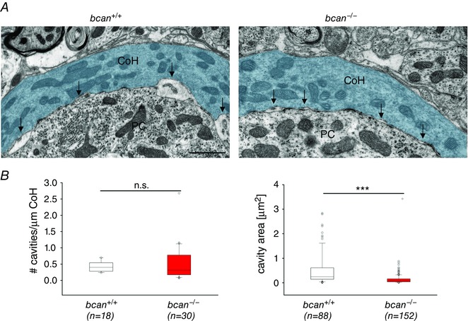
Key points: The proteoglycan brevican is a major component of the extracellular matrix of perineuronal nets and is highly enriched in the perisynaptic space suggesting a role for synaptic transmission. We have introduced the calyx of Held in the auditory brainstem as a model system to study the impact of brevican on dynamics and reliability of synaptic transmission. In vivo extracellular single-unit recordings at the calyx of Held in brevican-deficient mice yielded a significant increase in the action potential (AP) transmission delay and a prolongation of pre- and postsynaptic APs. The changes in dynamics of signal transmission were accompanied by the reduction of presynaptic vGlut1 and ultrastructural changes in the perisynaptic space. These data show that brevican is an important mediator of fast synaptic transmission at the calyx of Held.

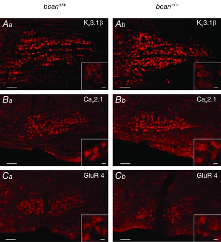
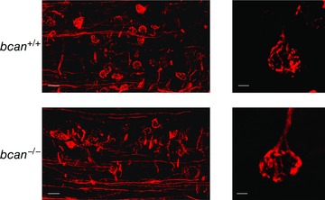
Abstract: The extracellular matrix is an integral part of the neural tissue. Its most conspicuous manifestation in the brain are the perineuronal nets (PNs) which surround somata and proximal dendrites of distinct neuron types. The chondroitin sulfate proteoglycan brevican is a major component of PNs. In contrast to other PN-comprising proteoglycans (e.g. aggrecan and neurocan), brevican is mainly expressed in the perisynaptic space closely associated with both the pre- and postsynaptic membrane. This specific localization prompted the hypothesis that brevican might play a role in synaptic transmission. In the present study we specifically investigated the role of brevican in synaptic transmission at a central synapse, the calyx of Held in the medial nucleus of the trapezoid body, by the use of in vivo electrophysiology, immunohistochemistry, biochemistry and electron microscopy. In vivo extracellular single-unit recordings were acquired in brevican-deficient mice and the dynamics and reliability of synaptic transmission were compared to wild-type littermates. In knockout mice, the speed of pre-to-postsynaptic action potential (AP) transmission was reduced and the duration of the respective pre- and postsynaptic APs increased. The reliability of signal transmission, however, was not affected by the lack of brevican. The changes in dynamics of signal transmission were accompanied by the reduction of (i) presynaptic vGlut1 and (ii) the size of subsynaptic cavities. The present results suggest an essential role of brevican for the functionality of high-speed synaptic transmission at the calyx of Held.


Figures

Figure 1
Figure 1. Complex structure of PNs is maintained in the MNTB in bcan−/− mice
The structure of PNs in brevican wild‐type (bcan +/+, Aa, Ba, Ca) and knockout mice (bcan −/−, Ab, Bb, Cb) by means of proteoglycan immunohistochemistry. A, in wild‐type mice, brevican (BCAN) is prominently expressed and covers nearly every principal neuron (Aa), while in bcan −/− mice brevican was completely absent (Ab). B and C, no obvious differences in the immunoreactivity of the proteoglycans aggrecan (ACAN) (B) and neurocan (NCAN) (C) are apparent between both genotypes. Overall, ACAN and NCAN immunohistochemistry shows that the complex structure of the PNs in the MNTB is maintained in bcan −/− mice. Scale bar in all MNTB overviews = 100 μm; scale bar in all high magnification insets = 10 μm. D, Western blot analysis of major proteoglycans. Protein extracts (35 μg per lane) from homogenized brainstems of 5 bcan +/+ and 5 bcan −/− mice were compared. Samples were subjected to SDS PAGE in a 6% gel. Blots were probed by use of respective antibodies against ACAN and NCAN (left panel). Intensity of chemiluminescence was quantified in relation to the bands of HSP90. Neither aggrecan (P = 0.7) nor neurocan (P = 0.62) protein levels showed significant differences between bcan +/+ and bcan −/− mice (right panel). Data are given as means ± SD.
Figure 2
Figure 2. Reliability of synaptic transmission and spontaneous discharge rates are not affected in bcan−/− mice
A, extracellular in vivo single‐unit recordings of MNTB neurons in bcan +/+ and bcan −/− mice. Complex voltage signals were composed of the discharge of the calyx of Held (preAP) and the postsynaptic action potential (postAP, see enlargement of the voltage trace in the inset to the right). Significant numbers of transmission failures, defined as preAPs not followed by postAPs, were detected in neither bcan +/+ mice nor bcan −/− mice. B, exemplary voltage traces (5 s, upper panel) of spontaneous activity in a unit of a bcan +/+ mouse (Ba) and a bcan −/− mouse (Bb). The respective spontaneous discharge rates (SR) were 37 Hz and 30 Hz. Blow‐ups of 2–3 individual complex waveforms are shown in the lower panel. C, overlay of 25 consecutive signals of a bcan+/+ unit and a bcan−/− unit exemplify the uniformity of the signal waveforms. D, spontaneous discharge rates of MNTB principal neurons did not differ between bcan +/+ and bcan −/ mice. E, the distribution of spontaneous discharge rates was similar in both genotypes. Forty‐one per cent of bcan +/+ units (black) and 39% of bcan −/− units (red) had spontaneous discharge rates <20 Hz. Also, in both genotypes the spontaneous discharge rates were found to cover a broad range (bcan +/+: 0.2–184 Hz; bcan −/−: 0.1–144 Hz). Data were obtained from 27 MNTB units in bcan +/+ mice and 28 MNTB units in bcan −/ mice. In D, data are presented as median [1st quartile, 3rd quartile]. Abbreviations: ANF, auditory nerve fibre; AVCN, anterior ventral cochlear nucleus; LSO, lateral superior olive; MNTB, medial nucleus of the trapezoid body.
Figure 3
Figure 3. Sound‐evoked response properties of MNTB principal cells are altered in bcan−/− mice
A, pure tone stimulation at characteristic frequency and 60 dB SPL in an exemplary MNTB unit of a bcan +/+ (top) and a bcan −/− mouse (bottom). The blow‐up of individual compound waveforms reveals that in both genotypes all presynaptic potentials (indicated by arrows) are followed by a postsynaptic action potential (indicated by stars), confirming the highly reliable synaptic transmission also under acoustic stimulation. B, frequency response areas (20 × 10 frequency–intensity combinations) of two exemplary MNTB principal neurons with similar characteristic frequency; top: bcan +/+, bottom: bcan −/−. A greyscale was used to encode different discharge rates. C, maximum sound‐evoked firing rates are significantly reduced in bcan −/− mice compared to respective wild‐type animals (P < 0.001). D, median thresholds at characteristic frequency are significantly elevated in bcan −/− mice (P < 0.001). Data were obtained from 25 MNTB units in bcan +/+ mice and 26 MNTB units in bcan −/− mice. In C, data are presented as means ± SD and in D, data are given as median [1st quartile, 3rd quartile]. P values: ***P < 0.001.
Figure 4
Figure 4. The lack of brevican reduces the speed of AP transmission delay at the calyx of Held
A, average complex waveforms of two exemplary MNTB units (bcan +/+: black, n = 1137; bcan −/−: red, n = 917) aligned to the positive peak of the presynaptic AP. The Pre–Post delay, i.e. the time lag between the positive peaks of the preAP and the postAP (red and black arrows above the respective signals), was longer in bcan −/− mice compared to wild‐types. B, Pre–Post delay of all MNTB units yielded significantly higher values in bcan−/− mice (0.46 ± 0.06 ms) compared to bcan+/+ mice (0.4 ± 0.07 ms, P < 0.001). C, in both genotypes the Pre–Post delay correlates with the spontaneous discharge rates of the units (bcan +/+: r = 0.78, P < 0.001; bcan −/−: r = 0.72, P < 0.001). Still, at given spontaneous rates, units in bcan −/− mice have longer AP transmission delays than in bcan +/+ mice. D and E, AP transmission delay was subdivided into two distinct physiological events: the time between the preAP and the EPSP (Pre–EPSP delay, D) and the time from the EPSP to the postAP (EPSP–Post delay, E). The time point of the EPSP was defined as the point of inflection on the rising flank of the postsynaptic AP (green dot in the inset). Note that the Pre–EPSP delay was significantly longer in bcan −/− mice (D, bcan +/+: 0.31 ± 0.07 ms; bcan −/−: 0.37 ± 0.06 ms; P < 0.001), while the EPSP–Post delay only revealed a tendency for prolongation in knockout mice (E, bcan +/+: 0.087 ± 0.01 ms; bcan −/−: 0.094 ± 0.02 ms; P = 0.068). F, the ratio of the Pre–EPSP delay and the Pre–Post delay was increased in bcan −/− (0.8 ± 0.01) compared to bcan +/+ mice (0.77 ± 0.01, P < 0.05), indicating that brevican might have a stronger effect on the speed of Pre–EPSP transmission than on the speed of EPSP–Post transmission. Shown are the values for individual units and the mean ± SD. The analysis was based on the same units shown in Fig. 2. P values: ***P < 0.001, *P < 0.05.
Figure 5
Figure 5. The lack of brevican causes a broadening of pre‐ and postsynaptic action potentials
A and C, average complex waveforms of a bcan +/+ MNTB unit (black) and a bcan −/− MNTB unit (red, same units as in Fig. 3) either aligned to the positive peak of the preAP (A) or to the positive peak of the postAP (C). Differences in signal durations are evidenced from the longer decaying slopes (black vs. red; defined as the time difference between positive and negative peaks, indicated by red and black arrows). B and D, both the presynaptic (B) and postsynaptic APs (D) were significantly longer in bcan −/− mice (P < 0.001). Shown are the values for individual units and means ± SD; same data sample as in Fig. 2. P values: ***P < 0.001.
Figure 6
Figure 6. Ion channels and receptors that mediate high‐speed transmission are expressed in bcan−/− mice
Coronal sections of the ventral lower brainstem dimensioned to include the entire MNTB show the respective immunoreactivity in adult bcan +/+ (left) and bcan −/− mice (right). The rostrocaudal planes of sections in the two genotypes are matched; midline to the left. Kv3.1β (A), Cav2.1 (B), and GluR4 (C); positive immunoreactivity to the antibodies against all three cellular components is seen in the overview staining and in the blow‐ups in both genotypes. Scale bar in all MNTB overviews = 100 μm and scale bar in all high magnification inserts = 10 μm.
Figure 7
Figure 7. The morphology of the calyx of Held is not affected in bcan−/− mice
Neuronal tracing of the projection from VCN to MNTB and anterograde labelling of the calyx of Held synapses with the lipophilic dye NeuroVue Red. In both bcan +/+ (top) and bcan −/− mice (bottom) a large number of traced VCN‐derived projections could be targeted (left) showing virtually similar fenestration patterns and morphology of calyces of Held in both genotypes (right). Scale bar in left images = 50 μm, scale bar in right images = 5 μm.
Figure 8
Figure 8. Glutamate transporter vGlut1 is reduced in bcan −/− mice
A, vGlut1 immunohistochemistry in coronal sections of the MNTB (upper panel: bcan +/+, lower panel: bcan −/−) provides good visualization of the calyces of Held throughout the MNTB (left panels) and also disclosed the typical fenestration patterns in these large axosomatic terminals (right panels, blow‐ups). B, quantification of the area of the MNTB covered by vGlut1 labelling (bcan +/+: black; bcan −/−: red; three animals of each genotype, nuclear regions on both sides) yielded a significant reduction in the mean vGlut1 coverage of the MNTB in mutant mice (P < 0.001). C, respective vGlut2 immunoreactivity in coronal sections of the MNTB; upper panel: bcan +/+, lower panel: bcan −/−. D, quantification of vGlut2 labelling yielded no significant differences between the genotypes in the MNTB (P = 0.544). Data are given as means ± SEM. The sample size is given as follows: n = mice/MNTB. P values: ***P < 0.001. Scale bars in A and C: left, 100 μm; right, 10 μm.
Figure 9
Figure 9. The size of subsynaptic cavities is reduced in bcan−/− mice
A, ultrastructural analysis of subsynaptic cavities at the axosomatic calyx of Held in the MNTB of a bcan +/+ (left) and a bcan −/− mouse (right). Subsynaptic, extracellular cavities (arrows) are formed between the opposing membranes of the calyx of Held (CoH, blue) and the principal neuron (PC). These cavities were significantly reduced in size in bcan −/− mice compared to bcan +/+ mice. B, quantification of the number (left) and the size (right) of the cavities in both genotypes. The number of cavities which was normalized to the length of the analysed calyx of Held profile was not affected (left, P = 0.617), while the size of the subsynaptic cavities was significantly reduced in bcan −/− mice (P < 0.001). Data were obtained from 18 MNTB sections taken of 2 bcan +/+ mice and from 28 MNTB sections of 2 bcan −/− animals. The analysis yielded 18 calyx of Held profiles with 88 subsynaptic cavities in bcan +/+ mice and 30 calyx of Held profiles with 152 subsynaptic cavities in bcan −/− mice. Data are given as median [1st quartile, 3rd quartile], P values: ***P < 0.001, scale bar in left image = 1 μm (applies for both images).

References

    1. Baker CA, Montey KL, Pongstaporn T & Ryugo DK (2010). Postnatal development of the endbulb of held in congenitally deaf cats. Front Neuroanat 4, 19. - PMC - PubMed
    1. Bergles DE & Edwards RH (2008). The role of glutamate transporters in synaptic transmission In Structural and Functional Organization of the Synapse, ed. Hell JW, Ehlers & Michael D, pp. 23–61. Springer, New York.
    1. Billups B (2005). Colocalization of vesicular glutamate transporters in the rat superior olivary complex. Neurosci Lett 382, 66–70. - PubMed
    1. Blaesse P, Ehrhardt S, Friauf E & Nothwang HG (2005). Developmental pattern of three vesicular glutamate transporters in the rat superior olivary complex. Cell Tissue Res 320, 33–50. - PubMed
    1. Blosa M, Sonntag M, Brückner G, Jäger C, Seeger G, Matthews RT, Rübsamen R, Arendt T & Morawski M (2013). Unique features of extracellular matrix in the mouse medial nucleus of trapezoid body–implications for physiological functions. Neuroscience 228, 215–234. - PubMed

Publication types

LinkOut - more resources