

Chemically related 4,5-linked aminoglycoside antibiotics drive subunit rotation in opposite directions
- PMID: 26224058
- PMCID: PMC4522699
- DOI: 10.1038/ncomms8896
Chemically related 4,5-linked aminoglycoside antibiotics drive subunit rotation in opposite directions
Abstract
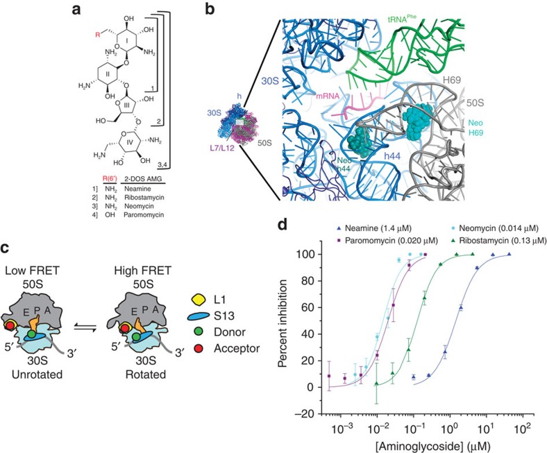
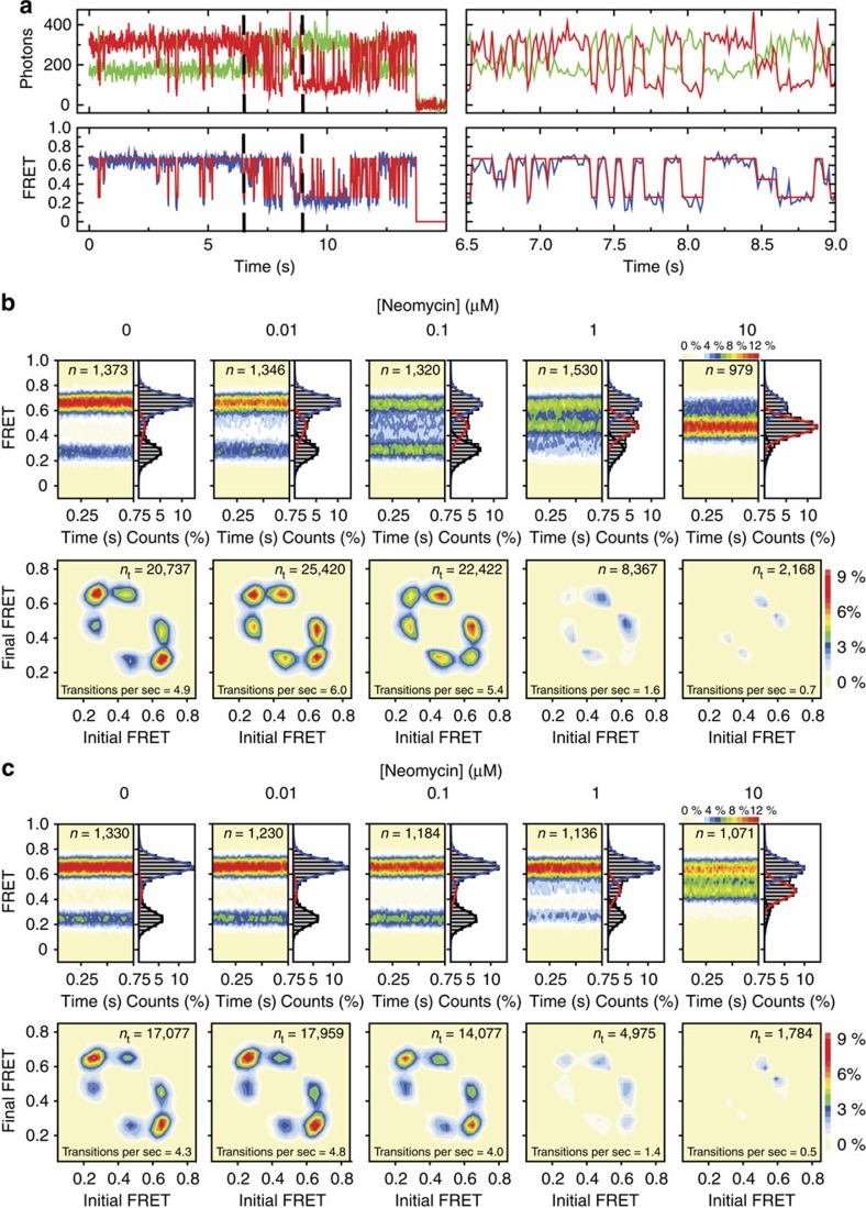
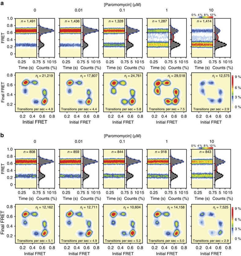
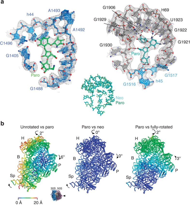
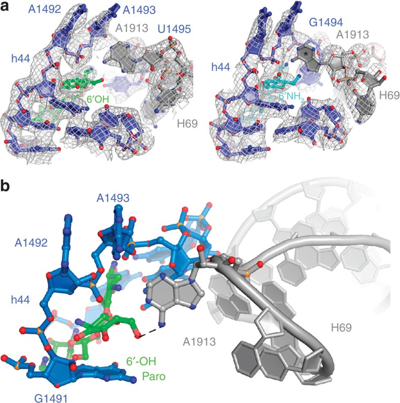
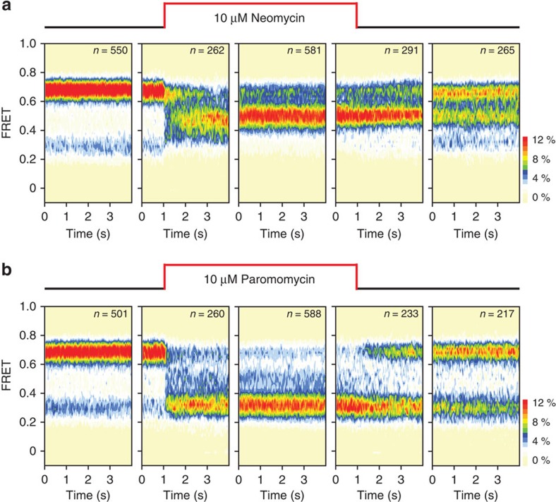
Dynamic remodelling of intersubunit bridge B2, a conserved RNA domain of the bacterial ribosome connecting helices 44 (h44) and 69 (H69) of the small and large subunit, respectively, impacts translation by controlling intersubunit rotation. Here we show that aminoglycosides chemically related to neomycin-paromomycin, ribostamycin and neamine-each bind to sites within h44 and H69 to perturb bridge B2 and affect subunit rotation. Neomycin and paromomycin, which only differ by their ring-I 6'-polar group, drive subunit rotation in opposite directions. This suggests that their distinct actions hinge on the 6'-substituent and the drug's net positive charge. By solving the crystal structure of the paromomycin-ribosome complex, we observe specific contacts between the apical tip of H69 and the 6'-hydroxyl on paromomycin from within the drug's canonical h44-binding site. These results indicate that aminoglycoside actions must be framed in the context of bridge B2 and their regulation of subunit rotation.
Conflict of interest statement
S.C.B and R.B.A have an equity interest in Lumidyne Technologies. The remaining authors declare no competing financial interests.
Figures
Similar articles
-
Allosteric control of the ribosome by small-molecule antibiotics.Nat Struct Mol Biol. 2012 Sep;19(9):957-63. doi: 10.1038/nsmb.2360. Epub 2012 Aug 19. Nat Struct Mol Biol. 2012. PMID: 22902368 Free PMC article.
-
Characterization of a 30S ribosomal subunit assembly intermediate found in Escherichia coli cells growing with neomycin or paromomycin.Arch Microbiol. 2008 May;189(5):441-9. doi: 10.1007/s00203-007-0334-6. Epub 2007 Dec 5. Arch Microbiol. 2008. PMID: 18060665
-
Binding of neomycin-class aminoglycoside antibiotics to the A-site of 16 S rRNA.J Mol Biol. 1998 Mar 27;277(2):347-62. doi: 10.1006/jmbi.1997.1552. J Mol Biol. 1998. PMID: 9514735
-
Effect of structural modifications on the biological properties of aminoglycoside antibiotics containing 2-deoxystreptamine.Adv Appl Microbiol. 1974;18(0):191-307. doi: 10.1016/s0065-2164(08)70572-0. Adv Appl Microbiol. 1974. PMID: 4613147 Review. No abstract available.
-
Aminoglycosides versus bacteria--a description of the action, resistance mechanism, and nosocomial battleground.J Biomed Sci. 2008 Jan;15(1):5-14. doi: 10.1007/s11373-007-9194-y. Epub 2007 Jul 27. J Biomed Sci. 2008. PMID: 17657587 Review.
Cited by
-
Geometric alignment of aminoacyl-tRNA relative to catalytic centers of the ribosome underpins accurate mRNA decoding.Nat Commun. 2023 Sep 11;14(1):5582. doi: 10.1038/s41467-023-40404-9. Nat Commun. 2023. PMID: 37696823 Free PMC article.
-
N6', N6''', and O4' Modifications to Neomycin Affect Ribosomal Selectivity without Compromising Antibacterial Activity.ACS Infect Dis. 2017 May 12;3(5):368-377. doi: 10.1021/acsinfecdis.6b00214. Epub 2017 Apr 6. ACS Infect Dis. 2017. PMID: 28343384 Free PMC article.
-
Structure of the bacterial ribosome at 2 Å resolution.Elife. 2020 Sep 14;9:e60482. doi: 10.7554/eLife.60482. Elife. 2020. PMID: 32924932 Free PMC article.
-
Hyper-swivel head domain motions are required for complete mRNA-tRNA translocation and ribosome resetting.Nucleic Acids Res. 2022 Aug 12;50(14):8302-8320. doi: 10.1093/nar/gkac597. Nucleic Acids Res. 2022. PMID: 35808938 Free PMC article.
-
Influence of ring size in conformationally restricted ring I analogs of paromomycin on antiribosomal and antibacterial activity.RSC Med Chem. 2021 Aug 5;12(9):1585-1591. doi: 10.1039/d1md00214g. eCollection 2021 Sep 23. RSC Med Chem. 2021. PMID: 34671740 Free PMC article.
References
-
- Puglisi J. D. et al.. in The Ribosome: Structure, Function, Antibiotics and Cellular Interactions eds Garrett R. A., Douthwaite S. R., Liljas A., Matheson A. T., Moore P. B., Noller H. F. ASM Press (2000).
-
- Cabanas M. J., Vazquez D. & Modolell J. Inhibition of ribosomal translocation by aminoglycoside antibiotics. Biochem. Biophys. Res. Commun. 83, 991–997 (1978). - PubMed
-
- Davies J., Gorini L. & Davis B. D. Misreading of RNA codewords induced by aminoglycoside antibiotics. Mol. Pharmacol. 1, 93–106 (1965). - PubMed
-
- Peske F., Savelsbergh A., Katunin V. I., Rodnina M. V. & Wintermeyer W. Conformational changes of the small ribosomal subunit during elongation factor G-dependent tRNA-mRNA translocation. J. Mol. Biol. 343, 1183–1194 (2004). - PubMed
Publication types
MeSH terms
Substances
Grants and funding
LinkOut - more resources
Full Text Sources
Other Literature Sources
Medical
Molecular Biology Databases
