. 2015 Jul 30:12:137.
doi: 10.1186/s12974-015-0360-2.

HMGB1 promotes the activation of NLRP3 and caspase-8 inflammasomes via NF-κB pathway in acute glaucoma

Affiliations

HMGB1 promotes the activation of NLRP3 and caspase-8 inflammasomes via NF-κB pathway in acute glaucoma

Wei Chi et al. J Neuroinflammation. .

Abstract

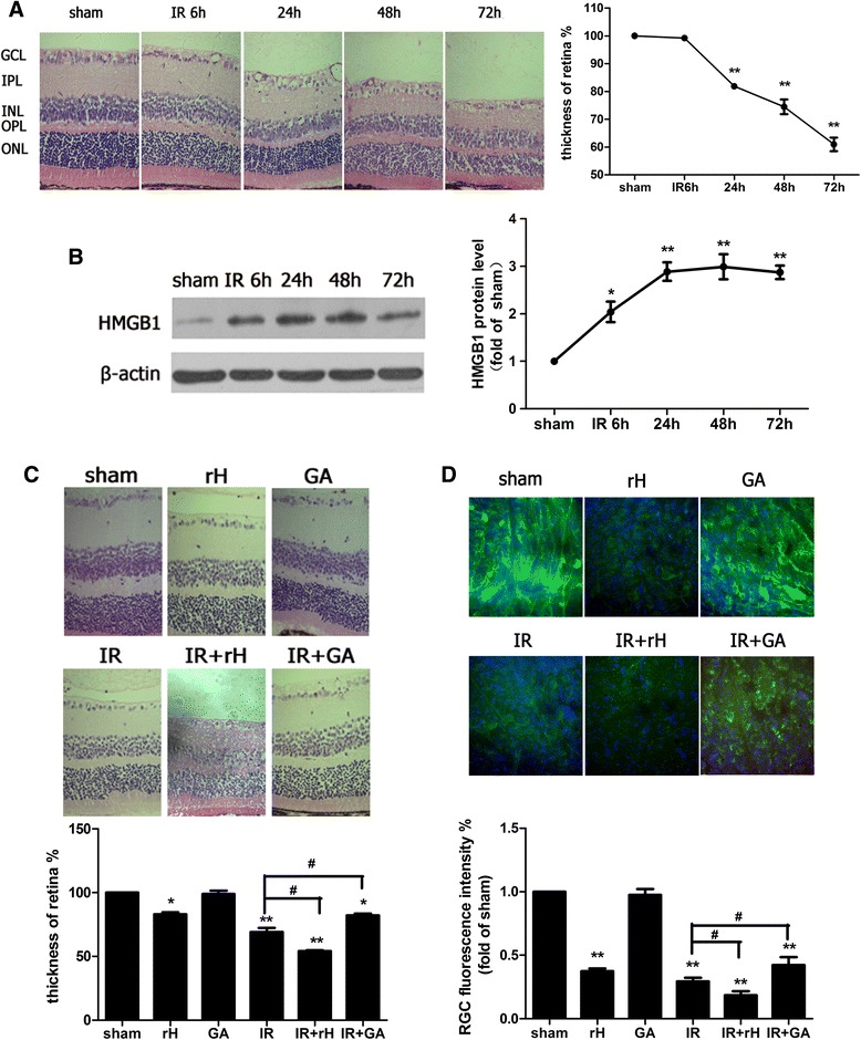
Background: Acute glaucoma is a significantly sight-threatening cause of irreversible blindness in the world characterized by a sudden and substantial intraocular pressure (IOP) increase and subsequent retinal ganglion cell (RGC) death. This study aims to explore the role of high-mobility group box 1 (HMGB1) in an acute glaucoma mouse model.

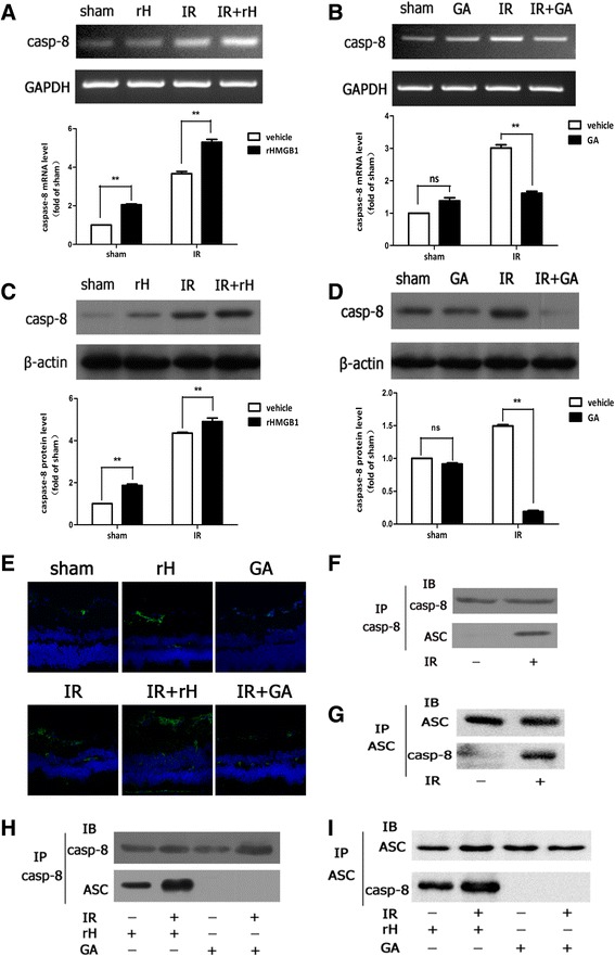
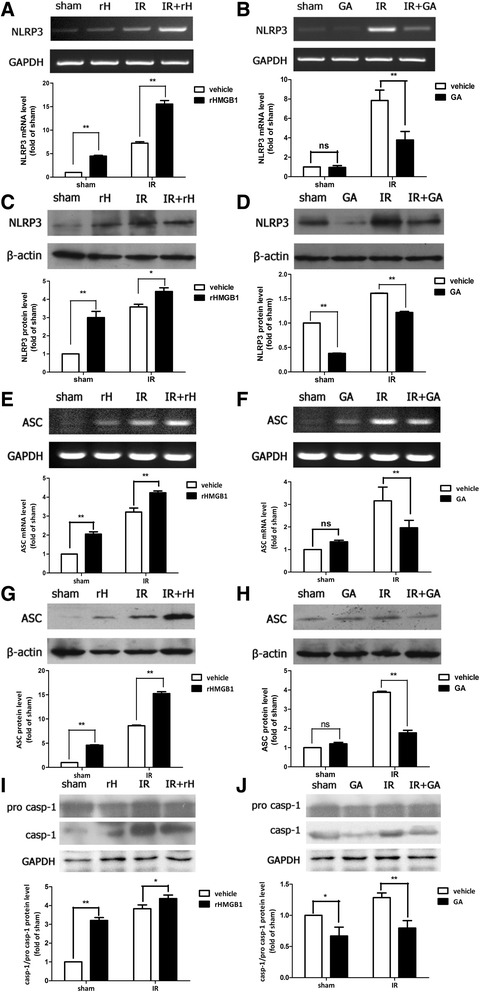
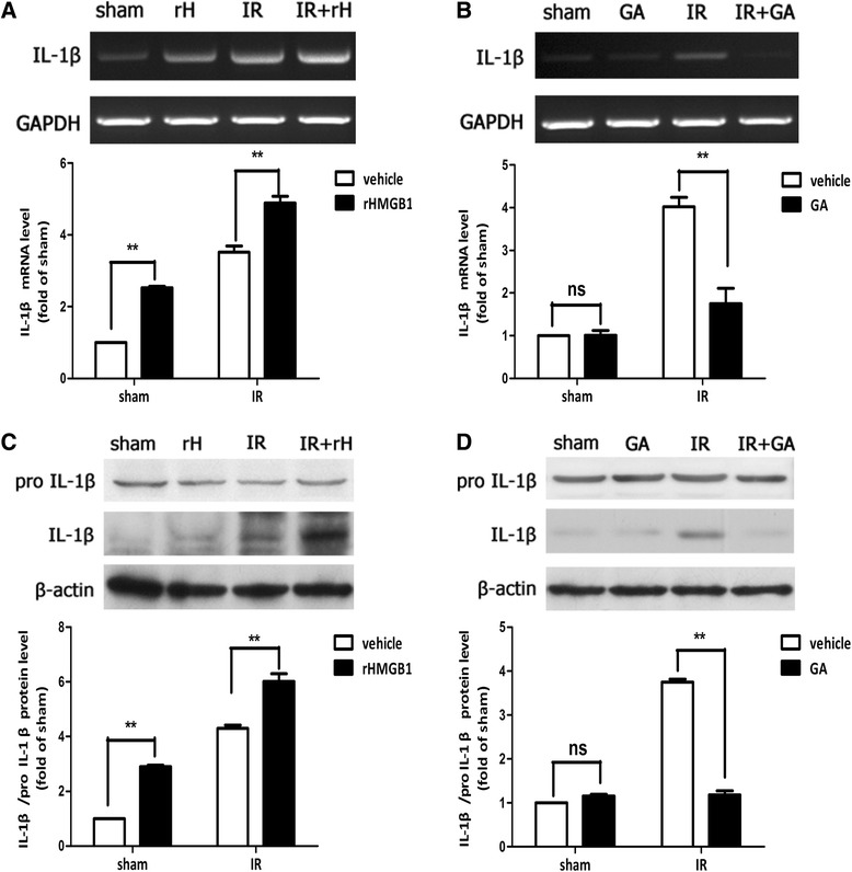
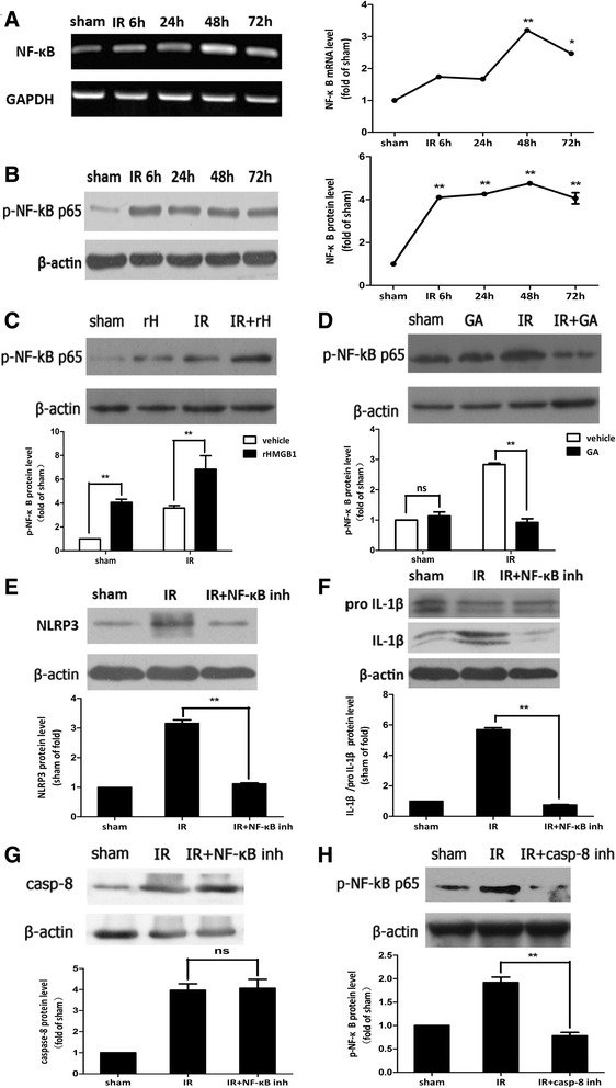
Methods: An acute glaucoma model was induced by a rapid and substantial increase IOP to 70 mmHg for 60 min via anterior chamber punctured and affused with Balance Salt Solution in C57BL/6 mice. Retinal tissue ischemic damage and loss of RGCs were assessed at 6, 24, 48, 72 h after high IOP treatment, and at 48 h, group with or without recombinant high-mobility group box 1 (rHMGB1), the HMGB1 inhibitor, glycyrrhizic acid (GA), and by HE and immunofluorescent staining. The nuclear factor κB (NF-κB) inhibitor, JSH-23, and caspase-8 inhibitor, Z-IETD-fmk, were injected into vitreous. Reverse transcription and semi-quantitative reverse transcription polymerase chain reaction (RT-PCR), western blotting, and immunoprecipitation were performed to evaluate the expression level of nucleotide-binding domain, leucine-rich repeat containing protein 3 (NLRP3), phosphor-NF-κB p65, caspase-8, caspase-1, apoptosis-associated speck-like protein containing a CARD (ASC), and interleukin-1β (IL-1β).

Results: HMGB1 was increased in ischemic retinal tissue during acute glaucoma as early as 6 h after rapid IOP elevation. Exogenous HMGB1 exacerbated retinal ischemic damage, RGC loss, and inhibition of endogenous HMGB1 significantly reduced the severity of disease. HMGB1 significantly induced the elevation of canonical NLRP3, ASC, caspase-1, and non-canonical capase-8-ASC inflammasome and promoted the processing of IL-1β. Furthermore, the effect of HMGB1 on NLRP3 inflammasome activation and IL-1β production was dependent on NF-κB pathway. Thus, HMGB1/caspase-8 pathway promoted the processing of IL-1β via NF-κB pathway.

Conclusion: The findings of this study identified a novel signaling pathway in which HMGB1, in response to acutely elevated intraocular pressure, activated the canonical NLRP3 and non-canonical caspase-8 inflammasomes and production of IL-1β during acute glaucoma development. These results provide new insights to the understanding of the innate response that contributes to pathogenesis of acute glaucoma.


Figures

Fig. 1
Fig. 1
HMGB1 was actively involved in the pathogenesis of retinal IR injury. a Hematoxylin and eosin staining of retina showed the degeneration of RGCs and mean thickness decrease of ischemic retinal tissue at different time points after reperfusion. GCL ganglion cell layer, IPL inner plexiform layer, INL inner nuclear layer, ONL outer nuclear layer, OPL, outer plexiform layer. b Western blot analysis of the cytoplasmic protein levels of HMGB1 at different time points after reperfusion. Each protein expression level is shown relative to that of controls. c The function of HMGB1 on the retinal tissue damage in retinal IR injury. The sham procedure was performed without elevating the container in contralateral eyes as control groups and retinal ischemia reperfusion (IR) injury was observed at 48 h after reperfusion. # P < 0.05 (IR vs IR + rH, IR vs IR + GA). d RGCs marker β3-tubulin detected RGCs damage by immunofluorescent staining in experimental groups. Data are shown as mean ± SD or percentage. *P < 0.05, **P < 0.01 vs sham group
Fig. 2
Fig. 2
HMGB1 promoted non-canonical caspase-8 inflammasome assembled in retinal IR injury. a, b RT-PCR analyzed caspase-8 mRNA expression. c, d Western blot detected expression of cleaved caspase-8 protein. e rHMGB1 could stimulate the activation of caspase-8 in RGCs, while inhibition of HMGB1 significantly suppressed the activation by confocal microscope. f, g ASC immunoprecipitated with caspase-8 and assembled caspase-8-ASC complex in IR model. h, i HMGB1 promoted caspase-8-ASC protein complex formation. Data are shown as mean ± SD. *P < 0.05, **P < 0.01 vs sham group
Fig. 3
Fig. 3
HMGB1 promoted the activation of canonical NLRP3 inflammasome in retinal IR injury. The intravitreal injection of exogenous rHMGB1 significantly promoted the mRNA and protein production of NLRP3 (a, c) and ASC (e, g) and activation of caspase-1 (i). Thus, the intravitreal injection of HMGB1 inhibitor, GA, significantly suppressed the production of NLRP3 (b, d) and ASC (f, h) and activation of caspase-1 (j). Data are shown as mean ± SD. *P < 0.05, **P < 0.01 vs sham group
Fig. 4
Fig. 4
HMGB1 promoted the processing of IL-1β in retinal IR injury. ad The intravitreal injection of rHMGB1 or HMGB1 inhibitor significantly enhanced or attenuated the processing of IL-1β in retinal IR injury. Data are shown as mean ± SD. *P < 0.05, **P < 0.01 vs sham group
Fig. 5
Fig. 5
HMGB1 regulated the activation of NLRP3, rather than the activation of caspase-8, via NF-κB pathway. a, b NF-κB was activated in ischemic retina at the early stage after reperfusion. c, d Intraocular injection rHMGB1 or GA could promote or suppress the production of phosphor-NF-κB p65. e, f Intravitreal injection of NF-κB p65 inhibitor, JSH-23, significantly reduced the activation of NLRP3 and decreased the processing of IL-1β, g rather than caspase-8. However, intravitreal injection of caspase-8 inhibitor, Z-IETD-fmk, decreased the production of phosphor-NF-κB p65, obviously (h). Data are shown as mean ± SD. *P < 0.05, **P < 0.01 vs sham group

References

    1. Quigley HA, Broman AT. The number of people with glaucoma worldwide in 2010 and 2020. Br J Ophthalmol. 2006;90:262–267. doi: 10.1136/bjo.2005.081224. - DOI - PMC - PubMed
    1. Weinreb RN, Aung T, Medeiros FA. The pathophysiology and treatment of glaucoma: a review. JAMA. 2014;311:1901–1911. doi: 10.1001/jama.2014.3192. - DOI - PMC - PubMed
    1. Andersson U, Tracey KJ. HMGB1 is a therapeutic target for sterile inflammation and infection. Annu Rev Immunol. 2011;29:139–162. doi: 10.1146/annurev-immunol-030409-101323. - DOI - PMC - PubMed
    1. Yang H, Antoine DJ, Andersson U, Tracey KJ. The many faces of HMGB1: molecular structure-functional activity in inflammation, apoptosis, and chemotaxis. J Leukoc Biol. 2013;93:865–873. doi: 10.1189/jlb.1212662. - DOI - PMC - PubMed
    1. Schallenberg M, Prokosch V, Thanos S. Regulation of retinal proteome by topical antiglaucomatous eye drops in an inherited glaucoma rat model. PLoS One. 2012;7 doi: 10.1371/journal.pone.0033593. - DOI - PMC - PubMed

Publication types