. 2015 Oct 1;26(19):3489-503.
doi: 10.1091/mbc.E14-05-1020. Epub 2015 Jul 29.

Involvement of the Tyro3 receptor and its intracellular partner Fyn signaling in Schwann cell myelination

Affiliations

Involvement of the Tyro3 receptor and its intracellular partner Fyn signaling in Schwann cell myelination

Yuki Miyamoto et al. Mol Biol Cell. .

Abstract

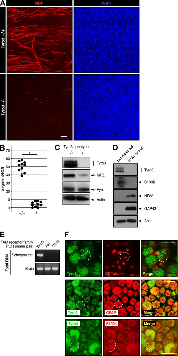
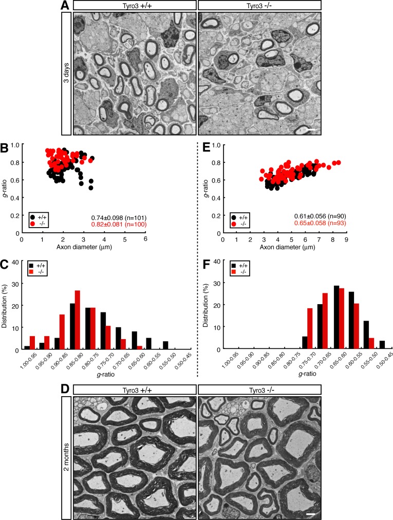
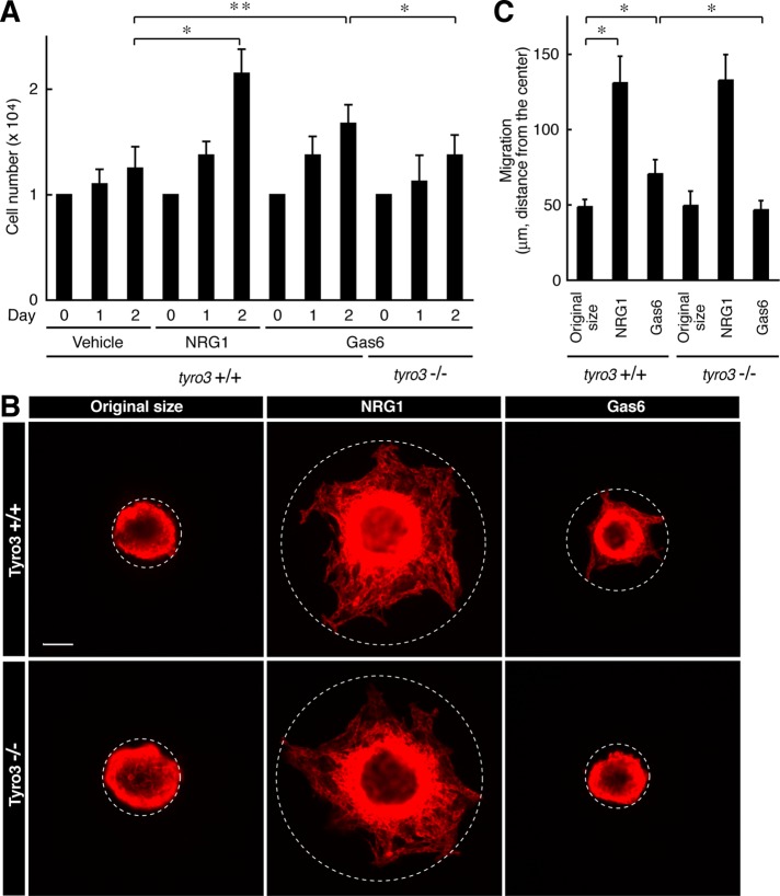
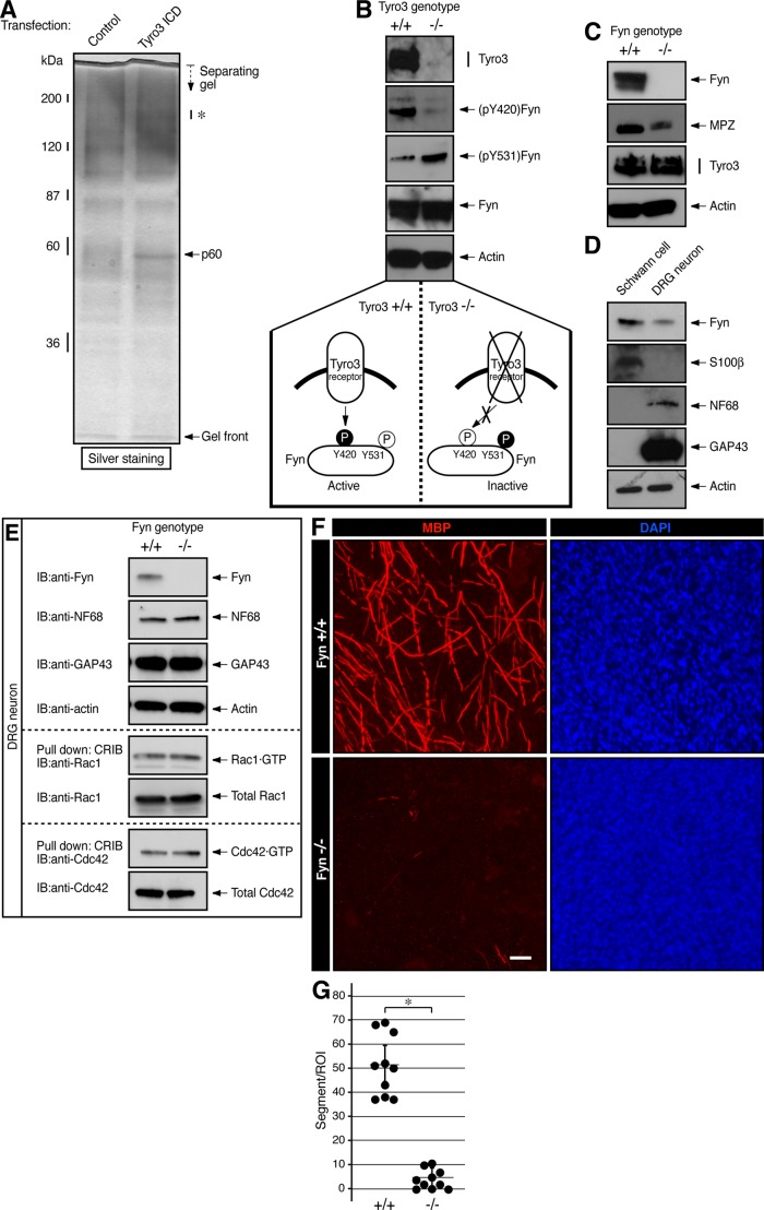
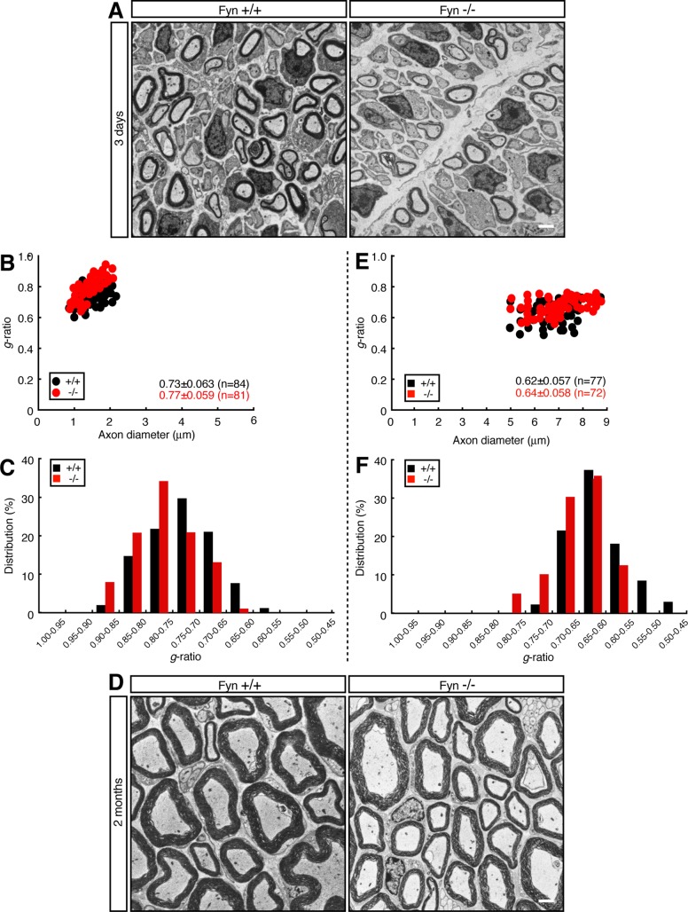
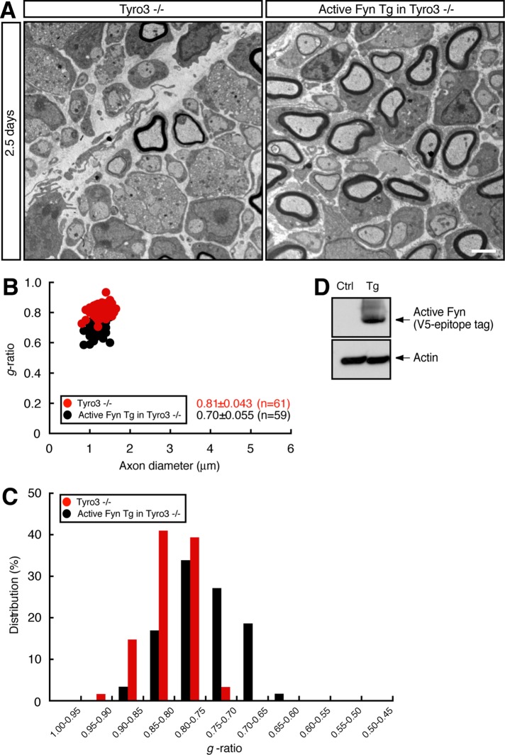
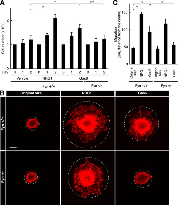
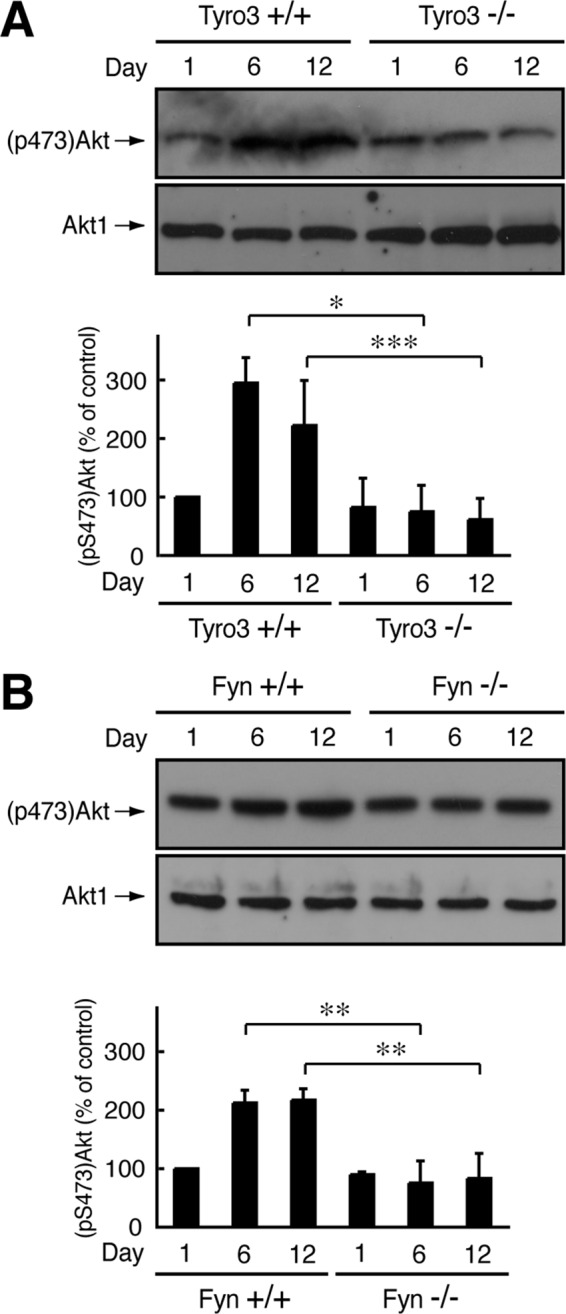
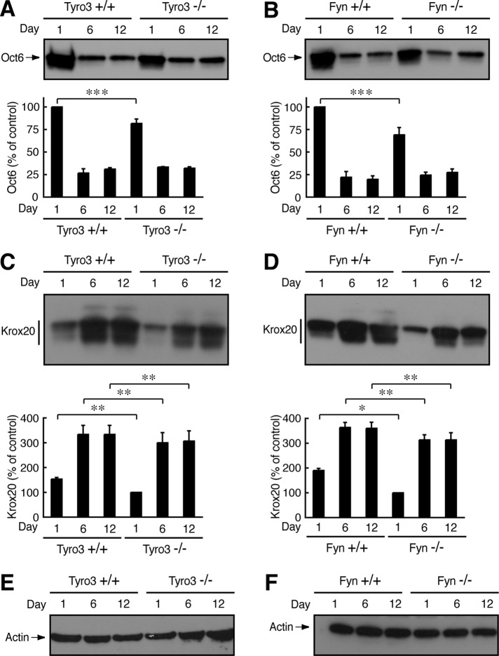
During early development of the peripheral nervous system, Schwann cell precursors proliferate, migrate, and differentiate into premyelinating Schwann cells. After birth, Schwann cells envelop neuronal axons with myelin sheaths. Although some molecular mechanisms underlying myelination by Schwann cells have been identified, the whole picture remains unclear. Here we show that signaling through Tyro3 receptor tyrosine kinase and its binding partner, Fyn nonreceptor cytoplasmic tyrosine kinase, is involved in myelination by Schwann cells. Impaired formation of myelin segments is observed in Schwann cell neuronal cultures established from Tyro3-knockout mouse dorsal root ganglia (DRG). Indeed, Tyro3-knockout mice exhibit reduced myelin thickness. By affinity chromatography, Fyn was identified as the binding partner of the Tyro3 intracellular domain, and activity of Fyn is down-regulated in Tyro3-knockout mice, suggesting that Tyro3, acting through Fyn, regulates myelination. Ablating Fyn in mice results in reduced myelin thickness. Decreased myelin formation is observed in cultures established from Fyn-knockout mouse DRG. Furthermore, decreased kinase activity levels and altered expression of myelination-associated transcription factors are observed in these knockout mice. These results suggest the involvement of Tyro3 receptor and its binding partner Fyn in Schwann cell myelination. This constitutes a newly recognized receptor-linked signaling mechanism that can control Schwann cell myelination.


Figures

FIGURE 1:
FIGURE 1:
Deletion of Tyro3 leads to decreased formation of myelin segments in cultures. (A, B) Schwann cell DRG neuronal cultures were established from Tyro3+/+ and Tyro3/ mouse embryonic DRGs and stained with an antibody against myelin marker protein MBP (red) or DAPI (blue). Scale bar, 20 μm. MBP-positive segments were counted in a 200-μm-square field (*p < 0.01; n = 10). (C) Tissue lysates from 6-d-old Tyro3+/+ and Tyro3/ mouse sciatic nerves were used for the respective immunoblottings. Expression levels of Tyro3, myelin marker protein MPZ, Fyn, and control actin. (D) Primary Schwann cells and neurons were isolated from wild-type mice, cultured, and used for the respective immunoblottings. Expression levels of Tyro3, Schwann cell marker protein S100β, neuronal cell marker proteins NF68 and GAP43, and control actin. (E) In Schwann cells, RT-PCR analysis detects the primary transcription of Tyro3. All TAM receptor members are detected in brain total RNA as the positive control. (F) Cross section of 6-d-old mouse sciatic nerve immunostained with antibodies for Tyro3 (green), and neuronal marker βIII tubulin (red), or Schwann cell marker S100β (red). A 7-d-old cross section was also immunostained with antibodies for Tyro3 (green) and Schwann cell marker GFAP (red). Dotted circle indicates a typical axon with Schwann cells. Scale bar, 5 μm.
FIGURE 2:
FIGURE 2:
Deletion of Tyro3 leads to decreased myelin thickness in mice. (A) Electron microscopic photographs of 3-d-old Tyro3+/+ and Tyro3/ mouse sciatic nerves (n = 101 and 100, respectively). Scale bar, 1 μm. (B) The g ratio—the numerical ratio between the diameter of the axon and the outer diameter of the myelinated fiber—plotted vs. axon diameter. The average g ratio is also given. (C) The data from 3-d-old mice in the form of the relative distribution of the g ratio. (D) Electron microscopic photographs of 2-mo-old Tyro3+/+ and Tyro3/ mouse sciatic nerves (n = 90 and 93, respectively). Scale bar, 1 μm. (E) The g ratio vs. axon diameter for these mice. The average g ratio is also given. (F) The relative distribution of the g ratio for these mice.
FIGURE 3:
FIGURE 3:
Deletion of Tyro3 inhibits Gas6-stimulated Schwann cell proliferation and migration. (A) Tyro3+/+ or Tyro3/ Schwann cells were cultured for 1 or 2 d in normal medium with or without 20 ng/ml Gas6 (*p < 0.01; **p < 0.025; n = 4). Treatment with 20 ng/ml NRG1 was used as the positive control. (B, C) Reaggregated Tyro3+/+ or Tyro3/ Schwann cells were allowed to migrate outward for 6 h in normal medium with or without 20 ng/ml Gas6 and stained with an antibody against S100β. Treatment with NRG1 was used as the positive control. The dotted circles indicate the maximum peripheries of Schwann cells migrating from the reaggregates. The distance furthest from the center was measured as the migrating distance (*p < 0.01; n = 4). Scale bar, 50 μm.
FIGURE 4:
FIGURE 4:
Tyro3 interacts with Fyn to regulate it in Schwann cells. (A) RT4-D6P2T cells were transfected with the control vector or myc-tagged Tyro3 intracellular tyrosine kinase domain (ICD). Coprecipitated proteins with Tyro3 ICD were collected with an anti-myc antibody and detected with silver staining. By mass spectrometric analysis, a Tyro3 ICD–interacting 60-kDa band (p60) was identified as a protein mixture of Fyn and Klrg1. In this experiment, the 150- to 180-kDa protein bands (indicated by an asterisk) were not identified. (B) Tissue lysates from Tyro3+/+ or Tyro3/ sciatic nerves were used for the respective immunoblottings. Phosphorylation levels of (pY420)Fyn and (pY531)Fyn in Fyn immunoprecipitates, as well as expression levels of Tyro3, Fyn, and control actin. Y420 phosphorylation in the kinase activation loop stimulates the activity of Fyn, whereas Y531 phosphorylation near the C-terminal position down-regulates Fyn (bottom schematic diagrams). (C) Tissue lysates from Fyn+/+ or Fyn/ sciatic nerves were used for the respective immunoblottings. Expression levels of Fyn, MPZ, Tyro3, and control actin. (D) Expression levels of Fyn, S100β, NF68, and GAP43, and control actin in Schwann cells and DRG neurons, (E) Cell lysates from Fyn+/+ or Fyn/ DRG neurons were used for the respective immunoblottings (Fyn, NF68, GAP43, control actin, and Rho GTPases Rac1 and Cdc42). They were also used for an affinity precipitation assay to detect with active, GTP-bound Rac1 or Cdc42, which are essential for forming neuronal structures. (F, G) Fyn+/+-or Fyn/-mouse Schwann cell DRG neuronal cultures were established from mouse embryonic DRG and stained with an antibody against MBP (red) or DAPI (blue). Scale bar, 20 μm. MBP-positive segments were counted in a 200-μm-square field (*p < 0.01; n = 10).
FIGURE 5:
FIGURE 5:
Electron microscopic analyses of sciatic nerves of Fyn-knockout mouse. (A) Electron microscopic photographs of 3-d-old Fyn+/+ and Fyn/ mouse sciatic nerves (n = 84 and 81, respectively). Scale bar, 1 μm. (B) The g ratio vs. axon diameter. The average g ratio is also given. (C) The relative distributions of the g ratio. (D) Electron microscopic photographs of 2-mo-old Fyn+/+ and Fyn/ mouse sciatic nerves (n = 77 and 72, respectively). Scale bar, 1 μm. (E) The g ratio vs. axon diameter for these mice. The average g ratio is also given. (F) The relative distribution of the g ratio for these mice.
FIGURE 6:
FIGURE 6:
Interbreeding of Tyro3-knockout mice with Fyn transgenic mice can rescue the Tyro3- knockout mouse thin myelin structure. (A) Electron microscopic photographs of 2.5-d-old mouse sciatic nerves for Tyro3/ mice and active Fyn transgenic (Tg) Tyro3/ mice (n = 61 and 59, respectively). Scale bar, 1 μm. (B) The g ratio vs. axon diameter. The average g ratio is also given. (C) The relative distribution of the g ratio. (D) Sciatic nerve extracts from active Fyn transgenic (Tg) and control (Ctrl) mice immunoblotted with an antibody for V5-epitope tag or actin.
FIGURE 7:
FIGURE 7:
Deletion of Fyn inhibits Gas6-stimulated Schwann cell proliferation and migration. (A) Fyn+/+ or Fyn/ Schwann cells were cultured for 1 or 2 d in normal medium with or without Gas6 (*p < 0.01; **p < 0.025; n = 4). (B, C) Reaggregated Fyn+/+ or Fyn/ Schwann cells were allowed to migrate outward for 6 h in normal medium with or without Gas6 and stained with an antibody against S100β. The dotted circles indicate the maximum peripheries of Schwann cells migrating from the reaggregates (*p < 0.01; n = 4). Scale bar, 50 μm.
FIGURE 8:
FIGURE 8:
Effect of Tyro3 or Fyn knockout on Akt phosphorylation. (A, B) Tissue lysates from Tyro3 or Fyn (+/+ or /) mouse sciatic nerves were used for immunoblotting with an antibody against Akt1 or (pS473)Akt and the band intensities analyzed (*p < 0.01; **p < 0.025; ***p < 0.05; n = 4).
FIGURE 9:
FIGURE 9:
Effect of Tyro3 or Fyn knockout on Oct6 or Krox20 expression. (A, B) Tissue lysates from Tyro3 or Fyn (+/+ or /) mouse sciatic nerves were used for immunoblotting with an antibody against Oct6 and the band intensities analyzed (***p 0.05; n = 3). (C, D) Tissue lysates from Tyro3 or Fyn (+/+ or /) mouse sciatic nerves used for immunoblotting with an antibody against Krox20 (*p 0.01; **p < 0.025; n = 3). (E, F) Representative actin immunoblots in Tyro3 or Fyn (+/+ or /) nerves.

References

    1. Arimura N, Kaibuchi K. Neuronal polarity: from extracellular signals to intracellular mechanisms. Nat Rev Neurosci. 2007;8:194–205. - PubMed
    1. Binder MD, Cate HS, Prieto AL, Kemper D, Butzkueven H, Gresle MM, Cipriani T, Jokubaitis VG, Carmeliet P, Kilpatrick TJ. Gas6 deficiency increases oligodendrocyte loss and microglial activation in response to cuprizone-induced demyelination. J Neurosci. 2008;28:5195–5206. - PMC - PubMed
    1. Binder MD, Kilpatrick TJ. TAM receptor signalling and demyelination. Neurosignals. 2009;17:277–287. - PubMed
    1. Binder MD, Xiao J, Kemper D, Ma GZ, Murray SS, Kilpatrick TJ. Gas6 increases myelination by oligodendrocytes and its deficiency delays recovery following cuprizone-induced demyelination. PLoS One. 2011;6:e17727. - PMC - PubMed
    1. Bunge RP. Expanding roles for the Schwann cell: ensheathment, myelination, trophism and regeneration. Curr Opin Neurobiol. 1993;3:805–809. - PubMed

Publication types

Substances

LinkOut - more resources