

Assessment of urethral support using MRI-derived computational modeling of the female pelvis
- PMID: 26224383
- PMCID: PMC5519823
- DOI: 10.1007/s00192-015-2804-8
Assessment of urethral support using MRI-derived computational modeling of the female pelvis
Abstract
Introduction and hypothesis: This study aimed to assess the role of individual anatomical structures and their combinations to urethral support function.
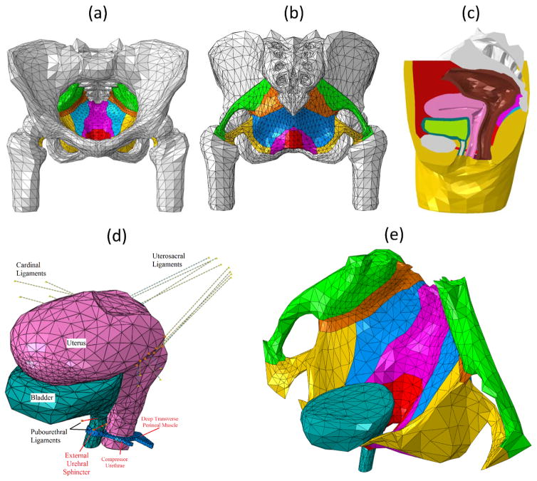
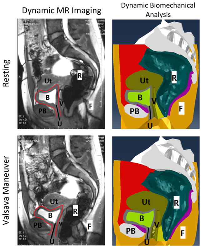
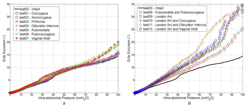
Methods: A realistic pelvic model was developed from an asymptomatic female patient's magnetic resonance (MR) images for dynamic biomechanical analysis using the finite element method. Validation was performed by comparing simulation results with dynamic MR imaging observations. Weaknesses of anatomical support structures were simulated by reducing their material stiffness. Urethral mobility was quantified by examining urethral axis excursion from rest to the final state (intra-abdominal pressure = 100 cmH2O). Seven individual support structures and five of their combinations were studied.
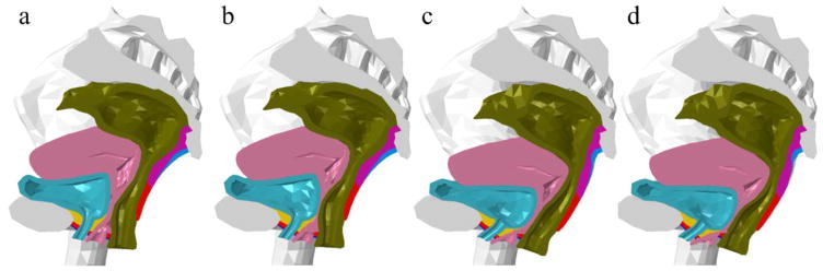
Result: Among seven urethral support structures, we found that weakening the vaginal walls, puborectalis muscle, and pubococcygeus muscle generated the top three largest urethral excursion angles. A linear relationship was found between urethral axis excursions and intra-abdominal pressure. Weakening all three levator ani components together caused a larger weakening effect than the sum of each individually weakened component, indicating a nonlinearly additive pattern. The pelvic floor responded to different weakening conditions distinctly: weakening the vaginal wall developed urethral mobility through the collapsed vaginal canal, while weakening the levator ani showed a more uniform pelvic floor deformation.
Conclusions: The computational modeling and dynamic biomechanical analysis provides a powerful tool to better understand the dynamics of the female pelvis under pressure events. The vaginal walls, puborectalis, and pubococcygeus are the most important individual structures in providing urethral support. The levator ani muscle group provides urethral support in a well-coordinated way with a nonlinearly additive pattern.
Keywords: Finite element method; Magnetic resonance imaging; Pelvic muscle; Stress urinary incontinence; Urethral hypermobility.
Conflict of interest statement
Yun Peng FINANCIAL DISCLAIMER/CONFLICT OF INTEREST: NONE Rose Khavari FINANCIAL DISCLAIMER/CONFLICT OF INTEREST: NONE Nissrine A. Nakib FINANCIAL DISCLAIMER/CONFLICT OF INTEREST: NONE Timothy B. Boone FINANCIAL DISCLAIMER/CONFLICT OF INTEREST: NONE Yingchun Zhang FINANCIAL DISCLAIMER/CONFLICT OF INTEREST: NONE
Figures
References
-
- Pirpiris A, Shek K, Dietz H. Urethral mobility and urinary incontinence. Ultrasound in Obstetrics & Gynecology. 2010;36(4):507–511. - PubMed
-
- Schick E, Jolivet-Tremblay M, Tessier J, Dupont C, Bertrand PE. Observations on the function of the female urethra: III: An overview with special reference to the relation between urethral hypermobility and urethral incompetence. Neurourology and urodynamics. 2004;23(1):22–26. - PubMed
-
- Delancey JOL. STRUCTURAL SUPPORT OF THE URETHRA AS IT RELATES TO STRESS URINARY-INCONTINENCE - THE HAMMOCK HYPOTHESIS. American Journal of Obstetrics and Gynecology. 1994;170(6):1713–1723. - PubMed
-
- Sendag F, Vidinli H, Kazandi M, Itil IM, Askar N, Vidinli B, Pourbagher A. Role of perineal sonography in the evaluation of patients with stress urinary incontinence. The Australian & New Zealand journal of obstetrics & gynaecology. 2003;43(1):54–57. - PubMed
-
- Fielding JR, Dumanli H, Schreyer AG, Okuda S, Gering DT, Zou KH, Kikinis R, Jolesz FA. MR-based three-dimensional modeling of the normal pelvic floor in women: quantification of muscle mass. American Journal of Roentgenology. 2000;174(3):657–660. - PubMed
Publication types
MeSH terms
Grants and funding
LinkOut - more resources
Full Text Sources
Other Literature Sources
Medical
