

Biomechanics of oral mucosa
- PMID: 26224566
- PMCID: PMC4535403
- DOI: 10.1098/rsif.2015.0325
Biomechanics of oral mucosa
Abstract
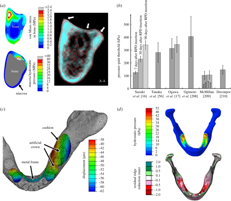
The prevalence of prosthodontic treatment has been well recognized, and the need is continuously increasing with the ageing population. While the oral mucosa plays a critical role in the treatment outcome, the associated biomechanics is not yet fully understood. Using the literature available, this paper provides a critical review on four aspects of mucosal biomechanics, including static, dynamic, volumetric and interactive responses, which are interpreted by its elasticity, viscosity/permeability, apparent Poisson's ratio and friction coefficient, respectively. Both empirical studies and numerical models are analysed and compared to gain anatomical and physiological insights. Furthermore, the clinical applications of such biomechanical knowledge on the mucosa are explored to address some critical concerns, including stimuli for tissue remodelling (interstitial hydrostatic pressure), pressure-pain thresholds, tissue displaceability and residual bone resorption. Through this review, the state of the art in mucosal biomechanics and their clinical implications are discussed for future research interests, including clinical applications, computational modelling, design optimization and prosthetic fabrication.
Keywords: hydrostatic pressure; hyperelastic; oral mucosa; pressure–pain threshold; residual ridge resorption; viscoelastic.
© 2015 The Authors.
Figures
Similar articles
-
Determination of oral mucosal Poisson's ratio and coefficient of friction from in-vivo contact pressure measurements.Comput Methods Biomech Biomed Engin. 2016;19(4):357-65. doi: 10.1080/10255842.2015.1028925. Epub 2015 May 29. Comput Methods Biomech Biomed Engin. 2016. PMID: 26024011
-
A comparative study on complete and implant retained denture treatments: a biomechanics perspective.J Biomech. 2015 Feb 5;48(3):512-9. doi: 10.1016/j.jbiomech.2014.11.043. Epub 2014 Dec 3. J Biomech. 2015. PMID: 25560272
-
Relationship between buccal mucosa ridging and viscoelastic behaviour of oral mucosa.J Oral Rehabil. 2011 Jun;38(6):429-33. doi: 10.1111/j.1365-2842.2010.02167.x. Epub 2010 Nov 5. J Oral Rehabil. 2011. PMID: 21054483
-
State-of-the-art research in lower-limb prosthetic biomechanics-socket interface: a review.J Rehabil Res Dev. 2001 Mar-Apr;38(2):161-74. J Rehabil Res Dev. 2001. PMID: 11392649 Review.
-
Computational and experimental characterization of skin mechanics: identifying current challenges and future directions.Wiley Interdiscip Rev Syst Biol Med. 2013 Sep-Oct;5(5):539-56. doi: 10.1002/wsbm.1228. Epub 2013 Jun 11. Wiley Interdiscip Rev Syst Biol Med. 2013. PMID: 23757148 Review.
Cited by
-
Effects of wearing removable dentures and aging on palatal mucosa blood flow by laser doppler.J Indian Prosthodont Soc. 2022 Apr-Jun;22(2):161-168. doi: 10.4103/jips.jips_292_21. J Indian Prosthodont Soc. 2022. PMID: 36511027 Free PMC article.
-
Functional comparison of pacifiers using finite element analysis.BMC Oral Health. 2022 Mar 2;22(1):49. doi: 10.1186/s12903-022-02087-4. BMC Oral Health. 2022. PMID: 35236336 Free PMC article.
-
Impact of Aging and Pathologies on Human Oral Mucosa: Preliminary Investigation of Biophysical Markers from Thermal and Vibrational Analyses.Biomolecules. 2025 Jul 8;15(7):978. doi: 10.3390/biom15070978. Biomolecules. 2025. PMID: 40723850 Free PMC article.
-
Nonlinear Biomechanical Characteristics of the Schneiderian Membrane: Experimental Study and Numerical Modeling.Biomed Res Int. 2018 Jun 21;2018:2829163. doi: 10.1155/2018/2829163. eCollection 2018. Biomed Res Int. 2018. PMID: 30035119 Free PMC article.
-
Effect of Number and Location on Stress Distribution of Mini Dental Implant-Assisted Mandibular Kennedy Class I Removable Partial Denture: Three-Dimensional Finite Element Analysis.Int J Dent. 2022 Mar 26;2022:4825177. doi: 10.1155/2022/4825177. eCollection 2022. Int J Dent. 2022. PMID: 35378727 Free PMC article.
References
-
- Ostlund SG. 1958. The effect of complete dentures on the gum tissues. Acta Odontol. Scand. 16, 1–41. (10.3109/00016355809028181) - DOI
Publication types
MeSH terms
LinkOut - more resources
Full Text Sources
Other Literature Sources
