. 2015 Aug 6;12(109):20150512.
doi: 10.1098/rsif.2015.0512.

Cell shape regulation through mechanosensory feedback control

Affiliations

Cell shape regulation through mechanosensory feedback control

Krithika Mohan et al. J R Soc Interface. .

Abstract

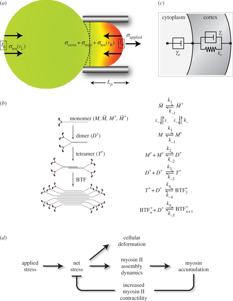
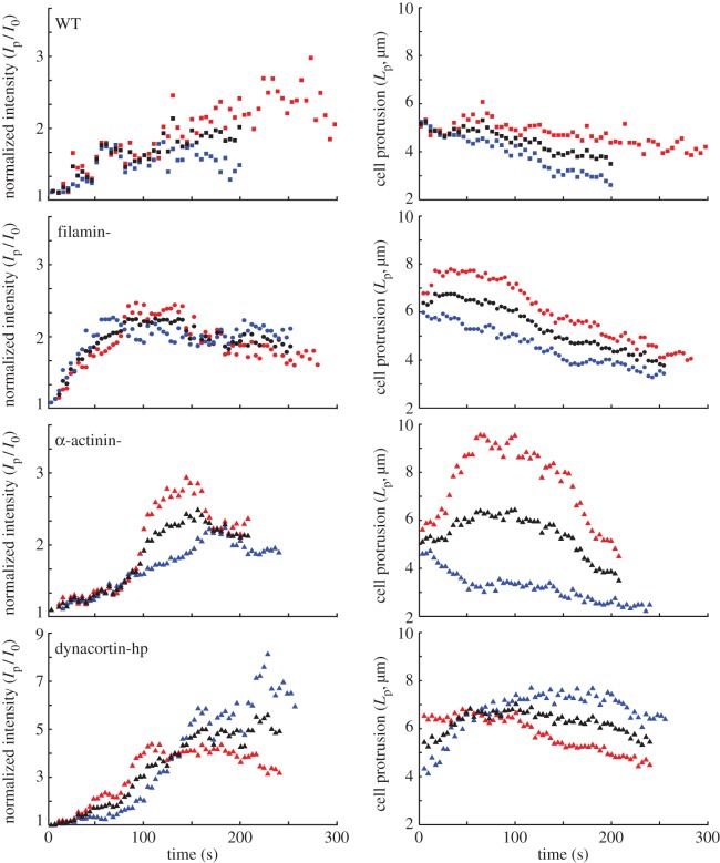
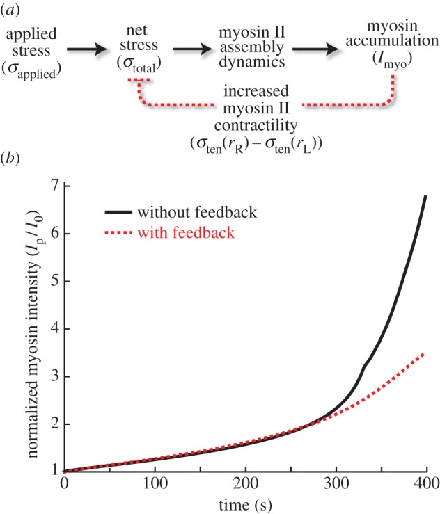
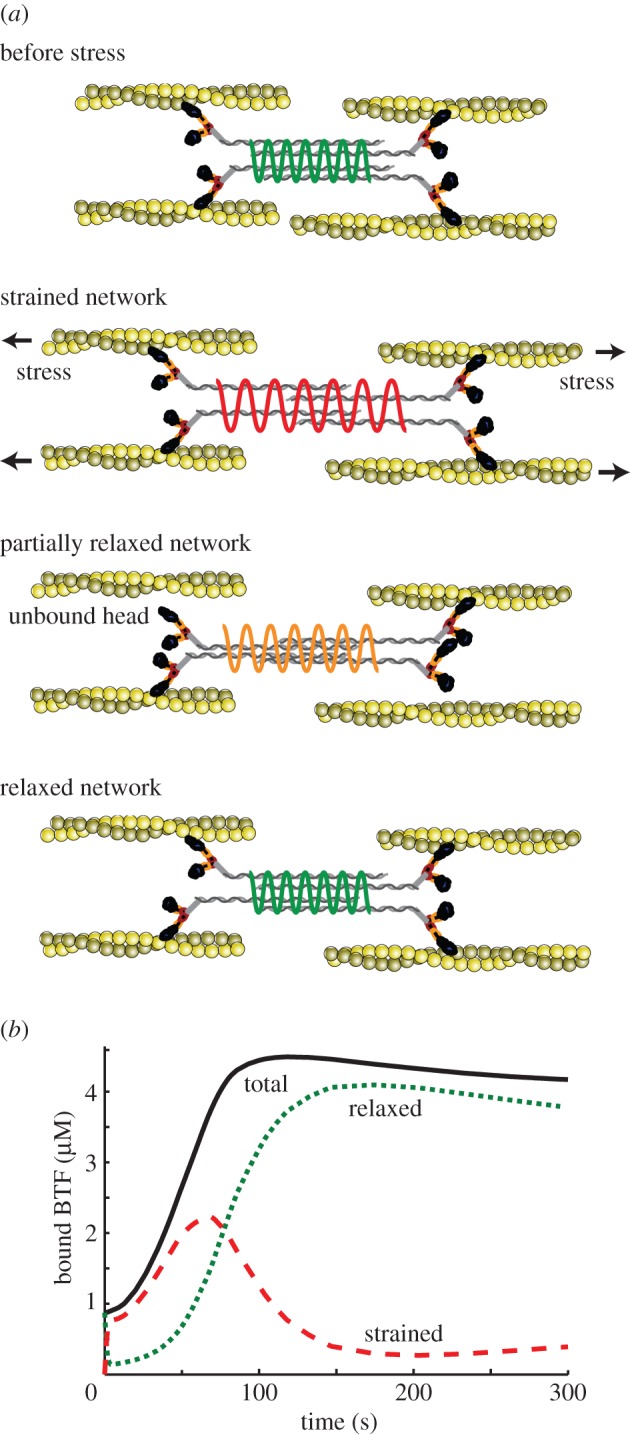
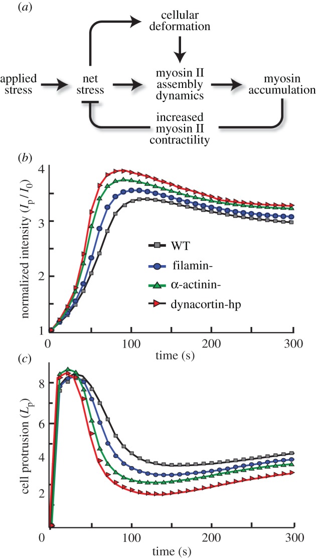
Cells undergo controlled changes in morphology in response to intracellular and extracellular signals. These changes require a means for sensing and interpreting the signalling cues, for generating the forces that act on the cell's physical material, and a control system to regulate this process. Experiments on Dictyostelium amoebae have shown that force-generating proteins can localize in response to external mechanical perturbations. This mechanosensing, and the ensuing mechanical feedback, plays an important role in minimizing the effect of mechanical disturbances in the course of changes in cell shape, especially during cell division, and likely in other contexts, such as during three-dimensional migration. Owing to the complexity of the feedback system, which couples mechanical and biochemical signals involved in shape regulation, theoretical approaches can guide further investigation by providing insights that are difficult to decipher experimentally. Here, we present a computational model that explains the different mechanosensory and mechanoresponsive behaviours observed in Dictyostelium cells. The model features a multiscale description of myosin II bipolar thick filament assembly that includes cooperative and force-dependent myosin-actin binding, and identifies the feedback mechanisms hidden in the observed mechanoresponsive behaviours of Dictyostelium cells during micropipette aspiration experiments. These feedbacks provide a mechanistic explanation of cellular retraction and hence cell shape regulation.

Keywords: cell shape regulation; force feedback; mechanosensing; myosin II.


Figures

Figure 1.
Figure 1.
Force feedback model. (a) Cartoon describing the stresses acting in an aspirated cell. (b) Assembly scheme for BTF formation (adapted from [13,14]). (c) Viscoelastic model used to simulate cell deformations. (d) Force feedback model incorporating the feedback effects arising from myosin accumulation by coupling BTF assembly dynamics to viscoelastic model for cellular deformation. (Online version in colour.)
Figure 2.
Figure 2.
Biphasic myosin accumulation and ensuing retraction observed in WT and mutant Dictyostelium cells depleted of different actin cross-linking proteins. Left panels show myosin intensity at the site of aspiration (Ip) normalized against the intensity at the cortex opposite to the site of aspiration (Io). Right panels show length of cell protrusion (Lp) into the pipette measured during the course of aspiration for different Dictyostelium strains. The blue and red data presented in this figure are measurements of single cells; the black markers denote their average. Some of these data were published in [13]. (Online version in colour.)
Figure 3.
Figure 3.
Simulations comparing effect of force feedback on myosin accumulation kinetics. (a) Diagram showing (b) comparison of BTF accumulation in response to externally applied stress σapplied, with (solid) and without (dotted) feedback from mechanosensitive accumulation of myosin and actin cross-linkers σtotal. (Online version in colour.)
Figure 4.
Figure 4.
Strained myosin–actin unbinding theory can explain multiple phases of myosin accumulation. (a) The cartoon depicts myosin II interactions with actin before, during, and after perturbation of the cytoskeletal network with external force. In this model, myosin II initially strained by the external stress relaxes over time. (b) Simulation of the stress induced strain and relaxation of myosin II. (Online version in colour.)
Figure 5.
Figure 5.
Simulations of the complete biomechanical feedback model. (a) Complete biomechanical feedback model. (b) Normalized myosin II intensity and (c) cellular protrusion after the application of a micropipette stress for various cellular strains. (Online version in colour.)

Similar articles

Cited by

References

    1. Effler JC, Iglesias PA, Robinson DN. 2007. A mechanosensory system controls cell shape changes during mitosis. Cell Cycle 6, 30 (10.4161/cc.6.1.3674) - DOI - PMC - PubMed
    1. Sampathkumar A, Yan A, Krupinski P, Meyerowitz EM. 2014. Physical forces regulate plant development and morphogenesis. Curr. Biol. 24, R475–R483. (10.1016/j.cub.2014.03.014) - DOI - PMC - PubMed
    1. Majkut S, Dingal P, Discher DE. 2014. Stress sensitivity and mechanotransduction during heart development. Curr. Biol. 24, R495–R501. (10.1016/j.cub.2014.04.027) - DOI - PMC - PubMed
    1. Levayer R, Lecuit T. 2012. Biomechanical regulation of contractility: spatial control and dynamics. Trends Cell Biol. 22, 61–81. (10.1016/j.tcb.2011.10.001) - DOI - PubMed
    1. Mohan K, Iglesias PA, Robinson DN. 2012. Separation anxiety: stress, tension and cytokinesis. Exp. Cell Res. 318, 1428–1434. (10.1016/j.yexcr.2012.03.028) - DOI - PMC - PubMed

Publication types

MeSH terms

Substances

LinkOut - more resources