. 2015 Sep 11;290(37):22460-73.
doi: 10.1074/jbc.M115.675595. Epub 2015 Jul 29.

ETS-related transcription factors ETV4 and ETV5 are involved in proliferation and induction of differentiation-associated genes in embryonic stem (ES) cells

Affiliations

ETS-related transcription factors ETV4 and ETV5 are involved in proliferation and induction of differentiation-associated genes in embryonic stem (ES) cells

Tadayuki Akagi et al. J Biol Chem. .

Abstract

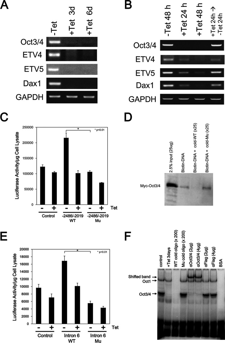
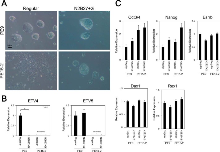
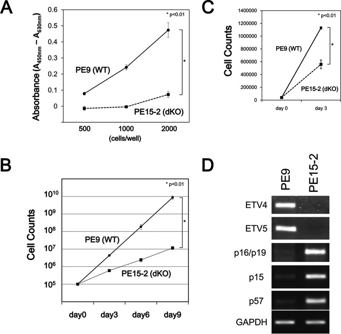
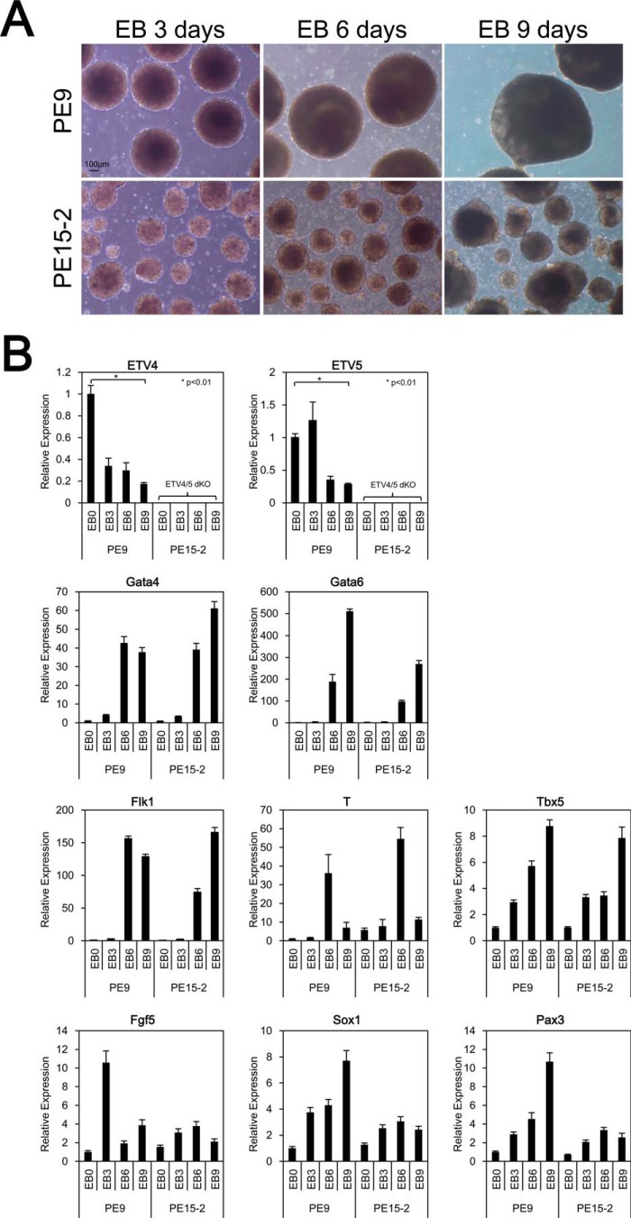
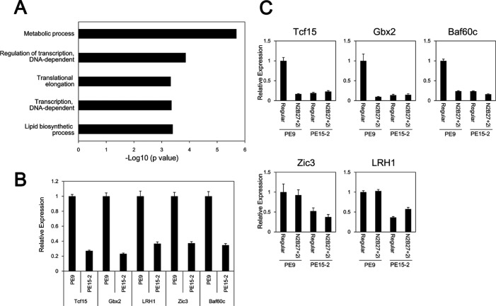
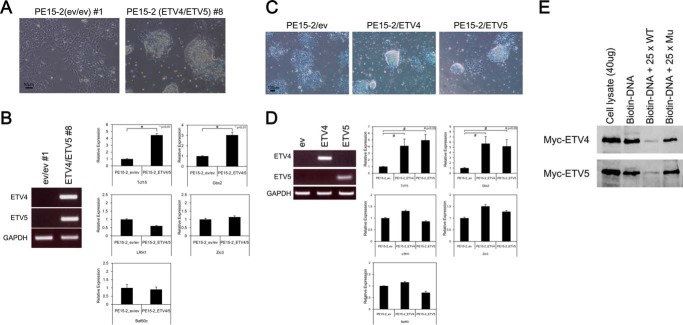
The pluripotency and self-renewal capacity of embryonic stem (ES) cells is regulated by several transcription factors. Here, we show that the ETS-related transcription factors Etv4 and Etv5 (Etv4/5) are specifically expressed in undifferentiated ES cells, and suppression of Oct3/4 results in down-regulation of Etv4/5. Simultaneous deletion of Etv4 and Etv5 (Etv4/5 double knock-out (dKO)) in ES cells resulted in a flat, epithelial cell-like appearance, whereas the morphology changed into compact colonies in a 2i medium (containing two inhibitors for GSK3 and MEK/ERK). Expression levels of self-renewal marker genes, including Oct3/4 and Nanog, were similar between wild-type and dKO ES cells, whereas proliferation of Etv4/5 dKO ES cells was decreased with overexpression of cyclin-dependent kinase inhibitors (p16/p19, p15, and p57). A differentiation assay revealed that the embryoid bodies derived from Etv4/5 dKO ES cells were smaller than the control, and expression of ectoderm marker genes, including Fgf5, Sox1, and Pax3, was not induced in dKO-derived embryoid bodies. Microarray analysis demonstrated that stem cell-related genes, including Tcf15, Gbx2, Lrh1, Zic3, and Baf60c, were significantly repressed in Etv4/5 dKO ES cells. The artificial expression of Etv4 and/or Etv5 in Etv4/5 dKO ES cells induced re-expression of Tcf15 and Gbx2. These results indicate that Etv4 and Etv5, potentially through regulation of Gbx2 and Tcf15, are involved in the ES cell proliferation and induction of differentiation-associated genes in ES cells.

Keywords: cell proliferation; differentiation; embryonic stem cell; gene regulation; oncogene; transcription factor.


Figures

FIGURE 1.
FIGURE 1.
Expression of Etv4 and Etv5 mRNA in ZHBTc4 ES cells. A, reduction of Etv4 and Etv5 mRNA expression after repression of Oct3/4 expression. ZHBTc4 ES cells were cultured with or without 1 μg/ml Tet for 3 or 6 days. Expression of the indicated genes was examined by RT-PCR. B, induction of Etv4 and Etv5 mRNA expression by Oct3/4 expression. ZHBTc4 ES cells were cultured with or without 1 μg/ml Tet for 24–48 h. To restore Oct3/4 expression, the culture medium of Tet-treated cells was changed to a Tet-free medium, and the cells were cultured for another 24 h. Dax1 was used for a positive control as an OCT3/4 target gene. Gapdh was used as an internal control. All results are representative of three independent experiments. C, determination of an OCT3/4-responsive element in the Etv4 gene by a luciferase assay. The promoter region of the Etv4 gene contains a consensus sequence for an OCT3/4 binding site (ATTTGCAT). ZHBTc4 ES cells were transfected with either pGL4-promoter (control), pGL4-promoter-Etv4 (−2486/−2019)-wild type (−2486/−2019 WT), or pGL4-promoter-Etv4 (−2486/−2019)-mutant (−2486/−2019 Mu). ZHBTc4 ES cells were divided into two dishes 24 h after transfection and cultured with (+) or without (−) 1 μg/ml Tet for 24 h, and luciferase activity was measured. The sequence of the OCT3/4-responsive element was changed from ATTTGCAT to GACGTGGG. Shown are means (bars) and S.D. (error bars) of triplicate assays. D, OCT3/4 directly binds to the putative binding sequences. The association of DNA and OCT3/4 was examined by a biotin-labeled DNA pull-down assay. Biotin-labeled oligonucleotide containing the putative binding site was incubated with cell extracts from Myc-Oct3/4-transfected HEK293 cells either with or without 25-fold non-labeled WT or mutated (Mu) oligonucleotide. The result shown is representative of three separate experiments. E, determination of an OCT3/4-responsive element in the Etv5 gene by a luciferase assay. The intron 6 region of the Etv5 gene contains the consensus sequence of an OCT3/4 binding site (ATGCAAAT). ZHBTc4 ES cells were transfected with either pGL4-promoter (control), pGL4-promoter-Etv5 intron 6-wild type (Intron 6 WT), or pGL4-promoter-ETV5 intron 6-mutant (Intron 6 Mu). ZHBTc4 ES cells were divided into two dishes 24 h after transfection and cultured with (+) or without (−) 1 μg/ml Tet for 24 h, and luciferase activity was measured. The sequence of the OCT3/4-responsive element was changed from ATGCAAAT to GTGCCGCT. Shown are the means (bars) and S.D. (error bars) of triplicate assays. F, OCT3/4 directly binds to the putative binding sequences. An association of DNA and OCT3/4 was examined by an EMSA. Radiolabeled oligonucleotide containing the putative binding site was incubated with nuclear extracts from ZHBTc4 ES cells either with or without 200-fold cold WT or cold mutated (Mu) oligonucleotides, an anti-OCT3/4 antibody, or an anti-FLAG antibody. The result shown is representative of three separate experiments. Note that a band of OCT1-DNA complex can be detected in this assay as described before (66).
FIGURE 2.
FIGURE 2.
Etv4/5 dKO ES cells express self-renewal marker genes. A, morphological signatures of PE9 (WT) and PE15-2 (Etv4/5 dKO) ES cells. PE9 and PE15-2 ES cells were cultured in the presence or absence of LIF for 3 days. Morphological signatures were observed. B, expression of self-renewal marker genes in PE9 and PE15-2 ES cells. Expression of the indicated self-renewal marker genes in the conditions of A in PE9 and PE15-2 ES cells was examined by quantitative RT-PCR. Expression of Etv4 and Etv5 mRNA was only shown in PE9 ES cells. All samples were analyzed in triplicate, and the data were normalized to Gapdh expression. C–E, immunofluorescent stain in PE9 and PE15-2 ES cells. Protein expression of the self-renewal marker genes, including NANOG (C), OCT3/4 (D), and ESRRB (E), in PE9 and PE15-2 ES cells was examined by immunofluorescent stain. Nuclei were stained with the Hoechst stain. F, Western blot analysis in PE9 and PE15-2 ES cells. Protein expression of the self-renewal marker genes (NANOG, OCT3/4, and ESRRB) was examined by Western blot analysis. α-Tubulin was used as a loading control. G, determination of alkaline phosphatase activity. Relative activity of alkaline phosphatase was determined in PE9 and PE15-2 ES cells. Shown are the means (bars) and S.D. (error bars) of triplicate plates. H, alkaline phosphatase stain in PE9 and PE15-2 ES cells. Alkaline phosphatase of ES cells was detected by the VECTOR Blue alkaline phosphatase substrate kit. Morphological signatures were observed. All results are representative of several independent experiments.
FIGURE 3.
FIGURE 3.
Etv4/5 dKO ES cells maintain the undifferentiated state in N2B27 serum-free medium plus two inhibitors. A, morphological signatures of PE9 (WT) and PE15-2 (Etv4/5 dKO) ES cells. PE9 and PE15-2 ES cells were cultured in either a regular serum- and LIF-containing medium or a chemically defined medium (DMEM/F-12 with N2B27 supplement plus 2i (mitogen-activated protein kinase/extracellular signal-regulated kinase (PD0325901) and glycogen synthase kinase 3 (CHIR99021) inhibitors) (2i condition)). Morphological signatures were observed. B, expression of Etv4 and Etv5 in the 2i condition. Expression of Etv4 and Etv5 in the conditions of A in PE9 and PE15-2 ES cells was examined by quantitative RT-PCR. C, expression of self-renewal marker genes in PE9 and PE15-2 ES cells. Expression of the indicated self-renewal marker genes in the conditions of A in PE9 and PE15-2 ES cells was examined by quantitative RT-PCR. All samples were analyzed in triplicate, and the data were normalized to Gapdh expression. Error bars, S.D.
FIGURE 4.
FIGURE 4.
ETV4 and ETV5 are involved in the proliferation of ES cells. A, proliferation of PE9 (WT) and PE15-2 (Etv4/5 dKO) ES cells. Either PE9 or PE15-2 ES cells were seeded at 500, 1000, or 2000 cells/well in a 96-well dish and subjected to a WST-1 assay. The results represent the means and S.D. (error bars) of triplicate assays. B, growth curves of PE9 and PE15-2 ES cells. PE9 and PE15-2 ES cells were cultured in the presence of LIF, and the cells were counted at the indicated days. The results represent the means and S.D. of four independent assays. C, direct cell counts of PE9 and PE15-2 ES cells in the 2i condition. PE9 and PE15-2 ES cells were cultured in the 2i condition, and the cells were counted at the indicated days. The results represent the means and S.D. of triplicate experiments. D, enhanced expression of cyclin-dependent kinase inhibitors in Etv4/5 dKO ES cells. Expression of cyclin-dependent kinase inhibitors, p16/p19, p15, and p57 mRNA in PE9 and PE15-2 ES cells was examined by RT-PCR. Gapdh was used as an internal control. The results are representative of three independent experiments.
FIGURE 5.
FIGURE 5.
Etv4/5 dKO ES cells have impaired abilities to induce ectoderm marker gene expression. A, morphological signatures of EBs derived from PE9 (WT) and PE15-2 (Etv4/5 dKO) ES cells. PE9 and PE15-2 ES cells were cultured in suspension without LIF for 3–9 days. Morphological signatures were observed. Scale bar, 100 μm. B, expression of differentiation marker genes in PE9 and PE15-2 EBs. Expression of the indicated differentiation marker genes in the conditions of A in PE9 and PE15-2 EBs was examined by quantitative RT-PCR. Expression of Etv4 and Etv5 mRNA was only shown in PE9 ES cells. All samples were analyzed in triplicate, and the data were normalized to Gapdh expression. Error bars, S.D.
FIGURE 6.
FIGURE 6.
Several transcription factors are down-regulated in Etv4/5 dKO ES cells. A, GO term analysis of down-regulated candidates in Etv4/5 dKO ES cells. Genes involved in metabolic processes; regulation of transcription, DNA-dependent; translation elongation; transcription, DNA-dependent; and lipid biosynthetic are enriched significantly. GO categories are ranked according to their p value. B, expression of several transcription-related genes in PE9 and PE15-2 ES cells. Expression of transcription-related genes, including Tcf15, Gbx2, Lrh1, Zic3, and Baf60c, is down-regulated in PE15-2 ES cells, as shown by microarray analysis. Expression of the indicated genes in PE9 and PE15-2 ES cells was examined by quantitative RT-PCR. C, expression of several transcription-related genes in PE9 and PE15-2 ES cells in the 2i condition. PE9 and PE15-2 ES cells were cultured in the 2i condition, and expression of the indicated genes was examined by quantitative RT-PCR. All samples were analyzed in triplicate, and the data were normalized to Gapdh expression. Error bars, S.D.
FIGURE 7.
FIGURE 7.
Both ETV4 and ETV5 are able to rescue the epithelial like morphology and expression of Tcf15 and Gbx2 in dKO ES cells. A, overexpression of both ETV4 and ETV5 in PE15-2 (Etv4/5 dKO) ES cells. ETV4/5-expressing PE15-2 ES cells and control cells were established as described under “Experimental Procedures.” Morphological signatures were observed. Scale bar, 50 μm. B, induction of Tcf15 and Gbx2 expression in ETV4/5-expressing PE15-2 ES cells. Expression of Tcf15 and Gbx2 mRNA, as well as Lrh1, Zic3, and Baf60c mRNA, in the PE15-2 ES cell lines was examined by quantitative RT-PCR. All samples were analyzed in triplicate, and the data were normalized to Gapdh expression. Expression of Etv4 and Etv5 mRNA was examined by regular RT-PCR. C, overexpression of either ETV4 or ETV5 in PE15-2 (Etv4/5 dKO) ES cells. ETV4- or ETV5-expressing PE15-2 ES cells and control cells were established as described under “Experimental Procedures.” Morphological signatures were observed. Scale bar, 100 μm. D, induction of Tcf15 and Gbx2 expression in ETV4- and ETV5-expressing PE15-2 ES cells. Expression of Tcf15 and Gbx2 mRNA as well as Lrh1, Zic3, and Baf60c mRNA in the PE15-2 ES cell lines was examined by quantitative RT-PCR. All samples were analyzed in triplicate, and the data were normalized to Gapdh expression. Expression of Etv4 and Etv5 mRNA was examined by regular RT-PCR. E, ETV4 and ETV5 directly bind to the putative binding sequences of the Gbx2 gene. Biotin-labeled oligonucleotide containing the putative binding site was incubated with cell extracts from Myc-ETV4- or Myc-ETV5-transfected HEK293 cells either with or without 25-fold non-labeled WT or mutated (Mu) oligonucleotide. The result shown is representative of three separate experiments. Error bars, S.D.

Similar articles

Cited by

References

    1. Ying Q. L., Nichols J., Chambers I., Smith A. (2003) BMP induction of Id proteins suppresses differentiation and sustains embryonic stem cell self-renewal in collaboration with STAT3. Cell 115, 281–292 - PubMed
    1. Smith A. G., Heath J. K., Donaldson D. D., Wong G. G., Moreau J., Stahl M., Rogers D. (1988) Inhibition of pluripotential embryonic stem cell differentiation by purified polypeptides. Nature 336, 688–690 - PubMed
    1. Williams R. L., Hilton D. J., Pease S., Willson T. A., Stewart C. L., Gearing D. P., Wagner E. F., Metcalf D., Nicola N. A., Gough N. M. (1988) Myeloid leukaemia inhibitory factor maintains the developmental potential of embryonic stem cells. Nature 336, 684–687 - PubMed
    1. Niwa H., Ogawa K., Shimosato D., Adachi K. (2009) A parallel circuit of LIF signalling pathways maintains pluripotency of mouse ES cells. Nature 460, 118–122 - PubMed
    1. Matsuda T., Nakamura T., Nakao K., Arai T., Katsuki M., Heike T., Yokota T. (1999) STAT3 activation is sufficient to maintain an undifferentiated state of mouse embryonic stem cells. EMBO J. 18, 4261–4269 - PMC - PubMed

Publication types

MeSH terms