. 2015 Oct 2;290(40):24051-65.
doi: 10.1074/jbc.M115.660357. Epub 2015 Jul 29.

Evidence that the DNA mismatch repair system removes 1-nucleotide Okazaki fragment flaps

Affiliations

Evidence that the DNA mismatch repair system removes 1-nucleotide Okazaki fragment flaps

Lyudmila Y Kadyrova et al. J Biol Chem. .

Abstract

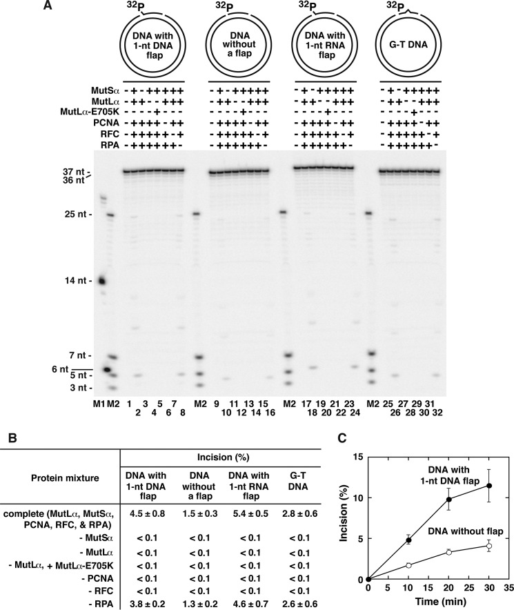
The DNA mismatch repair (MMR) system plays a major role in promoting genome stability and suppressing carcinogenesis. In this work, we investigated whether the MMR system is involved in Okazaki fragment maturation. We found that in the yeast Saccharomyces cerevisiae, the MMR system and the flap endonuclease Rad27 act in overlapping pathways that protect the nuclear genome from 1-bp insertions. In addition, we determined that purified yeast and human MutSα proteins recognize 1-nucleotide DNA and RNA flaps. In reconstituted human systems, MutSα, proliferating cell nuclear antigen, and replication factor C activate MutLα endonuclease to remove the flaps. ATPase and endonuclease mutants of MutLα are defective in the flap removal. These results suggest that the MMR system contributes to the removal of 1-nucleotide Okazaki fragment flaps.

Keywords: DNA endonuclease; DNA mismatch repair; DNA replication; Okazaki fragment maturation; cancer; genomic instability; mutL homolog 1 (MLH1).


Figures

FIGURE 1.
FIGURE 1.
CAN1 mutation rates and can1 mutation spectra in the wild-type, msh2Δ, rad27Δ, and msh2Δ rad27Δ strains. A, CAN1 mutation rates. Each of the mutants was made in the two different wild-type backgrounds: E134 and BY4742. The numbers above the bars are the relative mutation rates. B, can1 mutation spectra in the wild-type strain E134 and its mutant derivatives. The relative mutation rates are in parentheses. a, all 1-bp insertions were formed in mononucleotide runs that were ≥N2.
FIGURE 2.
FIGURE 2.
Human and yeast MutSα proteins recognize 1-nt DNA flaps. The gel mobility shift assays with the oligonucleotide-based DNA substrates and calculations of the apparent Kd values were performed as described under “Experimental Procedures.” All six substrates had the same bottom strand. The DNA sequences of the homoduplex and nicked DNA substrates were identical to each other and to the his7-2 sequence, in which the majority of +1 frameshifts are formed. Compared with the top strand of the homoduplex or nicked substrate, the top strands of the flapped and 1-nt insertion substrates each contained an extra nucleotide residue, which was necessary to produce the 1-nt flap or 1-nt insertion. A, representative images showing binding of yeast MutSα to the different DNA substrates. Each DNA-binding reaction was carried out in the mixture containing the indicated concentration of yeast MutSα and the indicated DNA substrate (2 nm). B and C, apparent Kd values for binding of yeast MutSα (B) and human MutSα (C) to the indicated DNA substrates. The apparent Kd values were calculated using the data that were obtained by quantification of images, including those shown in A. The numbers above the bars are the apparent Kd values.
FIGURE 3.
FIGURE 3.
Human MutSα recognizes 1-nt DNA and RNA flaps on 2-kb circular DNA molecules. A, diagrams of the 2-kb circular DNAs used in the DNA-binding reactions. Each diagram also shows the relative position of the hybridization probe (bar with an asterisk). The hybridization probe is complementary to the continuous strand. B, apparent Kd values for binding of human MutSα to the indicated circular substrates. The numbers above the bars are the apparent Kd values. The gel mobility shift assays and calculations of the apparent Kd values were carried out as detailed under “Experimental Procedures.”
FIGURE 4.
FIGURE 4.
1-nt DNA and RNA flaps activate MutLα endonuclease to incise the discontinuous strands in the presence of MutSα, PCNA, RFC, and RPA. Each DNA incision reaction was carried out in the mixture containing the indicated human proteins and DNA substrate (1.5 nm). When MutSα, MutLα, MutLα-E705K, PCNA, RFC, and RPA were present in the reaction mixtures, their concentrations were 40, 16, 16, 24, 4, and 40 nm, respectively. After a 10-min incubation, the reactions were stopped by the addition of NaOH and EDTA to the final concentrations of 40 and 5 mm, respectively. The reaction products were separated on alkaline 1.2% agarose gels, transferred onto nylon membranes, hybridized with 32P-labeled oligonucleotide 16, and visualized by phosphorimaging. A, representative images showing incision of the discontinuous strands in the presence of MutLα, MutSα, PCNA, RFC, and RPA. The diagrams outline the circular DNA substrates. Each diagram also shows the relative position of the hybridization probe (bar with an asterisk). The hybridization probe is complementary to the discontinuous strand. B, summary of incision of the discontinuous strands of the indicated DNA substrates at sites that are 4-nt 3′ to the flap or control nick. The data were obtained by quantification of images, including those shown in A, and are presented as averages ± 1 S.D., n ≥3.
FIGURE 5.
FIGURE 5.
MutLα endonuclease incises the discontinuous strand four nucleotides downstream from a 1-nt DNA or RNA flap. The 37-nt fragments of the 1-nt DNA and RNA flap-containing substrates and the 36-nt fragments of the control flap-free and G-T substrates were labeled at their 5′ ends with 32P. Each DNA incision reaction was performed in the mixture containing the indicated human proteins and 32P-labeled DNA substrate (1.5 nm). When MutSα, MutLα, MutLα-E705K, PCNA, RFC, and RPA were present in the reaction mixtures, their concentrations were 40, 16, 16, 24, 4, and 40 nm, respectively. The DNA incision reactions were stopped and analyzed as described under “Experimental Procedures.” A, representative image showing MutLα endonuclease-dependent incision of the discontinuous strand 4 nt downstream from the 1-nt flap. The incision reactions were incubated for 10 min. The diagrams outline the circular DNA substrates. B, summary of incision of the discontinuous strands of the indicated substrates at sites that are 4-nt 3′ to the flap or control nick. The DNA incision reactions were incubated for 10 min. C, time course of incision of the discontinuous strands of the indicated substrates at sites that are 4-nt 3′ to the flap or control nick. The incision reactions were carried out in the mixtures containing MutSα (40 nm), MutLα (16 nm), PCNA (24 nm), RFC (4 nm), RPA (40 nm), and the indicated DNA substrate (1.5 nm). The data in B and C are averages ± 1 S.D. (B, n ≥4; C, n ≥3) and were obtained by quantification of images, including the one shown in A.
FIGURE 6.
FIGURE 6.
CAF-1-dependent histone H3-H4 deposition stimulates the removal of 1-nt flaps by the activated MutLα endonuclease. The 37-nt fragment of the 1-nt DNA flap-containing substrate and the 36-nt fragment of the control flap-free substrate were labeled at their 5′ ends with 32P. Each DNA incision reaction was performed in the mixture containing the indicated human proteins and 32P-labeled DNA substrate (1.5 nm). When MutSα, MutLα, MutLα-D699N, MutLα-EA, PCNA, RFC, RPA, CAF-1, and the histone H3-H4 heterodimer were present in the reaction mixtures, their concentrations were 40, 16, 16, 16, 24, 4, 40, 24, and 88 nm, respectively. The reactions were incubated for 30 min and then stopped and analyzed as described under “Experimental Procedures.” A, representative image showing the effects of the indicated protein combinations on incision of the discontinuous strands of the indicated substrates at sites that are 4-nt 3′ from the flap or control nick. The diagrams outline the circular DNA substrates. B, graphical representation of the effects of the indicated protein combinations on incision of the discontinuous strands of the indicated substrates at sites that are 4-nt 3′ from the flap or control nick. The data were obtained by quantification of images, including the one shown in A and are averages ± 1 S.D., n ≥4. C, dependence of the incision on the presence of the 1-nt DNA flap. The flap dependence values were calculated from the data shown in B. The presence of a statistically significant difference between the flap dependences of the two indicated reactions was identified by unpaired t test.
FIGURE 7.
FIGURE 7.
CAF-1-dependent histone H3-H4 deposition protects the remote sites from incision by MutLα endonuclease. Each DNA incision reaction was performed in the mixture containing the indicated human proteins and DNA substrate (1.5 nm). When MutSα, MutLα, PCNA, RFC, RPA, and CAF-1 were present in the reaction mixtures, their concentrations were 40, 16, 24, 4, 40, and 24 nm, respectively. After a 30-min incubation, the incision reactions were stopped and analyzed as described in Fig. 4. A, image showing the effects of the different protein combinations on incision of the discontinuous strands of the 1-nt flap-containing and flap-free DNA substrates. The diagrams outline the DNA substrates. Each diagram also shows the relative position of the hybridization probe (bar with an asterisk), which is complementary to the discontinuous strand. B and C, incision of the discontinuous strands of the 1-nt flap-containing and flap-free DNA substrates as a function of concentration of histone H3-H4 heterodimers. The data were obtained by quantification of images, including the one shown in A, and are presented as averages ± 1 S.D., n = 2.
FIGURE 8.
FIGURE 8.
Flap removal in a reconstituted human system containing FEN1 and MutLα endonucleases. The DNA incision reactions were carried out in the mixtures containing the indicated human proteins and 32P-labeled circular DNA substrate (1.5 nm). When MutSα, MutLα, PCNA, RFC, RPA, CAF-1, and the histone H3-H4 heterodimer were present in the reaction mixtures, their concentrations were 40, 16, 24, 4, 40, 24, and 88 nm, respectively. After incubation for 10 min, the DNA incision reactions were stopped and analyzed as described under “Experimental Procedures.” A, representative image showing the effects of the different protein combinations on the removal of the 1-nt DNA flaps. The arrows indicate the positions of the 1- and 5-nt cleavage products generated by FEN1 and MutLα, respectively. The diagram outlines the circular DNA substrate. B, graphical representation of the effects of the different FEN1 concentrations on the yield of the product of MutLα endonuclease-dependent flap removal in the eight-protein system. The eight-protein system contained MutLα (16 nm), MutSα (40 nm), PCNA (24 nm), RFC (4 nm), RPA (40 nm), CAF-1 (24 nm), histone H3-H4 heterodimer (88 nm), and FEN1 (0.3, 0.6, 1.2, or 2.4 nm). C, graphical representation of the effects of the different FEN1 concentrations on the yield of the product of FEN1-dependent flap removal in the one-protein and eight-protein systems. The one-protein system contained FEN1 (0.3, 0.6, 1.2, or 2.4 nm). The data in B and C were obtained by quantification of images, including the one shown in A and are averages ± 1 S.D., n ≥4.
FIGURE 9.
FIGURE 9.
Role for the MMR system in DNA replication. The model suggests that the MMR system supports DNA replication by removing 1-nt Okazaki fragment flaps. The process of the removal of a 1-nt Okazaki fragment flap by the MMR system is initiated by the recognition of the flap by MutSα. In the next step, MutSα acts in conjunction with PCNA and RFC to activate MutLα endonuclease. The activated MutLα endonuclease then removes the flap.

Similar articles

Cited by

References

    1. Modrich P. (1995) Mismatch repair, genetic stability and tumour avoidance. Philos. Trans. R. Soc. Lond. B Biol. Sci. 347, 89–95 - PubMed
    1. Harfe B. D., Jinks-Robertson S. (2000) DNA mismatch repair and genetic instability. Annu. Rev. Genet. 34, 359–399 - PubMed
    1. Kunkel T. A., Erie D. A. (2005) DNA mismatch repair. Annu. Rev. Biochem. 74, 681–710 - PubMed
    1. Li G. M. (2008) Mechanisms and functions of DNA mismatch repair. Cell Res. 18, 85–98 - PubMed
    1. Jiricny J. (2013) Postreplicative mismatch repair. Cold Spring Harb. Perspect. Biol. 5, a012633. - PMC - PubMed

Publication types

MeSH terms

LinkOut - more resources