Case Reports
. 2015 Sep 24;126(13):1527-35.
doi: 10.1182/blood-2014-12-611012. Epub 2015 Jul 29.

Immunodeficiency and severe susceptibility to bacterial infection associated with a loss-of-function homozygous mutation of MKL1

Affiliations
Case Reports

Immunodeficiency and severe susceptibility to bacterial infection associated with a loss-of-function homozygous mutation of MKL1

Julien Record et al. Blood. .

Abstract

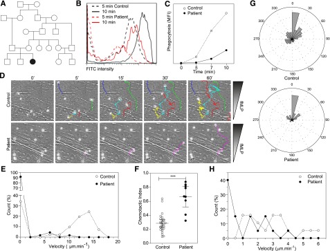
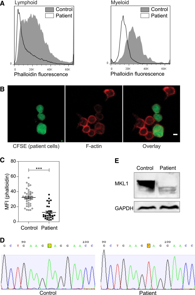
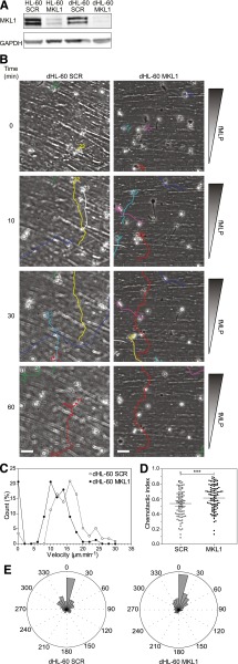
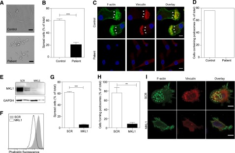
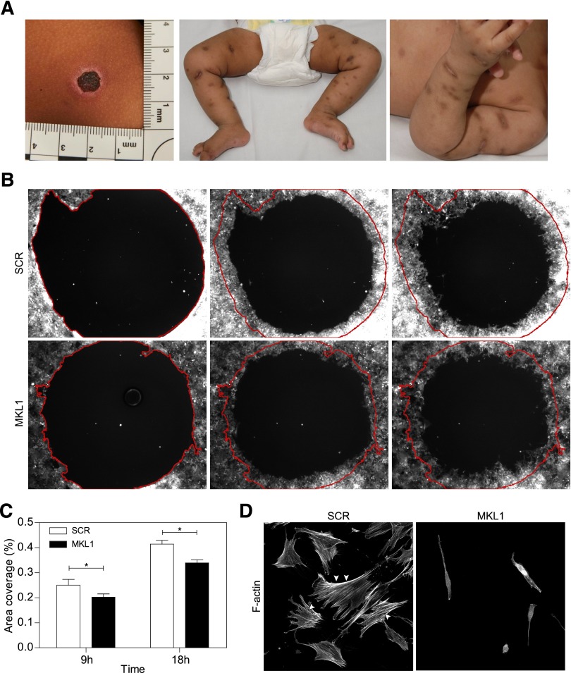
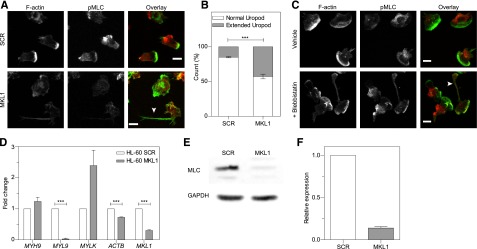
Megakaryoblastic leukemia 1 (MKL1), also known as MAL or myocardin-related transcription factor A (MRTF-A), is a coactivator of serum response factor, which regulates transcription of actin and actin cytoskeleton-related genes. MKL1 is known to be important for megakaryocyte differentiation and function in mice, but its role in immune cells is unexplored. Here we report a patient with a homozygous nonsense mutation in the MKL1 gene resulting in immunodeficiency characterized predominantly by susceptibility to severe bacterial infection. We show that loss of MKL1 protein expression causes a dramatic loss of filamentous actin (F-actin) content in lymphoid and myeloid lineage immune cells and widespread cytoskeletal dysfunction. MKL1-deficient neutrophils displayed reduced phagocytosis and almost complete abrogation of migration in vitro. Similarly, primary dendritic cells were unable to spread normally or to form podosomes. Silencing of MKL1 in myeloid cell lines revealed that F-actin assembly was abrogated through reduction of globular actin (G-actin) levels and disturbed expression of multiple actin-regulating genes. Impaired migration of these cells was associated with failure of uropod retraction likely due to altered contractility and adhesion, evidenced by reduced expression of the myosin light chain 9 (MYL9) component of myosin II complex and overexpression of CD11b integrin. Together, our results show that MKL1 is a nonredundant regulator of cytoskeleton-associated functions in immune cells and fibroblasts and that its depletion underlies a novel human primary immunodeficiency.


Figures

Figure 1
Figure 1
Patient neutrophils display phagocytosis and migration defects. (A) Family tree. (B-C) Fluorescein isothiocyanate (FITC) –positive Escherichia coli uptake by neutrophils analyzed by flow cytometry. Representative data from 2 independent experiments. (D) Migration of control and patient neutrophils in a Dunn chamber in response to an fMLP gradient. Tracks show migration path over time. The number of cells analyzed was 41 in the control and 110 and patient sample in 1 experiment. Scale bar = 10 μm. (E) Frequency distribution of the velocity of patient and control neutrophils. (F) Chemotactic index of the migrating neutrophils. (G) Distribution of instantaneous angle between the gradient and the cell path during migration. (H) Frequency distribution of the velocity of control and patient B lymphoblastoid cell lines migrating in collagen gel. Twenty cells were analyzed in each population in 1 experiment. Data are representative of 2 independent experiments. *** P < .001. MFI, mean fluorescent intensity.
Figure 2
Figure 2
Effect of MKL1 mutation on F-actin. (A) F-actin (Alexa Fluor 647 phalloidin) content in lymphoid and myeloid cells was analyzed by flow cytometry. Patient cells were stained with carboxyfluorescein diacetate succinimidyl ester (CFSE) and mixed with unstained control cells before fixation and staining. Representative data from 2 experiments. (B) Confocal images of patient cells (CFSE, green), F-actin (Alexa Fluor 647 phalloidin, red), and mixed with control cells (unstained). Scale bar = 5 μm. Data were analyzed from 1 experiment. (C) Quantification of the phalloidin MFI observed in (B). (D) Sequence analysis of control and patient samples leading to the identification of the MKL1 mutation. (E) MKL1 expression visualized by western blot in control and patient peripheral blood mononuclear cells (PBMCs). ***P < .001. GAPDH, glyceraldehyde-3-phosphate dehydrogenase.
Figure 3
Figure 3
Knockdown of MKL1 in HL-60 neutrophil-like cells mimics patient phenotype. HL-60 cells were differentiated into neutrophil-like cells for 5 days in culture media containing 1.3% dimethylsulfoxide. (A) Western blot of the MKL1 expression levels in undifferentiated (HL-60) or differentiated (dHL-60) cells carrying SCR or the shRNA against MKL1 (MKL1). (B) Migration of SCR and MKL1 neutrophils in a Dunn chamber in response to an fMLP gradient. Tracks show migration path over time. Scale bar = 20 μm. (C) Frequency distribution of the velocity of dHL-60 SCR and MKL1 cells during migration in a Dunn chamber in response to fMLP. The number of cells analyzed was 115 for SCR and 156 for MKL1 cells in 3 independent experiments. (D) Chemotactic index of dHL-60 SCR and dHL-60 MKL1 cells during migration in a Dunn chamber. (E) Instant angle distribution during the migration of SCR and MKL1 dHL-60 cells. ***P < .001.
Figure 4
Figure 4
Patient PBMC-derived DCs have morphologic abnormalities and severely reduced F-actin content. DCs were left to adhere to fibronectin-coated glass coverslips for 4 hours and were then fixed in 4% paraformaldehyde. (A) Phase contrast imaging shows poor spreading of patient DCs compared with controls. Scale bar = 20 μm. (B) Quantification of the percent of cells that had spread. (C-D) Cells were stained for DNA (4,6 diamidino-2-phenylindole, blue), F-actin (Alexa Fluor 488 phalloidin, green), and vinculin (red), and images were acquired by confocal microscopy. Podosome structures were clearly present in control cells (F-actin cores surrounded by vinculin in 22 of 29 cells; white arrows) but completely absent in patient cells (0 of 121 cells). Data were acquired in a single experiment. Scale bar = 10 μm. (E) Western blot of MKL1 in THP1 cells expressing SCR (THP1 SCR) and against MKL1 (THP1 MKL1). (F) F-actin content in THP1 SCR and THP1 MKL1 DCs analyzed by flow cytometry. (G) Quantification of the spreading of THP1 SCR and THP1 MKL1 DCs. (H-I) Confocal images of F-actin- and vinculin-stained cells demonstrate the lack of podosomes in THP1 MKL1 DCs. Scale bar = 10 μm. THP1 MKL1 DC data were acquired in 3 independent experiments. **P < 0.01; ***P < .001.
Figure 5
Figure 5
Knockdown of MKL1 alters fibroblast morphology and migration. (A) Clinical photos showing skin scarring and hyperpigmentation after treating skin abscesses with antibiotics. (B) Migration of fibroblasts expressing SCR and MKL1 shRNA at 0, 9, and 18 hours. (C) Quantification of the area of migration observed in (B). (D) Confocal images of fibroblasts expressing SCR and MKL1 shRNA. Cells were stained with Alexa Fluor 647 phalloidin. White arrows indicate stress fibers. *P < .05.
Figure 6
Figure 6
MKL1-deficient dHL-60 cells show lower content in F-actin. (A) Analysis of the F-actin content in dHL-60 MKL1 cells by flow cytometry. dHL-60 wild-type (WT) cells and dHL-60 cells carrying a scrambled or anti-MKL1 shRNA and coexpressing green fluorescent protein were mixed before fixation and staining for F-actin with Alexa Fluor 647 phalloidin. For clarity, only SCR and MKL1 populations are displayed on the flow cytometry graph. (B-C) Evaluation of the F-actin content in dHL-60 cells after stimulation with 100 nM fMLP for 1 minute in SCR and MKL1 populations, respectively. Data from 3 independent experiments were analyzed. (D) G-actin and F-actin content in dHL-60 SCR and MKL1 cells. (E-F) Analysis of the F-actin content in dHL-60 cells after stimulation with 100 nM fMLP for 1 minute in WT, MKL1, and MKL1 cells expressing mCherry G-actin. *P < .05; **P < .01; ***P < .001.
Figure 7
Figure 7
MKL1-deficient dHL-60 cells show impaired uropod retraction associated with downregulation of MYL9. (A) Knockdown of MKL1 induces a uropod retraction defect. dHL-60 cells were left to adhere to fibronectin-coated glass coverslips for 20 minutes, washed, then uniformly stimulated with fMLP for 2 minutes and fixed in 4% paraformaldehyde. Cells were then stained for F-actin and phosphorylation of MLC (pMLC). The dHL-60 MKL1 F-actin staining intensity was increased threefold in the merged (overlay) image. Images were taken with an ×40 lens. White arrow indicates extended uropod. (B) Quantification of the frequency of uropod extension in dHL-60 MKL1 cells observed in (A). More than 250 cells were analyzed in each condition in 2 independent experiments. (C) Inhibition of myosin II interaction with F-actin using blebbistatin (100 µM) induced a uropod retraction defect in dHL-60 WT cells. Images were taken with a ×63 lens. Scale bars = 10 μm. (D) mRNA expression in HL-60 SCR and MKL1 cells analyzed by qRT-PCR. (E-F) Myosin light chain expression was evaluated by western blot. ***P < .001.

Comment in

References

    1. Bouma G, Ancliff PJ, Thrasher AJ, Burns SO. Recent advances in the understanding of genetic defects of neutrophil number and function. Br J Haematol. 2010;151(4):312–326. - PubMed
    1. Ambruso DR, Knall C, Abell AN, et al. Human neutrophil immunodeficiency syndrome is associated with an inhibitory Rac2 mutation. Proc Natl Acad Sci USA. 2000;97(9):4654–4659. - PMC - PubMed
    1. Olson EN, Nordheim A. Linking actin dynamics and gene transcription to drive cellular motile functions. Nat Rev Mol Cell Biol. 2010;11(5):353–365. - PMC - PubMed
    1. Kalita K, Kuzniewska B, Kaczmarek L. MKLs: co-factors of serum response factor (SRF) in neuronal responses. Int J Biochem Cell Biol. 2012;44(9):1444–1447. - PubMed
    1. Miralles F, Posern G, Zaromytidou A-I, Treisman R. Actin dynamics control SRF activity by regulation of its coactivator MAL. Cell. 2003;113(3):329–342. - PubMed

Publication types

MeSH terms