. 2015 Jul 29;35(30):10731-49.
doi: 10.1523/JNEUROSCI.0614-15.2015.

Trib3 Is Elevated in Parkinson's Disease and Mediates Death in Parkinson's Disease Models

Affiliations

Trib3 Is Elevated in Parkinson's Disease and Mediates Death in Parkinson's Disease Models

Pascaline Aimé et al. J Neurosci. .

Abstract

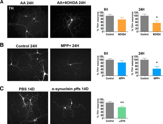
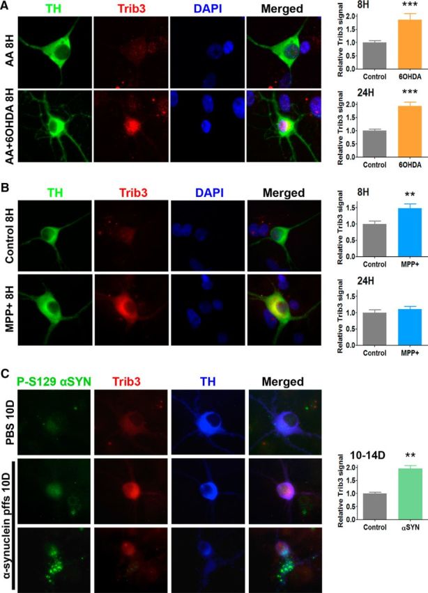
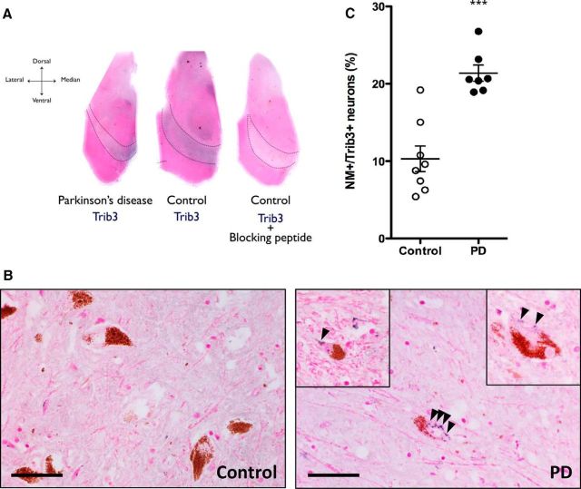
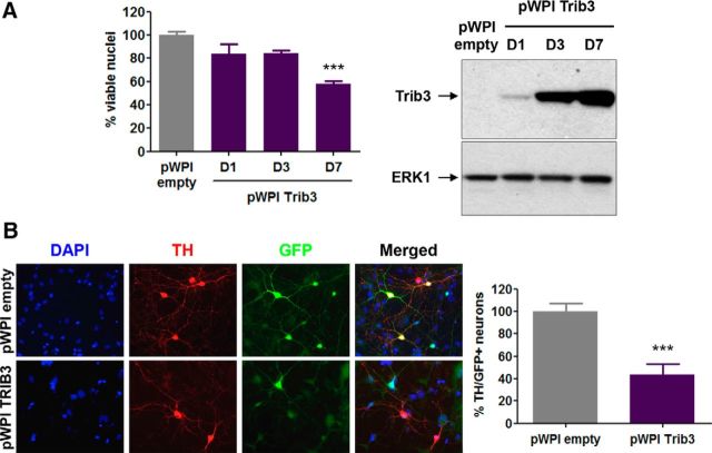
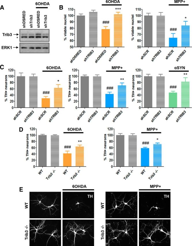
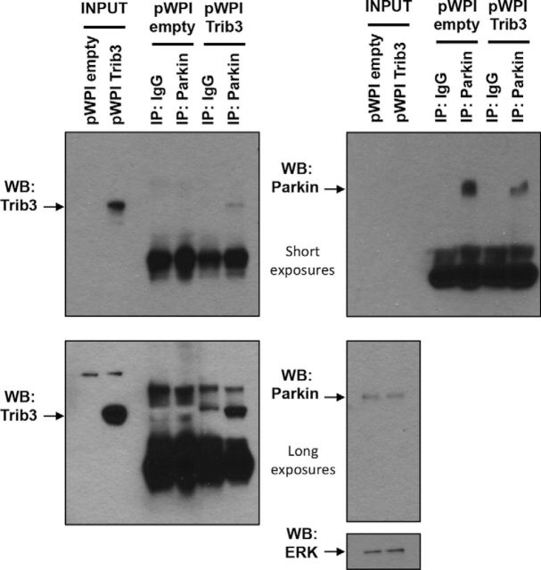
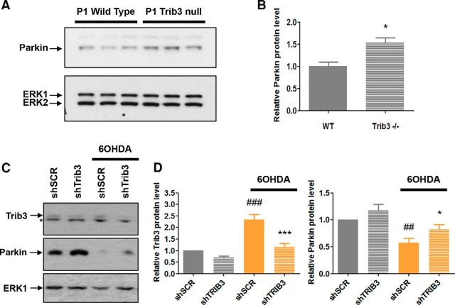
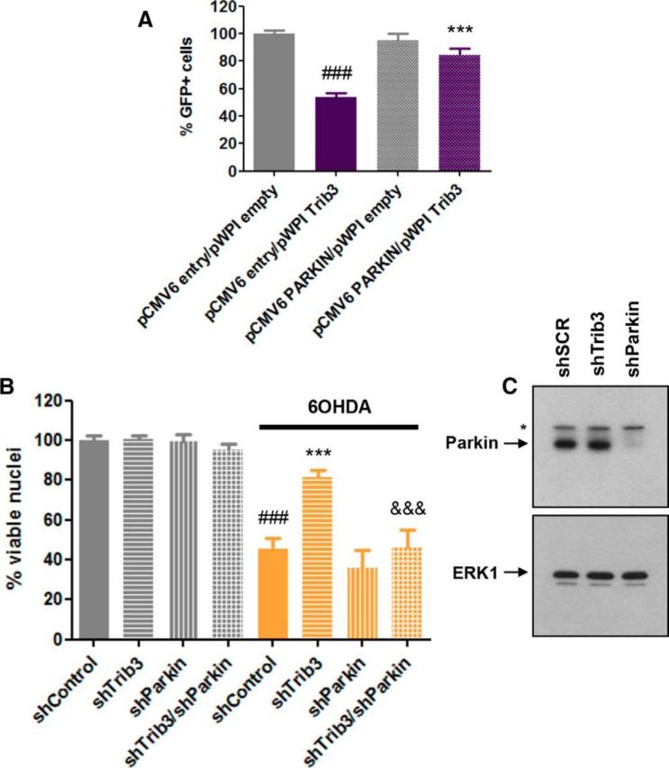
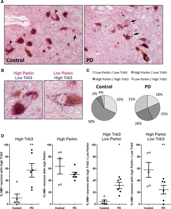
Parkinson's disease (PD) is characterized by the progressive loss of select neuronal populations, but the prodeath genes mediating the neurodegenerative processes remain to be fully elucidated. Trib3 (tribbles pseudokinase 3) is a stress-induced gene with proapoptotic activity that was previously described as highly activated at the transcriptional level in a 6-hydroxydopamine (6-OHDA) cellular model of PD. Here, we report that Trib3 immunostaining is elevated in dopaminergic neurons of the substantia nigra pars compacta (SNpc) of human PD patients. Trib3 protein is also upregulated in cellular models of PD, including neuronal PC12 cells and rat dopaminergic ventral midbrain neurons treated with 6-OHDA, 1-methyl-4-phenylpyridinium (MPP+), or α-synuclein fibrils (αSYN). In the toxin models, Trib3 induction is substantially mediated by the transcription factors CHOP and ATF4. Trib3 overexpression is sufficient to promote neuronal death; conversely, Trib3 knockdown protects neuronal PC12 cells as well as ventral midbrain dopaminergic neurons from 6-OHDA, MPP+, or αSYN. Mechanism studies revealed that Trib3 physically interacts with Parkin, a prosurvival protein whose loss of function is associated with PD. Elevated Trib3 reduces Parkin expression in cultured cells; and in the SNpc of PD patients, Parkin levels are reduced in a subset of dopaminergic neurons expressing high levels of Trib3. Loss of Parkin at least partially mediates the prodeath actions of Trib3 in that Parkin knockdown in cellular PD models abolishes the protective effect of Trib3 downregulation. Together, these findings identify Trib3 and its regulatory pathways as potential targets to suppress the progression of neuron death and degeneration in PD.

Significance statement: Parkinson's disease (PD) is the most common neurodegenerative movement disorder. Current treatments ameliorate symptoms, but not the underlying neuronal death. Understanding the core neurodegenerative processes in PD is a prerequisite for identifying new therapeutic targets and, ultimately, curing this disease. Here, we describe a novel pathway involving the proapoptotic protein Trib3 in neuronal death associated with PD. These findings are supported by data from multiple cellular models of PD and by immunostaining of postmortem PD brains. Upstream, Trib3 is induced by the transcription factors ATF4 and CHOP; and downstream, Trib3 interferes with the PD-associated prosurvival protein Parkin to mediate death. These findings establish this new pathway as a potential and promising therapeutic target for treatment of PD.

Keywords: ATF4; CHOP; Parkin; Parkinson's disease; Trib3; cell death.


Figures

Figure 1.
Figure 1.
Trib3 mRNA and protein are induced before cell death in cellular models of PD. A, Time course of cell death elicited by 6-OHDA and MPP+. Survival assay shows the remaining viable nuclei in neuronal PC12 cell cultures untreated (control) or treated with 100–150 μm 6-OHDA (left) or 1000 μm MPP+ (right) for 2, 8, 16, or 24 h. Cell death becomes evident by 16 h. **p < 0.005 (ANOVA with Student-Newman-Keuls tests). ***p < 0.0005 (ANOVA with Student-Newman-Keuls tests). Values are mean ± SEM from five independent experiments performed in triplicate. B, Trib3 mRNA is upregulated in cellular PD models before cell death. qPCR analysis of Trib3 mRNA levels in neuronal PC12 cells either untreated (control) or treated with 100–150 μm 6-OHDA (left) or 1000 μm MPP+ (right) for 2, 8, 16, and 24 h. 6-OHDA induces an increase in Trib3 mRNA starting at 8 h that is maintained at 16 and 24 h. ***p < 0.0005 (ANOVA with Student-Newman-Keuls tests). Trib3 mRNA level starts rising at 8 h and keeps increasing at 16 and 24 h with MPP+. **p < 0.005 (ANOVA with Student-Newman-Keuls tests). ***p < 0.0005 (ANOVA with Student-Newman-Keuls tests). Trib3 mRNA levels are normalized against those of α-tubulin and are mean ± SEM from at least three independent experiments performed in triplicate. C, Trib3 protein levels are elevated before cell death in cellular models of PD. Western blot quantification of Trib3 protein levels in neuronal PC12 cells untreated (control) or treated with 100–150 μm 6-OHDA or 1000 μm MPP+ for the indicated times. *p < 0.05 (ANOVA with Student-Newman-Keuls tests). Top portion of the panel shows representative Western blots. Trib3 corresponds to the top band; the bottom band noted with an asterisk is nonspecific. Densitometric quantification of Trib3 protein levels is shown at the bottom of the panel. Trib3 values are normalized against ERK1 and are mean ± SEM from at least six independent experiments.
Figure 2.
Figure 2.
6-OHDA, MPP+, and preformed α-synuclein fibrils induce death of cultured postnatally derived ventral midbrain dopaminergic neurons. A–C, Representative overview images of dopaminergic cells (positive for tyrosine hydroxylase (TH, white, left panels) and quantifications (right panels) show a decrease in the numbers of surviving dopaminergic (TH+) neurons after treatment with (A) 6-OHDA, (B) MPP+, or (C) preformed α-synuclein fibrils. *p < 0.05 (t tests). **p < 0.005 (t tests). A, B, Cultures were either untreated (control) or treated as indicated with 40 μm 6-OHDA in 0.015% ascorbic acid (AA + 6-OHDA) or 0.015% AA for 8 or 24 h, or with 40 μm MPP+ for 8 or 24 h and then immunostained for TH. C, Cultures were treated with PBS or 5 μg/ml of human WT α-synuclein-preformed fibrils (α-synuclein pffs) for 10–14 d and then immunostained for TH. Survival assay values are expressed as the mean ± SEM of the percentage of remaining TH+ neurons in cultures treated with 6-OHDA, MPP+, or α-synuclein, compared with the corresponding control conditions. Values were analyzed from at least three independent experiments performed in triplicate.
Figure 3.
Figure 3.
6-OHDA, MPP+, and preformed α-synuclein fibrils induce Trib3 expression in cultured postnatally derived ventral midbrain dopaminergic neurons. A–C, Representative images (left panels) and corresponding quantification (right panels) show an increase of Trib3 signal in dopaminergic cells (positive for TH) after treatment with (A) 6-OHDA, (B) MPP+, or (C) preformed α-synuclein fibrils. **p < 0.005 (t tests). ***p < 0.0005 (t tests). A, B, Cultures were either untreated (control) or treated as indicated with 40 μm MPP+ for 8 or 24 h; 0.015% ascorbic acid (AA) or 40 μm 6-OHDA in 0.015% AA (AA + 6-OHDA) for 8 or 24 h, and immunostained for TH (green) and Trib3 (red) expression. DAPI staining (blue) is shown as a nuclear marker. C, Cultures were treated with PBS or 5 μg/ml of human WT α-synuclein-preformed fibrils (α-synuclein pffs) for 10–14 d and then immunostained for phosphorylated α-synuclein (P-S129 α-syn, green) to show pathogenic intracellular α-synuclein aggregates, Trib3 (red) and TH (blue). To exemplify the diversity of α-synuclein aggregates induced by α-synuclein pffs treatment, images of two morphologically distinct dopaminergic neurons are shown in the two bottom rows. Trib3 relative protein levels correspond to the mean ± SEM of Trib3 densitometric signals measured in 40–85 individual neurons and expressed relative to the corresponding control conditions. Values were analyzed from 2 to 4 independent experiments performed in triplicate.
Figure 4.
Figure 4.
The proportion of Trib3+ dopaminergic neurons is increased in the substantia nigra of PD patients. A, Representative low-magnification images (4×) of postmortem midbrains from age-matched human control and PD patients immunostained for Trib3 (blue) and counterstained with Fast Red (pink) show basal expression of Trib3 in the substantia nigra (black dashed lines). Specificity of the Trib3 antibody was verified by a competition experiment performed by incubating the antibody with the corresponding immunizing peptide. Virtually no staining was observed in this control experiment. B, Representative high-magnification (40×) images of sections from the substantia nigra of a control and a PD patient brain. Dopaminergic neurons are identified by the presence of neuromelanin (NM) inclusions (brown). Examples of neurons with granular cytoplasmic Trib3 immunostaining are shown in the right panel (including insets). Black bar represents 25 μm. C, Quantification shows an increase in the percentage of NM+ neurons with Trib3 immunostaining in the substantia nigra of PD patients compared with control cases. ***p < 0.0005 (t test). These data are based on the observation of 8 control and 7 PD patients' brains. A total of 3923 NM+ neurons were scored in controls, and 1489 NM+ neurons were scored in PD cases.
Figure 5.
Figure 5.
Trib3 induces death of neuronal PC12 cells and postnatally derived ventral midbrain dopaminergic neurons. A, Left, Survival assay shows a decrease in the proportion of surviving neuronal PC12 cells after infection with a lentivirus carrying a Trib3-expressing vector (pWPI Trib3) for the indicated times (1, 3, or 7 d), compared with cells transduced with an empty control vector (pWPI empty). ***p < 0.005 (ANOVA with Dunnett's test). Right, Representative Western blot images show the level of Trib3 expression typically obtained during such experiments. B, Trib3 overexpression decreases survival of cultured postnatally derived ventral midbrain dopaminergic neurons. Left, Representative images of cultures infected with lentivirus expressing Trib3 and GFP (pWPI Trib3) or GFP alone (pWPI empty) and immunostained for TH (red) or GFP (green, lentiviral-infected cells) and stained with DAPI (blue, nuclear marker). Right, Corresponding quantifications of TH+-infected cells showing a decrease in the number of dopaminergic neurons (TH+/GFP+) infected with lentivirus carrying Trib3 for 6–9 d compared with dopaminergic neurons transduced with an empty control vector. ***p < 0.0005 (t test). Values are mean ± SEM and were obtained from at least three independent experiments done in triplicate.
Figure 6.
Figure 6.
Trib3 downregulation and Trib3 knock-out protect from cell death in PD cellular models. A, Representative Western blot images showing the extent of Trib3 knockdown obtained in neuronal PC12 cells treated with 150 μm 6-OHDA. Top band represents Trib3. Bottom band noted with an asterisk is nonspecific. B, Trib3 knockdown protects neuronal PC12 cells from death elicited by 6-OHDA and MPP+. Survival assay shows the proportion of surviving cells after treatment with 125–150 μm 6-OHDA (left) or 1000 μm MPP+ (right) for 24 h. Cells were infected as indicated with a control shRNA (against either DSRED, shDSRED) or a scrambled version of the shTRIB3 sequence (shSCR) or with an shRNA targeted against Trib3 (shTRIB3). C, Trib3 knockdown protects cultured rat ventral midbrain dopaminergic neurons from death elicited by 6-OHDA, MPP+, or preformed α-synuclein fibrils. Survival assays show the proportion of remaining rat dopaminergic (TH+) ventral midbrain neurons after treatment with 40 μm 6-OHDA for 24 h (left), 40 μm MPP+ for 24 h (middle), or 5 μg/ml of α-synuclein fibrils for 10 d (right). D, E, Dopaminergic (TH+) neurons cultured from Trib3−/− mouse ventral midbrain show decreased sensitivity to 6-OHDA and MPP+. D, Survival assays show the proportion of surviving wild-type (WT) and Trib3−/− mouse dopaminergic (TH+) ventral midbrain neurons after treatment for 24 h with 40 μm 6-OHDA (left) or 40 μm MPP+ (right). E, Representative images of TH+ neurons in cultures of WT and Trib3−/− ventral midbrain show that the absence of Trib3 protects processes from degeneration elicited by 6-OHDA and MPP+. A–D, Values are mean ± SEM from three or four independent experiments performed in triplicate. Multiple comparisons were performed using ANOVA with Student-Newman-Keuls post hoc tests: #Compared with untreated cells expressing a control shRNA or wild-type cells. *Compared with toxin-treated cells expressing a control shRNA or wild-type cells. *p < 0.05; **p < 0.005; ***p < 0.0005; ###p < 0.0005.
Figure 7.
Figure 7.
ATF4 and CHOP contribute to Trib3 induction by 6-OHDA and MPP+ in neuronal PC12 cells. A, ATF4 knockdown reduces induction of Trib3 mRNA in response to 6-OHDA and MPP+. Real-time qPCR analyses show the effects of lentivirally delivered ATF4 shRNA on levels of ATF4 mRNA (left) and Trib3 mRNA (right) after treatment with 150 μm 6-OHDA or 1000 μm MPP+ for 8 h. Control cultures were infected with a control shRNA (sh-MUTANT). B, CHOP knockdown reduces induction of Trib3 mRNA in response to 6-OHDA and MPP+. Real-time qPCR analyses show the effects of lentivirally delivered CHOP shRNA on levels of CHOP mRNA (left) and Trib3 mRNA (right) after treatment with 150 μm 6-OHDA or 1000 μm MPP+ for 8 h. Control cultures were infected with a control empty vector (empty). C, Combined knockdown of both ATF4 and CHOP does not enhance suppression of Trib3 mRNA induction by MPP+ over that achieved with knockdown of either alone. Experimental details are given in A and B, except that cells were infected with a mixture of both control vectors (control, empty vector, and sh-MUTANT), sh-ATF4, shCHOP, or both shRNAs (shATF4/shCHOP) as indicated. D, CHOP knockdown protects neuronal PC12 cells from 6-OHDA and MPP+. E, Knockdown of FOXO transcription factors does not significantly block Trib3 induction in PC12 cells treated with 6-OHDA. Neuronal PC12 cells were transduced with lentiviruses expressing either a control shRNA (shDSRED) or an shRNA targeted against FOXO family transcription factors (shFOXO) and treated with 6-OHDA. A–C, E, mRNA levels are normalized against α-tubulin or 18S rRNA. Values are mean ± SEM and were analyzed from at least three independent experiments done in duplicate or triplicate. Comparisons were performed using ANOVA with Student-Newman-Keuls post hoc tests: #Compared with untreated cells expressing a control shRNA. *Compared with toxin-treated cells expressing a control shRNA. *p < 0.05; **p < 0.005; ##p < 0.005; ***p < 0.0005; ###p < 0.0005.
Figure 8.
Figure 8.
Trib3 and Parkin physically interact in neuronal PC12 cells. Top, Short exposures. Bottom, Long exposures. Left panels, Western immunoblot images showing Trib3. Right panels, Parkin and ERK protein levels. Neuronal PC12 cells were transduced with an empty control vector (pWPI empty) or a Trib3-expressing vector (pWPI Trib3) for 48 h, and an immunoprecipitation (IP) experiment was performed with anti-Parkin antibody or control IgG.
Figure 9.
Figure 9.
Trib3 overexpression decreases Parkin levels. A, B, Trib3 overexpression decreases Parkin protein levels in neuronal PC12 cells. Representative Western blot images (A) and corresponding quantifications (B) of Trib3 (top) and Parkin (bottom) protein levels in neuronal PC12 cells transduced with an empty control vector (pWPI empty) or a Trib3-expressing vector (pWPI Trib3) for the indicated times (1, 3, or 7 d). Protein levels were quantified and normalized to ERK protein levels, and values are mean ± SEM from five independent experiments. Multiple comparisons were performed using ANOVA with Student-Newman-Keuls post hoc tests: **p < 0.005; ***p < 0.0005. C, qPCR analyses of Trib3 (left) and Parkin (right) mRNA levels in neuronal PC12 cells, transduced with pWPI empty or pWPI Trib3 for 1–2 d, shows that Trib3 overexpression does not affect Parkin mRNA levels. Trib3 and Parkin mRNA levels are normalized against 18S rRNA, and values are mean ± SEM and were analyzed from three independent experiments performed in triplicate. ***p < 0.0005 (t test). D, E, Trib3 overexpression reduces Parkin expression in cultured postnatally derived ventral midbrain dopaminergic neurons. D, Representative immunofluorescence images of cultured postnatally derived ventral midbrain dopaminergic neurons immunostained for TH (blue), GFP (green, lentiviral-infected cells), and Parkin (red) after 2 d of infection with pWPI empty or pWPI Trib3. E, Quantification of relative Parkin immunostaining signals in dopaminergic neurons expressing pWPI Trib3 for 1–4 d, compared with dopaminergic neurons expressing pWPI-empty. ***p < 0.0005 (t test). Trib3 relative protein levels correspond to the mean ± SEM of Trib3 densitometric signals measured in 45–58 individual neurons and normalized to the corresponding control conditions. Values were analyzed from three independent experiments done in triplicate.
Figure 10.
Figure 10.
Trib3 downregulation or deletion increases Parkin levels. A, B, Parkin level is increased in brains of Trib3-null mice. Representative Western immunoblot images (A) and corresponding quantifications (B) of Parkin protein levels in whole-brain lysates from postnatal day 1 (P1) wild-type and P1 Trib3-null mice. Values are normalized to ERK and expressed as mean ± SEM from 3 animals of each genotype. *p < 0.05 (t test). C, D, Trib3 knockdown suppresses the fall in Parkin levels that occurs in neuronal PC12 cells in response to 6-OHDA. C, Representative Western blot image showing Parkin and Trib3 protein levels in cells infected with lentivirus expressing either a control shRNA (shSCR) or shTrib3 and then treated for 8 h with 150 μm 6-OHDA. Top band represents Trib3. Bottom band noted with an asterisk is nonspecific. D, Quantification of relative Trib3 (left) and Parkin (right) protein levels under the conditions corresponding to those in C. Values are normalized to ERK and expressed as mean ± SEM from five independent experiments. #Compared with untreated cells expressing shControl. *Compared with cells treated with 6-OHDA and expressing shControl. ANOVA with Student-Newman-Keuls tests: *p < 0.05; ##p < 0.005; *** p < 0.0005; ###p < 0.0005.
Figure 11.
Figure 11.
Parkin overexpression protects from death caused by Trib3 overexpression, and Parkin knockdown abolishes the protective effect of Trib3 knockdown. A, Parkin overexpression protects neuronal PC12 cells from death caused by Trib3 overexpression. Data show the proportion of surviving GFP+ neuronal PC12 cells cotransfected with a Trib3-expressing vector (pWPI Trib3) and either a Parkin-expressing vector (pCMV6 Parkin) or an empty control vector (pWPI empty, pCMV6 entry). Values are mean ± SEM and were obtained from two independent experiments performed in quadruplicate. Multiple comparisons were performed using ANOVA with Student-Newman-Keuls post hoc tests. #Compared with cells cotransfected with pCMV6 entry and pWPI empty. *Compared with cells cotransfected with pCMV6 entry and pWPI TRIB3. ***p < 0.0005. ###p < 0.0005. B, Parkin knockdown abolishes the protective effect of Trib3 knockdown on 6-OHDA-treated neuronal PC12 cells. Replicate cultures of neuronal PC12 cells were infected with lentiviruses expressing a control shRNA (shControl), shTrib3, shParkin, or both shTrib3 and shParkin (shTrib3/shParkin) and then treated with 150 μm 6-OHDA for 24 h. Values show the relative proportion of viable cell nuclei after treatment for each condition and are mean ± SEM and were derived from three independent experiments performed in triplicate. Multiple comparisons were performed using ANOVA with Student-Newman-Keuls post hoc tests. #Compared with untreated cells expressing shControl. *Compared with cells treated with 6-OHDA and expressing shControl. &Compared with cells treated with 6-OHDA and expressing shTrib3. ***p < 0.0005. ###p < 0.0005. &&&p < 0.0005. C, Representative Western blot images showing the efficiency of Parkin knockdown in neuronal PC12 cells typically achieved by lentiviral-mediated delivery of shParkin used for these experiments. Parkin corresponds to the bottom band; the top band noted with an asterisk is nonspecific.
Figure 12.
Figure 12.
Trib3 and Parkin coexpression in the substantia nigra of control and PD patients. A, Representative images of dopaminergic neurons in postmortem midbrains from age-matched human control and PD patients immunostained for Trib3 (blue) and Parkin (violet). Dopaminergic neurons are identified by the presence of neuromelanin (NM) inclusions (brown). Arrows indicate NM+ neurons highly positive for Trib3 staining. Arrowheads indicate NM+ neurons highly positive for Parkin immunostaining. B, High-magnification images of two examples of dopaminergic neurons displaying high Parkin and low Trib3 levels (left) and low Parkin and high Trib3 levels (right). C, Pie charts showing the proportion of NM+ neurons with low Parkin/low Trib3, high Parkin/low Trib3, low Parkin/high Trib3, and high Parkin/high Trib3 staining observed in controls (left) and PD cases (right). D, Scatter plots showing, from left to right: the proportion of NM+ neurons with high Trib3, high Parkin, high Trib3/low Parkin, high Parkin/low Trib3 staining in controls and PD patients. *p < 0.05 (t tests). **p < 0.005 (t tests). These data are based on the blinded scoring of 5 control and 6 PD patients' brains. A total of 225 NM+ neurons were scored in controls, and 233 NM+ neurons were scored in PD cases.

Similar articles

Cited by

References

    1. Aynaud MM, Suspène R, Vidalain PO, Mussil B, Guétard D, Tangy F, Wain-Hobson S, Vartanian JP. Human Tribbles 3 protects nuclear DNA from cytidine deamination by APOBEC3A. J Biol Chem. 2012;287:39182–39192. doi: 10.1074/jbc.M112.372722. - DOI - PMC - PubMed
    1. Bi XP, Tan HW, Xing SS, Wang ZH, Tang MX, Zhang Y, Zhang W. Overexpression of TRB3 gene in adipose tissue of rats with high fructose-induced metabolic syndrome. Endocr J. 2008;55:747–752. doi: 10.1507/endocrj.K08E-049. - DOI - PubMed
    1. Braak H, Bohl JR, Müller CM, Rüb U, de Vos RA, Del Tredici K. Stanley Fahn Lecture 2005: the staging procedure for the inclusion body pathology associated with sporadic Parkinson's disease reconsidered. Mov Disord. 2006;21:2042–2051. doi: 10.1002/mds.21065. - DOI - PubMed
    1. Bromati CR, Lellis-Santos C, Yamanaka TS, Nogueira TC, Leonelli M, Caperuto LC, Gorjão R, Leite AR, Anhê GF, Bordin S. UPR induces transient burst of apoptosis in islets of early lactating rats through reduced AKT phosphorylation via ATF4/CHOP stimulation of TRB3 expression. Am J Physiol Regul Integr Comp Physiol. 2011;300:R92-R100. doi: 10.1152/ajpregu.00169.2010. - DOI - PubMed
    1. Carracedo A, Gironella M, Lorente M, Garcia S, Guzmán M, Velasco G, Iovanna JL. Cannabinoids induce apoptosis of pancreatic tumor cells via endoplasmic reticulum stress-related genes. Cancer Res. 2006;66:6748–6755. doi: 10.1158/0008-5472.CAN-06-0169. - DOI - PubMed

Publication types

MeSH terms