

Giant and thrombosed left ventricular aneurysm
- PMID: 26225205
- PMCID: PMC4513496
- DOI: 10.4330/wjc.v7.i7.431
Giant and thrombosed left ventricular aneurysm
Abstract
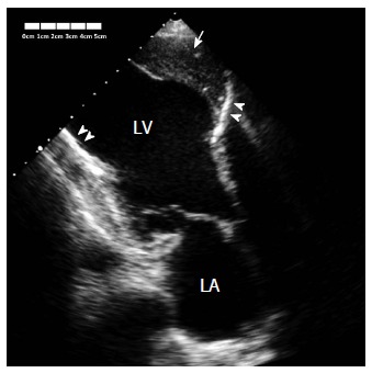
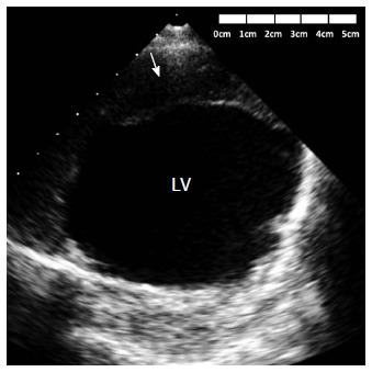
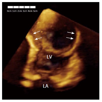
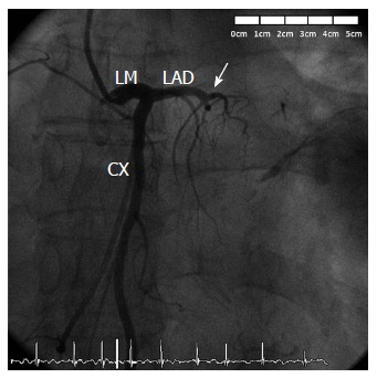
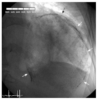
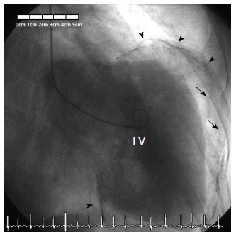
Left ventricular aneurysms are a frequent complication of acute extensive myocardial infarction and are most commonly located at the ventricular apex. A timely diagnosis is vital due to the serious complications that can occur, including heart failure, thromboembolism, or tachyarrhythmias. We report the case of a 78-year-old male with history of previous anterior myocardial infarction and currently under evaluation by chronic heart failure. Transthoracic echocardiogram revealed a huge thrombosed and calcified anteroapical left ventricular aneurysm. Coronary angiography demonstrated that the left anterior descending artery was chronically occluded, and revealed a big and spherical mass with calcified borders in the left hemithorax. Left ventriculogram confirmed that this spherical mass was a giant calcified left ventricular aneurysm, causing very severe left ventricular systolic dysfunction. The patient underwent cardioverter-defibrillator implantation for primary prevention.
Keywords: Coronary angiography; Coronary artery disease; Echocardiography; Left ventricular aneurysm; Myocardial infarction.
Figures
References
-
- Tikiz H, Atak R, Balbay Y, Genç Y, Kütük E. Left ventricular aneurysm formation after anterior myocardial infarction: clinical and angiographic determinants in 809 patients. Int J Cardiol. 2002;82:7–14; discussion 14-16. - PubMed
-
- Abrams DL, Edelist A, Luria MH, Miller AJ. Ventricular aneurysm. a reappraisal based on a study of sixty-five consecutive autopsied cases. Circulation. 1963;27:164–169. - PubMed
Publication types
LinkOut - more resources
Full Text Sources
Other Literature Sources
Miscellaneous
