

Desiccating Stress-Induced MMP Production and Activity Worsens Wound Healing in Alkali-Burned Corneas
- PMID: 26225631
- PMCID: PMC4525635
- DOI: 10.1167/iovs.15-16631
Desiccating Stress-Induced MMP Production and Activity Worsens Wound Healing in Alkali-Burned Corneas
Abstract
Purpose: To evaluate the effects of dry eye on ocular surface protease activity and sight threatening corneal complications following ocular surface chemical injury.
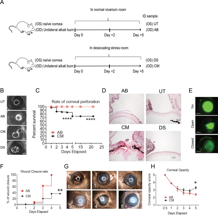
Methods: C57BL/6 mice were subjected to unilateral alkali burn (AB) with or without concomitant dry eye for 2 or 5 days. Mice were observed daily for appearance of corneal perforation. Whole corneas were harvested and lysed for RNA extraction. Quantitative real-time PCR was performed to measure expression of inflammation cytokines, matrix metalloproteinases (MMP). Matrix metalloproteinase-9 activity, gelatinase activity, and myeloperoxidase (MPO) activity were evaluated in corneal lysates. Presence of infiltrating neutrophils was evaluated by immunohistochemistry and flow cytometry.
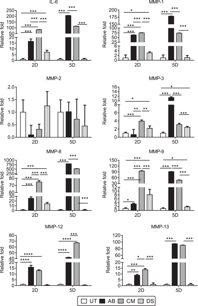
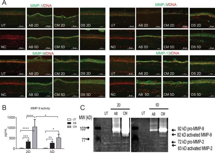
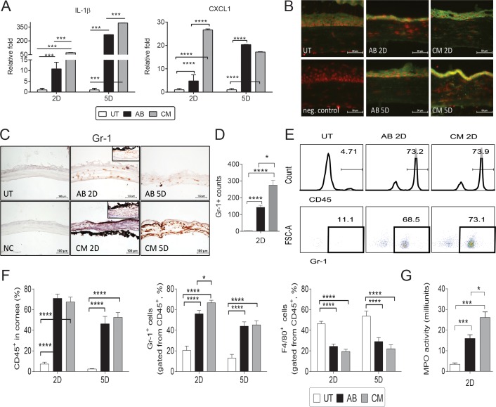
Results: Eyes subjected to the combined model of AB and dry eye (CM) had 20% sterile corneal perforation rate as soon as 1 day after the initial injury, which increased to 35% by 5 days, delayed wound closure and increased corneal opacity. Increased levels of IL-1β, -6, and MMPs-1, -3, -8, -9, and -13, and chemokine (C-X-C motif) ligand 1 (CSCL1) transcripts were found after 2 days in CM compared with AB corneas. Increased MMP-1, -3, -9, and -13 immunoreactivity and gelatinolytic activity were seen in CM corneas compared with AB. Increased neutrophil infiltration and MPO activity was noted in the CM group compared with AB 2 days post injury.
Conclusions: Desiccating stress worsens outcome of ocular AB, creating a cytokine and protease storm with greater neutrophil infiltration, increasing the risk of corneal perforation.
Figures
Similar articles
-
Differential Effects of Dexamethasone and Doxycycline on Inflammation and MMP Production in Murine Alkali-Burned Corneas Associated with Dry Eye.Ocul Surf. 2016 Apr;14(2):242-54. doi: 10.1016/j.jtos.2015.11.006. Epub 2016 Jan 6. Ocul Surf. 2016. PMID: 26772899 Free PMC article.
-
Dexamethasone Drug Eluting Nanowafers Control Inflammation in Alkali-Burned Corneas Associated With Dry Eye.Invest Ophthalmol Vis Sci. 2016 Jun 1;57(7):3222-30. doi: 10.1167/iovs.16-19074. Invest Ophthalmol Vis Sci. 2016. PMID: 27327581 Free PMC article.
-
LRG1 facilitates corneal fibrotic response by inducing neutrophil chemotaxis via Stat3 signaling in alkali-burned mouse corneas.Am J Physiol Cell Physiol. 2021 Sep 1;321(3):C415-C428. doi: 10.1152/ajpcell.00517.2020. Epub 2021 Jul 14. Am J Physiol Cell Physiol. 2021. PMID: 34260299
-
Accelerated wound healing of alkali-burned corneas in MRL mice is associated with a reduced inflammatory signature.Invest Ophthalmol Vis Sci. 2005 Nov;46(11):4097-106. doi: 10.1167/iovs.05-0548. Invest Ophthalmol Vis Sci. 2005. PMID: 16249486
-
MMP-8 Is Critical for Dexamethasone Therapy in Alkali-Burned Corneas Under Dry Eye Conditions.J Cell Physiol. 2016 Nov;231(11):2506-16. doi: 10.1002/jcp.25364. Epub 2016 Mar 28. J Cell Physiol. 2016. PMID: 26923552 Free PMC article.
Cited by
-
Alarmins from conjunctival fibroblasts up-regulate matrix metalloproteinases in corneal fibroblasts.Int J Ophthalmol. 2020 Jul 18;13(7):1031-1038. doi: 10.18240/ijo.2020.07.03. eCollection 2020. Int J Ophthalmol. 2020. PMID: 32685388 Free PMC article.
-
Short ragweed pollen promotes M2 macrophage polarization via TSLP/TSLPR/OX40L signaling in allergic inflammation.Mucosal Immunol. 2019 Sep;12(5):1141-1149. doi: 10.1038/s41385-019-0187-8. Epub 2019 Jul 26. Mucosal Immunol. 2019. PMID: 31350466 Free PMC article.
-
Relationship between dry eye and expressions of CXCR3 and CCR5 after ocular acid burn.BMC Ophthalmol. 2022 Dec 15;22(1):489. doi: 10.1186/s12886-022-02678-3. BMC Ophthalmol. 2022. PMID: 36522768 Free PMC article.
-
Tissue-derived microparticles reduce inflammation and fibrosis in cornea wounds.Acta Biomater. 2019 Feb;85:192-202. doi: 10.1016/j.actbio.2018.12.027. Epub 2018 Dec 19. Acta Biomater. 2019. PMID: 30579044 Free PMC article.
-
Differential Effects of Dexamethasone and Doxycycline on Inflammation and MMP Production in Murine Alkali-Burned Corneas Associated with Dry Eye.Ocul Surf. 2016 Apr;14(2):242-54. doi: 10.1016/j.jtos.2015.11.006. Epub 2016 Jan 6. Ocul Surf. 2016. PMID: 26772899 Free PMC article.
References
-
- Ward DL,, Gorie C. Occupational eye injuries in soldiers. J Occup Med. 1991; 33: 646–650. - PubMed
-
- Lau JJ,, Thach AB,, Burden JH,, Ward TP,, Hshieh PB,, Hollifield RD. Eye injuries in the U.S. Armed Forces. Mil Med. 2000; 165: 683–686. - PubMed
-
- Ari AB. Eye injuries on the battlefields of Iraq and Afghanistan: public health implications. Optometry. 2006; 77: 329–339. - PubMed
-
- Buglisi JA,, Knoop KJ,, Levsky ME,, Euwema M. Experience with bandage contact lenses for the treatment of corneal abrasions in a combat environment. Mil Med. 2007; 172: 411–413. - PubMed
-
- Seet B,, Wong TY. Military laser weapons: current controversies. Ophthalmic Epidemiol. 2001; 8: 215–226. - PubMed
Publication types
MeSH terms
Substances
Grants and funding
LinkOut - more resources
Full Text Sources
Other Literature Sources
Research Materials
Miscellaneous
