Review
. 2015 Dec;63(6):1491-501.
doi: 10.1016/j.jhep.2015.07.008. Epub 2015 Jul 28.

Emerging evidence on the role of the Hippo/YAP pathway in liver physiology and cancer

Affiliations
Review

Emerging evidence on the role of the Hippo/YAP pathway in liver physiology and cancer

Dean Yimlamai et al. J Hepatol. 2015 Dec.

Abstract

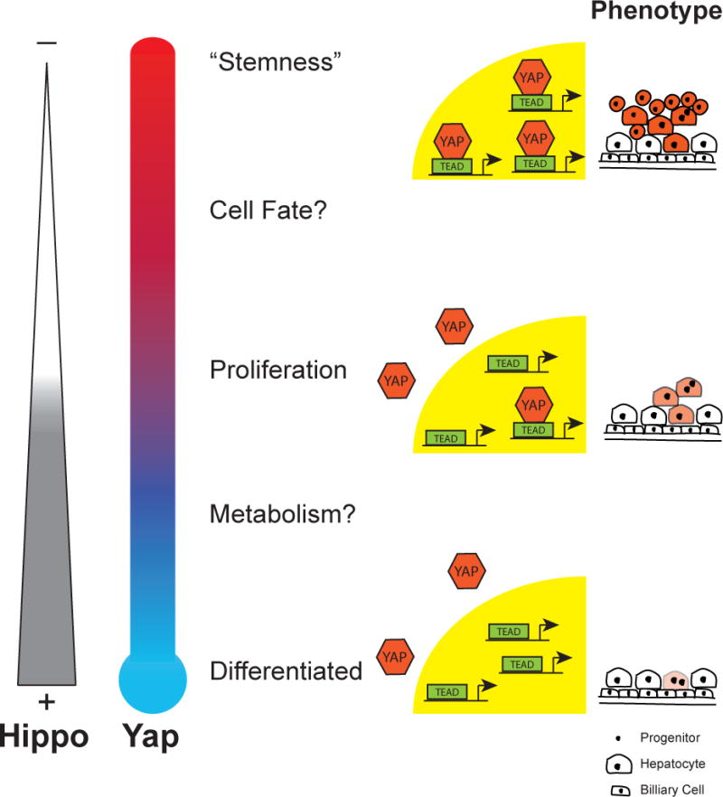
The Hippo pathway and its regulatory target, YAP, has recently emerged as an important biochemical signaling pathway that tightly governs epithelial tissue growth. Initially defined in Drosophilia, this pathway has shown remarkable conservation in vertebrate systems with many components of the Hippo/YAP pathway showing biochemical and functional conservation. The liver is particularly sensitive to changes in Hippo/YAP signaling with rapid increases in liver size becoming manifest on the order of days to weeks after perturbation. The first identified direct targets of Hippo/YAP signaling were pro-proliferative and anti-apoptotic gene programs, but recent work has now implicated this pathway in cell fate choice, stem cell maintenance/renewal, epithelial to mesenchymal transition, and oncogenesis. The mechanisms by which Hippo/YAP signaling is changed endogenously are beginning to come to light as well as how this pathway interacts with other signaling pathways, and important details for designing new therapeutic interventions. This review focuses on the known roles for Hippo/YAP signaling in the liver and promising avenues for future study.

Keywords: Hippo signaling; Liver regeneration; Stem cell biology.


Figures

Figure 1
Figure 1. The Hippo pathway in Drosophila and Mammals
Schematic of a subset of the Hippo pathway with known roles in the liver. The primary activity of the Hippo pathway is to restrict yorkie (YAP in mammals) to the cytoplasm. Reduced activity of the Hippo pathway results in translocation of yorkie to the nucleus where it binds to the TEAD family of transcription factors to upregulate gene expression. Known target genes of the pathway in Drosophila and Mammals are indicated after the arrowhead in the nucleus. Known roles for the pathway are indicated below the diagram of the transcriptional element. A. The Hippo pathway in Drosophila. Colored shapes represent known effectors of the Hippo pathway. Selected effectors have homologous partners in mammals that are associated with liver proliferation/cancer. These are color/shape-matched for clarity. B. The Hippo pathway in Mammals. Colored shapes represent known effectors of liver proliferation/cancer in the mammals. Open/dotted shapes represent potential/expected partners of the Hippo pathway that have yet to be functionally demonstrated in the liver, but have evidence from other tissues for their interaction.
Figure 2
Figure 2. The Effect of Liver YAP Expression
A. 2 weeks – Gross morphology of littermate control and YAP S127A overexpressing livers 2 weeks after high yield YAP induction in the adult. 5 month – Gross morphology of littermate control and a YAP S127A overexpressing liver with tumors (arrowheads) 5 months after low yield induction in the adult. H&E slides show histology of a highly undifferentiated HCC in the YAP overexpressing liver (dotted line). B. Linear versus Parallel models of YAP modulation. Colored shapes represent known effectors of the Hippo Pathway that directly modulate YAP expression. They are arranged in the two proposed models of YAP regulation, linear and parallel [22]. C. Sox9 staining demonstrates differential hepatocyte response to YAP S127A expression in periportal (PP) as compared to central venous (CV) areas 2 weeks after induction. Dashed boxes are enlarged in pictures noted below. Dashed lines in the PP/CV pictures indicate the borders of the blood vessel [23].
Figure 3
Figure 3. Hippo/YAP Levels Mediate Cellular Behavior
Hippo signaling and YAP activity is inversely related and determines particular cellular processes at various levels. High Hippo signaling activity is correlated with low levels of YAP protein and cytoplasmic YAP. Low Hippo activity is associated with high levels of YAP and a propensity for it to be localized to the nucleus. Proposed activities for varying levels of Hippo/YAP are depicted next to the YAP “thermometer”. At the far right, animations represent the proliferation and dedifferentiation of cells associated with the various levels of YAP expression. Darker shading suggests increased YAP expression.
Figure 4
Figure 4. Known Mutations in Hippo/YAP signaling in Liver Cancer
Colored shapes represent the known effectors of the Hippo pathway that, when knocked out or overexpressed, are associated with the development of the indicated form of cancer. References to the papers are indicated.

References

    1. Wu S, Huang J, Dong J, Pan D. hippo encodes a Ste-20 family protein kinase that restricts cell proliferation and promotes apoptosis in conjunction with salvador and warts. Cell. 2003;114:445–456. - PubMed
    1. Justice RW, Zilian O, Woods DF, Noll M, Bryant PJ. The Drosophila tumor suppressor gene warts encodes a homolog of human myotonic dystrophy kinase and is required for the control of cell shape and proliferation. Genes Dev. 1995;9:534–546. - PubMed
    1. Tapon N, Harvey KF, Bell DW, Wahrer DC, Schiripo TA, Haber D, et al. salvador Promotes both cell cycle exit and apoptosis in Drosophila and is mutated in human cancer cell lines. Cell. 2002;110:467–478. - PubMed
    1. Pan D. The Hippo Signaling Pathway in Development and Cancer. Developmental cell. 2010;19:491–505. - PMC - PubMed
    1. Zhao B, Lei Q-Y, Guan K-L. The Hippo-YAP pathway: new connections between regulation of organ size and cancer. Current opinion in cell biology. 2008;20:638–646. - PMC - PubMed

Publication types

MeSH terms