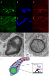
Absence of Dystrophin Related Protein-2 disrupts Cajal bands in a patient with Charcot-Marie-Tooth disease
- PMID: 26227883
- PMCID: PMC4920059
- DOI: 10.1016/j.nmd.2015.07.001
Absence of Dystrophin Related Protein-2 disrupts Cajal bands in a patient with Charcot-Marie-Tooth disease
Abstract
Using exome sequencing in an individual with Charcot-Marie-Tooth disease (CMT) we have identified a mutation in the X-linked dystrophin-related protein 2 (DRP2) gene. A 60-year-old gentleman presented to our clinic and underwent clinical, electrophysiological and skin biopsy studies. The patient had clinical features of a length dependent sensorimotor neuropathy with an age of onset of 50 years. Neurophysiology revealed prolonged latencies with intermediate conduction velocities but no conduction block or temporal dispersion. A panel of 23 disease causing genes was sequenced and ultimately was uninformative. Whole exome sequencing revealed a stop mutation in DRP2, c.805C>T (Q269*). DRP2 interacts with periaxin and dystroglycan to form the periaxin-DRP2-dystroglycan complex which plays a role in the maintenance of the well-characterized Cajal bands of myelinating Schwann cells. Skin biopsies from our patient revealed a lack of DRP2 in myelinated dermal nerves by immunofluorescence. Furthermore electron microscopy failed to identify Cajal bands in the patient's dermal myelinated axons in keeping with ultrastructural pathology seen in the Drp2 knockout mouse. Both the electrophysiologic and dermal nerve twig pathology support the interpretation that this patient's DRP2 mutation causes characteristic morphological abnormalities recapitulating the Drp2 knockout model and potentially represents a novel genetic cause of CMT.
Keywords: Charcot–Marie–Tooth disease; Hereditary motor and sensory neuropathy; Myelin; Nerve conduction studies; Whole exome sequencing.
Copyright © 2015 Elsevier B.V. All rights reserved.
Figures


References
-
- Sherman DL, Fabrizi C, Gillespie CS, Brophy PJ. Specific disruption of a schwann cell dystrophin-related protein complex in a demyelinating neuropathy. Neuron. 2001;30:677–87. - PubMed
-
- Nageotte J. Les étranglements de Ranvier et les espaces interannulaires des fibres nerveuses a myéline. Comptes Rendus de l'Association des Anatomistes. 1910:15.
-
- Nemiloff A. Über die Beziehung der sog. “Zellen der Schwannschen Scheide” zum Myelin in den Nervenfasen von Säugetieren. Archiv für Mikroskopische Anatomie und Entwicklungsgeschichte. 1910:19.
-
- Ramόn y Cajal S. El aparato endocelular de Golgi de la célula de Schwann y algunas observaciones sobre la estructura de los tubos nerviosos. Trabajos del Laboratorio de Investigaciones Biolόgicas de la Universidad de Madrid. 1912;10:26.
-
- Guilbot A, Williams A, Ravise N, et al. A mutation in periaxin is responsible for CMT4F, an autosomal recessive form of Charcot-Marie-Tooth disease. Human molecular genetics. 2001;10:415. - PubMed
Publication types
MeSH terms
Substances
Grants and funding
LinkOut - more resources
Full Text Sources
Other Literature Sources
Medical
Miscellaneous

